Model of Allergic Asthma
Nilesh Dharajiya1, Swapnil V. Vaidya2, Hiroki Murai2, Victor Cardenas2, Alexander Kurosky3, Istvan Boldogh4, Sanjiv A. Sur2*
1National Heart, Lung, and Blood Institute (NHLBI) Proteomics Center, Department of Biochemistry and Molecular Biology, University of Texas Medical Branch, Galveston, Texas, United States of America,2Department of Internal Medicine, University of Texas Medical Branch, Galveston, Texas, United States of America,3Department of Biochemistry and Molecular Biology, University of Texas Medical Branch, Galveston, Texas, United States of America,4Department of Microbiology, University of Texas Medical Branch, Galveston, Texas, United States of America
Abstract
Allergic asthma is characterized by airway eosinophilia, increased mucin production and allergen-specific IgE. Fc gamma receptor IIb (FccRIIb), an inhibitory IgG receptor, has recently emerged as a negative regulator of allergic diseases like anaphylaxis and allergic rhinitis. However, no studies to date have evaluated its role in allergic asthma. Our main objective was to study the role of FccRIIb in allergic lung inflammation. We used a murine model of allergic airway inflammation. Inflammation was quantified by BAL inflammatory cells and airway mucin production. FccRIIb expression was measured by qPCR and flow cytometry and the cytokines were quantified by ELISA. Compared to wild type animals, FccRIIb deficient mice mount a vigorous allergic lung inflammation characterized by increased bronchoalveolar lavage fluid cellularity, eosinophilia and mucin content upon ragweed extract (RWE) challenge. RWE challenge in sensitized mice upregulated FccRIIb in the lungs. Disruption of IFN-cgene abrogated this upregulation. Treatment of naı¨ve mice with the Th1-inducing agent CpG DNA increased FccRIIb expression in the lungs. Furthermore, treatment of sensitized mice with CpG DNA prior to RWE challenge induced greater upregulation of FccRIIb than RWE challenge alone. These observations indicated that RWE challenge upregulated FccRIIb in the lungs by IFN-c- and Th1-dependent mechanisms. RWE challenge upregulated FccRIIb on pulmonary CD14+/MHC II+mononuclear cells and CD11c+cells. FccRIIb deficient mice also exhibited an exaggerated RWE-specific IgE response upon sensitization when compared to wild type mice. We propose that FccRIIb physiologically regulates allergic airway inflammation by two mechanisms: 1) allergen challenge mediates upregulation of FccRIIb on pulmonary CD14+/MHC II+mononuclear cells and CD11c+cells by an IFN-cdependent mechanism; and 2) by attenuating the allergen specific IgE response during sensitization. Thus, stimulating FccRIIb may be a therapeutic strategy in allergic airway disorders.
Citation:Dharajiya N, Vaidya SV, Murai H, Cardenas V, Kurosky A, et al. (2010) FccRIIb Inhibits Allergic Lung Inflammation in a Murine Model of Allergic Asthma. PLoS ONE 5(2): e9337. doi:10.1371/journal.pone.0009337
Editor:Derya Unutmaz, New York University, United States of America
ReceivedNovember 12, 2009;AcceptedJanuary 25, 2010;PublishedFebruary 22, 2010
Copyright:ß2010 Dharajiya et al. This is an open-access article distributed under the terms of the Creative Commons Attribution License, which permits unrestricted use, distribution, and reproduction in any medium, provided the original author and source are credited.
Funding:This work was supported by grants from National Institutes of Health (NIH) RO1HL071163 (S.S.), National Institute of Allergy and Infectious Disease (NIAID), PO1AI062885-01 (I.B., S.S.), National Heart, Lung, and Blood Institute (NHLBI) Proteomics Initiative, NO1HV-28184 (S.S.) and National Institute of Environmental Health Sciences (NIEHS) Center Grant, EOS 006677 (I.B., S.S.). The funders had no role in study design, data collection and analysis, decision to publish or preparation of manuscript.
Competing Interests:The authors have declared that no competing interests exist. * E-mail: sasur@utmb.edu
Introduction
Allergic asthma is an airway inflammatory disease that is characterized by bronchial hyper-responsiveness, airway eosino-philia, goblet cell hyperplasia and production of allergen specific IgE. Cross-linking of the high affinity IgE receptor (FceRI) on mast cells by IgE, in the presence of allergen activates Btk, PLC-gamma and PI3K [1–4]. This ultimately leads to production and release of pro-inflammatory substances like histamine, leukotrienes and cytokines that promote allergic inflammation. In addition, cytokines produced by allergen specific Th2 cells such as IL4, IL5, IL9, IL13 and IL25 also promote allergen-specific IgE production and allergic airway inflammation [5–14].
There is considerable amount of data on pro-inflammatory mediators that contribute to the development of allergic inflammation. However, relatively little is known about negative regulatory mechanisms that attenuate allergic inflammation.
few studies have shown a regulatory role of this receptor in animal models of allergic diseases. One study showed that IgG antibodies can prevent IgE mediated anaphylaxisin vivothrough both antigen
interception and FccRIIb cross-linking [33]. Another study demonstrated a regulatory role of FccRIIb in a murine model of allergic rhinitis[34]. However, the role of this receptor in allergic lung inflammation has not been elucidated.
We recently showed in a gene micro-array analysis (GEO accession number GSE18083) that allergen challenge upregulated 352 genes in the lungs four hours after the challenge [35]. Careful review of that list revealed FccRIIb as one of the genes upregulated. Based on this observation, we hypothesized that FccRIIb may play a regulatory role in allergic airway inflamma-tion. Here we show that mice lacking FccRIIb have exaggerated allergic airway inflammation, suggesting its negative regulatory role in asthma. We further show that allergen challenge upregulates FccRIIb in the lungs in an IFN-c dependent mechanism. Our results indicate that FccRIIb upregulation physiologically reduces allergic airway inflammation.
Materials and Methods
Ethics Statement
All animal experiments were approved by the Institutional Animal Care and Use Committee of the University of Texas Medical Branch at Galveston.
Mice
Female BALB/c mice, 6–8 wk old, were purchased from the Harlan Laboratories (Indianapolis, IN). BALB/c IFN-c KO, C57Bl6 FccRIIb knock-out (KO) and C57Bl6 WT mice were purchased from Jackson laboratories (Bar Harbor, Maine). BALB/ c FccRIIb knock-out (KO) mice were purchased from Taconics (Albany, NY). All mice were maintained in a specific pathogen-free environment throughout the experiment.
Model of Allergic Sensitization and Challenge
WT Balb/c, IFN-cKO or FccRIIb KO mice were sensitized by two intraperitoneal (i.p.) injections of endotoxin-free RWE (150mg) and alum on days 0 and 4. On day 11, allergen challenge was performed by intranasal (i.n.) instillation of RWE (200mg) in
anesthetized mice. Mice were sacrificed at various time points as indicated after the challenge and bronchoalveolar lavage (BAL) fluid, blood, lung and spleen specimens were collected. Mice sensitized but not challenged served as the zero time point. For the Th1/CpG experiments, 35mg CpG or GpC oligonucleotides were administered intranasally in 50ml of sterile PBS [36].
Ragweed Extract
We have previously shown that endotoxin contamination alters the inflammatory cell recruitment following allergen challenge [37]. To avoid this problem endotoxin-free ragweed (lot XP56-D10-1320) was purchased from Greer Laboratories (Lenoir, NC).
Measurement of Allergic Airway Inflammation
For BAL fluid analyses, mice were anesthetized with an i.p. injection of ketamine and xylazine, tracheotomy performed and the trachea was cannulated. BAL of both lungs was performed twice with 0.7 ml of sterile PBS (pH 7.3) through the tracheal cannula with a syringe. Total cell counts were performed on BAL samples and differential cell counts were done on cytocentrifuge preparations (Cytospin 3; Thermo Shandon) stained with Wright-Giemsa, counting 200 cells from each animal. Mucin was quantified using mucin-binding lectin Jacalin (Calbiochem, La
jolla, CA) as described previously [38]. Aliquots of BAL fluid were diluted 1:100, 1:1000 and 1:10000, added in triplicate to individual wells of microtiter ELISA plates and incubated for 2 h at room temperature. Plates were washed and blocked with 5% BSA and 0.02% biotinylated jacalin was added. After 1 h incubation at room temperature, plates were washed extensively, then developed with alkaline phosphatase-conjugated avidin (Sigma) and nitrophenylphosphate (Sigma) and quantified by comparison with a mucin (Sigma) standard curve. The morpho-metric method we described previously was used to quantify mucin in lung epithelium[39]. Briefly, coronal sections of the 4% paraformaldehyde-fixed lungs were stained with PAS stain. Morphometirc analysis was done using MetamorphTM software (Version 5, Universal Imaging, Downingtown, Pennsylvania). Several images from five different levels per lung (three animals per group) were obtained and reassembled using the montage stage stitching algorithm of the MetamorphTM software. The integrated morphometric analysis function was used to transform total pixel area of the signal to mm2 per mm of peribronchial diameter.
Measurement of Enhanced Pause Index (PENH Index)
PENH was assessed by a method previously described [40] using a dual chamber whole body plethysmograph obtained from Buxco (Troy, NY). Mice were exposed for 3 min to nebulized PBS and subsequently to 37.5 mg/ml nebulized methacholine (Sigma Chemicals) in PBS using the AeroSonic ultrasonic nebulizer. After each nebulization, recordings were taken for 4 min. The PENH values measured during each 4 min sequence were averaged and expressed as the percentage of baseline PENH values after PBS exposure.
Quantification of Serum RWE-Specific IgE
Serum was collected from RWE-sensitized WT and FccRIIb KO mice 4 h after challenge with PBS (WT PBS and KO PBS) or RWE (WT RWE and KO RWE). RWE-specific IgE was quantified using standard sandwich ELISA technique and comparison with an IgE standard curve as described previous-ly[41].
Antigen Recall Assay
Splenocytes were obtained from sensitized WT and FccRIIb KO mice after crushing the spleens and making single cell suspensions. These were incubated with or without RWE for 3 d and Th2 cytokines (IL-4, IL-5, IL-9 and IL-13) were quantified in the supernatants using standard ELISA techniques as described previously [36,41,42].
Quantitative RT-PCR
Balb/c mice sensitized with RWE were challenged with either RWE or PBS. Mice were sacrificed and lungs collected at 1, 4, 24, 72 and 240 h post-challenge. RNA was extracted and quantitative PCR analyses were performed using the SYBR green real time PCR kit (Applied Biosystems) as described previously [35,43]. Transcript copy numbers for FccRIIb and beta-actin were quantified by comparing to a standard curve generated from serial log-dilutions of the target DNA [44,45]. FccRIIb signal was normalized to beta-actin. Table 1 shows the primers used.
Flow Cytometry
expression of FccRIIb on dendritic cells, 16106 cells were incubated with anti-CD11c PE (Pharmingen, Clone HL3) and anti-CD16/CD32-biotin (Pharmingen, Clone 2.4G2) for 30 min on ice protected from light. After three washes, cells were incubated with Streptavidin Cy-chrome (Pharmingen,#554062). Species and isotype matched antibodies were used as controls. FACS analysis was performed using analytical Flow cytometer (FACS Scan, Beckton Dickinson) with CellQuest software (San jose, CA). Further analyses were performed using FlowJo software (Tree Star Inc., Ashland, OR). Similarly, FccRIIb expression on macrophages (CD14; Clone rmC5-3, Pharmingen and anti-MHC class II-FITC; Miltenyi biotech,#130-081-601) and B cells (anti-B220, Clone RA3-6B2, Pharmingen) was studied.
Statistical Methods
There were 4–6 animals in each group and results are representative of at least two independent experiments. Statistical significance between groups was determined using Student’s T test.
Results
Disruption of the FccRIIb Gene Augments Allergic Airway Inflammation
We assessed the biological role of FccRIIb in a murine model of allergic asthma. C57Bl6 wild type (WT) and C57Bl6 FccRIIb knock-out (KO) mice were sensitized and then challenged with RWE. RWE challenge in WT mice recruited 3-fold more total inflammatory cells, 10-fold more eosinophils, 2-fold more
lymphocytes and macrophages (Figure 1A, 1B, 1C and 1D). Disrupting the FccRIIb gene further increased total inflammatory cells (5-fold), eosinophils (12-fold), lymphocytes (5-fold) and macrophages (3.6-fold) in the BAL (Figure 1A, 1B, 1C and 1D). To determine the reproducibility of this result in a different strain of mouse, we repeated this experiment in Balb/c mice. RWE challenge in WT Balb/c mice recruited 3-fold more total inflammatory cells, 32-fold more eosinophils and 3-fold more lymphocytes in BAL as compared to PBS challenge (Figure 2A, 2B and 2C) at 72 h post-challenge. Similar to our observations in C57Bl6 mice, RWE challenge in Balb/c FccRIIb KO mice further increased total cells (2.3-fold increase), eosinophils (5.2-fold increase) and lymphocytes (2-fold increase) in BAL fluid as compared to WT mice (Figure 2A, 2B and 2C). RWE challenge in WT mice increased mucin-containing cells in the airway (Figures 2D, 2E and 2G) and mucin levels in BAL fluid (Figure 2H). RWE challenge in mice that lacked FccRIIb further increased mucin-containing cells in the airway (Figures 2E, 2F and 2G) and mucin levels in BAL fluid (Figure 2H) as compared to WT mice. RWE challenge in mice that lacked FccRIIb induced greater increase in enhanced pause (PENH) index as compared to wild type mice (Figure 2I).
RWE Challenge Upregulates FccRIIb in the Lungs by an IFN-c-Dependent Mechanism
Since allergen challenge recruits inflammatory cells that express FccRIIb to the lungs, and lack of FccRIIb further increases this inflammation, we hypothesized that allergen challenge upregulates FccRIIb on pulmonary inflammatory cells. Quantitative PCR of lung mRNA confirmed that RWE challenge upregulated FccRIIb as early as 4 hours post-RWE challenge, and gene expression peaked at 24 h (Figure 3A). This upregulation was sustained till 10 d after challenge (Figure 3A). Prior studies have shown that IFN-c and Th1 response can inhibit allergic inflammation [39,47–50]. Since our studies suggested that FccRIIb inhibited allergic airway inflammation, we sought to determine whether its upregulation was Th1 or IFN-cdependent. RWE challenge upregulated FccRIIb in wild type mice but not in IFN-cKO mice (Figure 3B). IFN-c KO mice also exhibited greater allergic
Table 1.Primers used for quantitative PCR analyses.
Gene Forwad Primer Reverse Primer
b-actin ACACCTTCTACAATGAGCTG GGATCTTCATGAGGTAGTCC
FccRIIb ATCTTGCTGCTGGGACTCAT TGACTGTGGCCTTAAACGTG
doi:10.1371/journal.pone.0009337.t001
Figure 1. Role of FccRIIb in allergic airway inflammation.(A, B, C and D) Total inflammatory cells (A), eosinophils (B), macrophages (C) and
lymphocytes (D) were quantified in BAL of C57Bl6 RWE-sensitized WT and FccRIIb KO mice challenged with either PBS (WT PBS and KO PBS) or RWE (WT RWE and KO RWE). *, p,0.05.
airway inflammation when compared to WT mice (data not shown). Treatment of naı¨ve wild type mice with the Th1-inducing CpG DNA significantly upregulated FccRIIb; however, GpC control DNA (which does not induce IFN-c) failed to do so (Figure 3C). Furthermore, intra-nasal administration of CpG DNA, but not GpC DNA, 48 h prior to RWE challenge in wild type mice enhanced RWE-induced FccRIIb upregulation (Figure 3D). These findings indicated that RWE-challenge upregulated FccRIIb by an IFN-cand Th1-dependent mechanism.
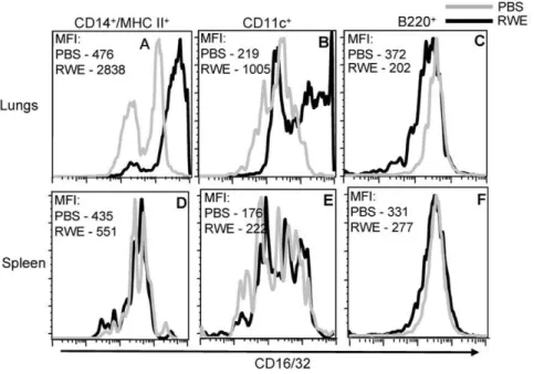
RWE Challenge Upregulates FccRIIb in CD14+MHC
II+Mononuclear Cells and CD11c+Cells in the Lungs
We verified the upregulation of FccRIIb in the lungs by flow cytometry measurements of single cell suspensions of whole lungs. RWE challenge upregulated FccRIIb on pulmonary CD14+/MHC II+ cells (Figure 4A) and on CD11c+ cells (Figure 4B), but not on B220+ cells (Figure 4C). Further-more, intrapulmonary RWE challenge failed to upregulate FccRIIb expression on these cells in the spleen (Figures 4D, 4E and 4F). This suggested that RWE challenge upregulated
FccRIIb expression on CD14+/MHC II+ and CD11c+ cells in the challenged organ (lungs) with no detectable systemic upregulation.
Disruption of the FccRIIb Gene Augments Serum RWE-Specific IgE Levels after Antigen Sensitization, but Does Not Affect Th2 Cytokine Production in Antigen Recall Assay
Building on the observation that FccRIIb regulated RWE challenge induced allergic lung inflammation, we examined its role in the sensitization process and antigen-driven Th2 cytokine production. As shown inFigure 5A, sensitized FccRIIb KO mice had significantly higher RWE-specific IgE levels when compared to WT mice. We hypothesized that this enhanced IgE response in FccRIIb KO mice was due to an exaggerated Th2 response. To test this hypothesis we performed an antigen recall assay using splenocytes from sensitized WT and FccRIIb KO mice. Importantly, IL-4, IL-5 and IL-13 production in response to RWE was similar in WT and FccRIIb KO mice (Figures 5B, 5C and 5D). Thus, disruption of FccRIIb increased antigen-specific
Figure 2. Role of FccRIIb in allergic airway inflammation.(A, B and C) Total inflammatory cells (A), eosinophils (B) and lymphocytes (C) were
quantified in BAL of Balb/c RWE-sensitized WT and FccRIIb KO mice challenged with either PBS (WT PBS) or RWE (WT RWE and KO RWE). (D, E and F) Lung sections were obtained from RWE-sensitized WT and FccRIIb KO mice challenged with either PBS (WT PBS) or RWE (WT RWE & KO RWE). These sections were stained with PAS to identify mucin containing cells. (G) Mucin containing cells in the lung sections were analyzed by morphometric analyses of PAS staining area. (H) Mucin was quantified in BAL samples by ELISA using biotinylated mucin binding lectin. (I) WT and FccRIIb KO mice were sensitized with RWE and challenged with either PBS (WT PBS) or RWE (WT & KO RWE). PENH was measured by Buxco whole body plethysmography. *, p,0.05.
IgE levelsin vivowithout increasing antigen-induced Th2 cytokine
production.
Discussion
FccRIIb is an inhibitory IgG receptor that can prevent BCR-, TCR- and FceRI-mediated activation of B-, T- and mast cells by recruitment of SHIP to its ITIM motif [17,51–54]. Multiple studies have looked at the role of FccRIIb in down regulating specific allergic inflammatory cells in vitro. However, only a few
studies have demonstrated its regulatory role in animal models of allergic disease. One study showed that disruption of FccRIIb increased nasal eosinophilia in mice sensitized and challenged with Schistosoma egg antigen (SEA)[34]. Another study suggested a role of upregulated FccRIIb in the inhibition of anaphylaxis[55]. In this study we demonstrated the role of FccRIIb in regulating allergen-induced eosinophilic inflammation in the lungs. We further showed for the first time that allergen challenge upregulated FccRIIb in the lungs.
The genes that regulate FccRIIb expression in the lungs have not been described. Here we demonstrate that IFNc plays a critical role in mediating allergen-induced FccRIIb upregulation. We recently showed that IFNc plays an important role in upregulating Th1-associated genes such as p47 and p65 GTPases, Socs1, Cxcl9 and Cxcl10 after allergen challenge [35]. Our observations in the current manuscript indicate that FccRIIb is another allergen-induced IFNc-dependent, CpG DNA inducible
gene. Other reports have demonstrated upregulation of FccRIIb on naı¨ve human blood-derived monocytes and dendritic cells by IL-4[56,57]. This apparent disparity between mice and humans in regulation of FccRIIb by Th1 and Th2 cytokines could be due to tissue-specific differences in the regulation of FccRIIb, or may reflect divergence in regulation of the gene in the two species.
RWE challenge upregulated FccRIIb on pulmonary CD14+/ MHC II+macrophages in this study. Alveolar macrophages have been shown to play a regulatory role in airway inflammation. Monocytes/macrophages account for a large number of cells in the airway in quiescent asthma. Removal of macrophages from the airways of patients with asthma by BAL enhances eosinophilic inflammation[58]. There could be several mechanisms by which alveolar macrophages contribute to this regulatory function. Macrophages express functional FceRI and cross-linking leads to activation and secretion of pro-inflammatory cytokines[59,60]. It is possible that the balance of expression of FccRIIb and FceRI by alveolar macrophages determines a pro-inflammatory versus anti-inflammatory role of these cells. In the present study, RWE challenge also upregulated CXCL9 and CXCL10 (data not shown), which are Th1-associated chemokines that have been shown to inhibit allergic airway inflammation[61,62]. It is possible that airway monocytes secrete these anti-inflammatory cytokines upon FccRIIb ligation, and mediate attenuation of allergic inflammation. Another possibility is that RWE challenge induces the anti-inflammatory PGE2 by macrophages in an FccRIIb dependent fashion[63].
Figure 3. Expression of FccRIIb in the lungs after RWE challenge.(A) Balb/c mice sensitized with RWE and challenged with either RWE (filled
squares) or PBS (open diamond). Mice were sacrificed 1, 4, 24, 72 and 240 h after challenge, lungs were collected and RNA was extracted. Quantitative PCR (qPCR) analysis for FccRIIb was performed on these RNA samples using SYBR green Real time PCR kit (Applied biosystems). (B) Wild-type and
INF-cdeficient BALB/c mice were sensitized with RWE, and challenged with PBS or RWE. 4 h later, the lungs were collected and qPCR for FccRIIb was performed. (C) Naı¨ve wild-type mice were challenged with PBS, CpG DNA or GpC DNA. 4 hours post-challenge lungs were collected and FccRIIb expression was quantified by qPCR. (D) Wild-type BALB/c mice were sensitized with RWE. The mice were pre-treated with PBS (PBS challenge or RWE challenge group) or 35mg CpG oligonucleotide intranasally (CpGRRWE) 48 h prior to RWE challenge. 4 h post-challenge, lungs were collected and qPCR for FccRIIb was performed. * = p,0.05.
Allergen challenge also upregulated FccRIIb on pulmonary CD11c+ cells, most likely dendritic cells. Myeloid dendritic cells have been shown to regulate allergic airway inflammation by
inducing a Th2 immune response[64,65]. FccRIIb on DCs can potentially inhibit the induction of the Th2 cytokine response. However in the present study, the antigen recall assay failed to
Figure 4. Identification of cells in the lungs that upregulate FccRIIb after RWE challenge.Single cell lung and spleen suspensions were
prepared from RWE-sensitized BALB/c mice that were challenged with PBS or RWE. A multi-color FACS analysis for FccRIIb and cell specific markers (CD14/MHC II for macrophages, CD11c for dendritic cells and B220 for B cells) was performed on these cells. FccRIIb expression is shown for PBS challenged (grey histogram) and RWE challenged (black histogram) mice. FccRIIb expression is increased on CD14+/MHC II+and CD11c+gated cells. Data from one representative animal in each group. MFI, Mean fluorescence intensity.
doi:10.1371/journal.pone.0009337.g004
Figure 5. Role of FccRIIb on serum IgE levels and antigen-induced Th2 cytokine production.(A) RWE-specific IgE levels in serum were
quantified in sensitized WT and FccRIIb KO mice. (B, C and D) Splenocytes from sensitized wild-type and FccRIIb KO mice were cultured with PBS or RWE for four days, and the cell supernatants were analyzed for IL-4, IL-5 and IL-13 levels by ELISA. *, P,0.05; NS, not significant.
show an increase in IL-4 and IL-5 production in FccRIIb knockout mice. These observations suggested that FccRIIb does not affect the antigen presenting and Th2 skewing properties of DCs.
A previous study showed a critical role of Fc receptor gamma chain in the sensitization phase of allergic airway inflammation [66]. In the present study, absence of FccRIIb increased levels of allergen specific IgE after sensitization. This indicated that FccRIIb can specifically attenuate IgE humoral responses, suggesting its specific regulatory role in allergic lung inflammation. IgE production by the differentiating B cell requires class switch recombination (CSR) to Ce that is CD40 and IL-4 depen-dent[67,68]. FccRIIb deficient splenocytes made similar amount of IL-4 as wild type splenocytes in allergen recall assay. Thus, T cell-secreted IL-4 might not be involved in the FccRIIb-mediated suppression of Ce class switch. One possibility is that FccRIIb suppresses CD40L expression on T cells thus reducing the stimulus for IgE class switch. Another mechanism might involve regulation of IgE production by DCs. CSR in B cells is regulated by the expression of BAFF (BLyS) and APRIL on DCs[69–71]. One report showed inhibition of B cell IgE production by DCs via direct cell-cell interaction as well as by soluble factors including TGF-b and IFN-c[72]. It is possible that FccRIIb expression affects the ability of DCs to regulate IgE production by B cells. Yet another possibility is that the enhanced IgE response in FccRIIb deficient mice is independent of the Th2 T cell response.
Upregulation of FccRIIb on mast cells after exposure to allergen can lead to co-ligation of FccRIIb and FceRI by allergen
and inhibit activation/degranulation of the mast cell. This concept was exploited in recent studies using two novel bio-engineered fusion proteins, one that consists of human Fc regions of IgG1 and IgE linked together and another a fusion protein made by linking an allergen to human IgG1 Fc region[73]. These proteins block pro-inflammatory mediator and cytokine release from allergic cells and prevent skin, lung and systemic allergic reactivity in a murine model[16,73–77]. Our study demonstrates that Fcc RIIb-depen-dent regulatory mechanism(s) control allergic airway inflamma-tion, making this inhibitory receptor a physiologically relevant therapeutic target in allergic asthma. FccRIIb appears to inhibit both allergic sensitization (possibly by attenuating the IgE response) as well as allergic inflammation from allergen exposure (possibly by upregulating FccRIIb expression on inflammatory cells in the target organ). Stimulating the inhibitory FccRIIb receptor is an elegant strategy because it is naturally upregulated by allergen exposure, and has the potential of controlling allergic inflammation by inhibiting multiple cells and mediators. In this manner it is likely to alter airway remodeling and disease progression.
Author Contributions
Conceived and designed the experiments: ND SS. Performed the experiments: ND HM. Analyzed the data: ND SV HM SS. Contributed reagents/materials/analysis tools: VC AK IB SS. Wrote the paper: ND SV SS.
References
1. Kalesnikoff J, Galli SJ (2008) New developments in mast cell biology. Nat Immunol 9: 1215–1223.
2. Laffargue M, Calvez R, Finan P, Trifilieff A, Barbier M, et al. (2002) Phosphoinositide 3-kinase gamma is an essential amplifier of mast cell function. Immunity 16: 441–451.
3. Kawakami Y, Kitaura J, Satterthwaite AB, Kato RM, Asai K, et al. (2000) Redundant and opposing functions of two tyrosine kinases, Btk and Lyn, in mast cell activation. J Immunol 165: 1210–1219.
4. Manetz TS, Gonzalez-Espinosa C, Arudchandran R, Xirasagar S, Tybulewicz V, et al. (2001) Vav1 regulates phospholipase cgamma activation and calcium responses in mast cells. Mol Cell Biol 21: 3763–3774.
5. Cohn L, Homer RJ, Marinov A, Rankin J, Bottomly K (1997) Induction of airway mucus production By T helper 2 (Th2) cells: a critical role for interleukin 4 in cell recruitment but not mucus production. J Exp Med 186: 1737–1747. 6. Corry DB, Folkesson HG, Warnock ML, Erle DJ, Matthay MA, et al. (1996)
Interleukin 4, but not interleukin 5 or eosinophils, is required in a murine model of acute airway hyperreactivity. J Exp Med 183: 109–117.
7. Grunig G, Warnock M, Wakil AE, Venkayya R, Brombacher F, et al. (1998) Requirement for IL-13 independently of IL-4 in experimental asthma. Science 282: 2261–2263.
8. Zhu Z, Homer RJ, Wang Z, Chen Q, Geba GP, et al. (1999) Pulmonary expression of interleukin-13 causes inflammation, mucus hypersecretion, subepithelial fibrosis, physiologic abnormalities, and eotaxin production. J Clin Invest 103: 779–788.
9. Foster PS, Hogan SP, Ramsay AJ, Matthaei KI, Young IG (1996) Interleukin 5 deficiency abolishes eosinophilia, airways hyperreactivity, and lung damage in a mouse asthma model. J Exp Med 183: 195–201.
10. Robinson DS, Hamid Q, Ying S, Tsicopoulos A, Barkans J, et al. (1992) Predominant TH2-like bronchoalveolar T-lymphocyte population in atopic asthma. N Engl J Med 326: 298–304.
11. Ray A, Cohn L (1999) Th2 cells and GATA-3 in asthma: new insights into the regulation of airway inflammation. J Clin Invest 104: 985–993.
12. Herrick CA, Bottomly K (2003) To respond or not to respond: T cells in allergic asthma. Nat Rev Immunol 3: 405–412.
13. Passalacqua G, Ciprandi G (2008) Allergy and the lung. Clin Exp Immunol 153 Suppl 1: 12–16.
14. Goswami S, Angkasekwinai P, Shan M, Greenlee KJ, Barranco WT, et al. (2009) Divergent functions for airway epithelial matrix metalloproteinase 7 and retinoic acid in experimental asthma. Nat Immunol.
15. Ravetch JV, Bolland S (2001) IgG Fc receptors. Annu Rev Immunol 19: 275–290.
16. Zhang K, Kepley CL, Terada T, Zhu D, Perez H, et al. (2004) Inhibition of allergen-specific IgE reactivity by a human Ig Fcgamma-Fcepsilon bifunctional fusion protein. J Allergy Clin Immunol 114: 321–327.
17. Daeron M, Malbec O, Latour S, Arock M, Fridman WH (1995) Regulation of high-affinity IgE receptor-mediated mast cell activation by murine low-affinity IgG receptors. J Clin Invest 95: 577–585.
18. Kepley CL, Cambier JC, Morel PA, Lujan D, Ortega E, et al. (2000) Negative regulation of FcepsilonRI signaling by FcgammaRII costimulation in human blood basophils. J Allergy Clin Immunol 106: 337–348.
19. Rabinovitch N, Gelfand EW (2004) Expression of functional activating and inhibitory Fcgamma receptors on human B cells. Int Arch Allergy Immunol 133: 285–294.
20. Muta T, Kurosaki T, Misulovin Z, Sanchez M, Nussenzweig MC, et al. (1994) A 13-amino-acid motif in the cytoplasmic domain of Fc gamma RIIB modulates B-cell receptor signalling. Nature 368: 70–73.
21. Bruhns P, Vely F, Malbec O, Fridman WH, Vivier E, et al. (2000) Molecular basis of the recruitment of the SH2 domain-containing inositol 5-phosphatases SHIP1 and SHIP2 by fcgamma RIIB. J Biol Chem 275: 37357–37364. 22. Jensen WA, Marschner S, Ott VL, Cambier JC (2001) FcgammaRIIB-mediated
inhibition of T-cell receptor signal transduction involves the phosphorylation of SH2-containing inositol 5-phosphatase (SHIP), dephosphorylation of the linker of activated T-cells (LAT) and inhibition of calcium mobilization. Biochem Soc Trans 29: 840–846.
23. Qin D, Wu J, Vora KA, Ravetch JV, Szakal AK, et al. (2000) Fc gamma receptor IIB on follicular dendritic cells regulates the B cell recall response. J Immunol 164: 6268–6275.
24. Phillips NE, Parker DC (1983) Fc-dependent inhibition of mouse B cell activation by whole anti-mu antibodies. J Immunol 130: 602–606.
25. Nakamura A, Yuasa T, Ujike A, Ono M, Nukiwa T, et al. (2000) Fcgamma receptor IIB-deficient mice develop Goodpasture’s syndrome upon immuniza-tion with type IV collagen: a novel murine model for autoimmune glomerular basement membrane disease. J Exp Med 191: 899–906.
26. Yuasa T, Kubo S, Yoshino T, Ujike A, Matsumura K, et al. (1999) Deletion of fcgamma receptor IIB renders H-2(b) mice susceptible to collagen-induced arthritis. J Exp Med 189: 187–194.
27. Bolland S, Yim YS, Tus K, Wakeland EK, Ravetch JV (2002) Genetic modifiers of systemic lupus erythematosus in FcgammaRIIB(-/-) mice. J Exp Med 195: 1167–1174.
28. Ujike A, Ishikawa Y, Ono M, Yuasa T, Yoshino T, et al. (1999) Modulation of immunoglobulin (Ig)E-mediated systemic anaphylaxis by low-affinity Fc receptors for IgG. J Exp Med 189: 1573–1579.
29. Pritchard NR, Cutler AJ, Uribe S, Chadban SJ, Morley BJ, et al. (2000) Autoimmune-prone mice share a promoter haplotype associated with reduced expression and function of the Fc receptor FcgammaRII. Curr Biol 10: 227–230. 30. Stefanescu RN, Olferiev M, Liu Y, Pricop L (2004) Inhibitory Fc gamma
31. Suzuki Y, Shirato I, Okumura K, Ravetch JV, Takai T, et al. (1998) Distinct contribution of Fc receptors and angiotensin II-dependent pathways in anti-GBM glomerulonephritis. Kidney Int 54: 1166–1174.
32. Takai T, Ono M, Hikida M, Ohmori H, Ravetch JV (1996) Augmented humoral and anaphylactic responses in Fc gamma RII-deficient mice. Nature 379: 346–349.
33. Strait RT, Morris SC, Finkelman FD (2006) IgG-blocking antibodies inhibit IgE-mediated anaphylaxis in vivo through both antigen interception and Fc gamma RIIb cross-linking. J Clin Invest 116: 833–841.
34. Watanabe T, Okano M, Hattori H, Yoshino T, Ohno N, et al. (2004) Roles of FcgammaRIIB in nasal eosinophilia and IgE production in murine allergic rhinitis. Am J Respir Crit Care Med 169: 105–112.
35. Dharajiya N, Vaidya S, Sinha M, Luxon B, Boldogh I, et al. (2009) Allergen challenge induces Ifng dependent GTPases in the lungs as part of a Th1 transcriptome response in a murine model of allergic asthma. PLoS One 4: e8172.
36. Sur S, Wild JS, Choudhury BK, Sur N, Alam R, et al. (1999) Long term prevention of allergic lung inflammation in a mouse model of asthma by CpG oligodeoxynucleotides. J Immunol 162: 6284–6293.
37. Hunt LW, Gleich GJ, Ohnishi T, Weiler DA, Mansfield ES, et al. (1994) Endotoxin contamination causes neutrophilia following pulmonary allergen challenge. Am J Respir Crit Care Med 149: 1471–1475.
38. Lee SH, Kiss A, Xu J, Qian Y, Bashoura L, et al. (2004) Airway glycoprotein secretion parallels production and predicts airway obstruction in pulmonary allergy. J Allergy Clin Immunol 113: 72–78.
39. Boldogh I, Bacsi A, Choudhury BK, Dharajiya N, Alam R, et al. (2005) ROS generated by pollen NADPH oxidase provide a signal that augments antigen-induced allergic airway inflammation. J Clin Invest 115: 2169–2179. 40. Justice JP, Shibata Y, Sur S, Mustafa J, Fan M, et al. (2001) IL-10 gene knockout
attenuates allergen-induced airway hyperresponsiveness in C57BL/6 mice. Am J Physiol Lung Cell Mol Physiol 280: L363–L368.
41. Wild JS, Sigounas A, Sur N, Siddiqui MS, Alam R, et al. (2000) IFN-gamma-inducing factor (IL-18) increases allergic sensitization, serum IgE, Th2 cytokines, and airway eosinophilia in a mouse model of allergic asthma. J Immunol 164: 2701–2710.
42. Choudhury BK, Wild JS, Alam R, Klinman DM, Boldogh I, et al. (2002) In vivo role of p38 mitogen-activated protein kinase in mediating the anti-inflammatory effects of CpG oligodeoxynucleotide in murine asthma. J Immunol 169: 5955–5961.
43. Dharajiya N, Choudhury BK, Bacsi A, Boldogh I, Alam R, et al. (2007) Inhibiting pollen reduced nicotinamide adenine dinucleotide phosphate oxidase-induced signal by intrapulmonary administration of antioxidants blocks allergic airway inflammation. J Allergy Clin Immunol 119: 646–653.
44. Rohr UP, Wulf MA, Stahn S, Steidl U, Haas R, et al. (2002) Fast and reliable titration of recombinant adeno-associated virus type-2 using quantitative real-time PCR. J Virol Methods 106: 81–88.
45. Scheurer ME, Dillon LM, Chen Z, Follen M, dler-Storthz K (2007) Absolute quantitative real-time polymerase chain reaction for the measurement of human papillomavirus E7 mRNA in cervical cytobrush specimens. Infect Agent Cancer 2: 8.
46. Hoffmann PR, Gurary A, Hoffmann FW, Jourdan-Le SC, Teeters K, et al. (2007) A new approach for analyzing cellular infiltration during allergic airway inflammation. J Immunol Methods 328: 21–33.
47. Flaishon L, Topilski I, Shoseyov D, Hershkoviz R, Fireman E, et al. (2002) Cutting edge: anti-inflammatory properties of low levels of IFN-gamma. J Immunol 168: 3707–3711.
48. Fulkerson PC, Zimmermann N, Brandt EB, Muntel EE, Doepker MP, et al. (2004) Negative regulation of eosinophil recruitment to the lung by the chemokine monokine induced by IFN-gamma (Mig, CXCL9). Proc Natl Acad Sci U S A 101: 1987–1992.
49. Gavett SH, O’Hearn DJ, Li X, Huang SK, Finkelman FD, et al. (1995) Interleukin 12 inhibits antigen-induced airway hyperresponsiveness, inflamma-tion, and Th2 cytokine expression in mice. J Exp Med 182: 1527–1536. 50. Huang TJ, MacAry PA, Eynott P, Moussavi A, Daniel KC, et al. (2001)
Allergen-specific Th1 cells counteract efferent Th2 cell-dependent bronchial hyperresponsiveness and eosinophilic inflammation partly via IFN-gamma. J Immunol 166: 207–217.
51. Bruhns P, Vely F, Malbec O, Fridman WH, Vivier E, et al. (2000) Molecular basis of the recruitment of the SH2 domain-containing inositol 5-phosphatases SHIP1 and SHIP2 by fcgamma RIIB. J Biol Chem 275: 37357–37364. 52. Jensen WA, Marschner S, Ott VL, Cambier JC (2001) FcgammaRIIB-mediated
inhibition of T-cell receptor signal transduction involves the phosphorylation of SH2-containing inositol 5-phosphatase (SHIP), dephosphorylation of the linker of activated T-cells (LAT) and inhibition of calcium mobilization. Biochem Soc Trans 29: 840–846.
53. Phillips NE, Parker DC (1983) Fc-dependent inhibition of mouse B cell activation by whole anti-mu antibodies. J Immunol 130: 602–606.
54. Qin D, Wu J, Vora KA, Ravetch JV, Szakal AK, et al. (2000) Fc gamma receptor IIB on follicular dendritic cells regulates the B cell recall response. J Immunol 164: 6268–6275.
55. Strait RT, Morris SC, Finkelman FD (2006) IgG-blocking antibodies inhibit IgE-mediated anaphylaxis in vivo through both antigen interception and Fc gamma RIIb cross-linking. J Clin Invest 116: 833–841.
56. Tridandapani S, Siefker K, Teillaud JL, Carter JE, Wewers MD, et al. (2002) Regulated expression and inhibitory function of Fcgamma RIIb in human monocytic cells. J Biol Chem 277: 5082–5089.
57. Pricop L, Redecha P, Teillaud JL, Frey J, Fridman WH, et al. (2001) Differential modulation of stimulatory and inhibitory Fc gamma receptors on human monocytes by Th1 and Th2 cytokines. J Immunol 166: 531–537.
58. Hunt LW, Gleich GJ, Kita H, Weiler DA, Schroeder DR, et al. (2002) Removal of bronchoalveolar cells augments the late eosinophilic response to segmental allergen challenge. Clin Exp Allergy 32: 210–216.
59. Williams J, Johnson S, Mascali JJ, Smith H, Rosenwasser LJ, et al. (1992) Regulation of low affinity IgE receptor (CD23) expression on mononuclear phagocytes in normal and asthmatic subjects. J Immunol 149: 2823–2829. 60. Joseph M, Tonnel AB, Torpier G, Capron A, Arnoux B, et al. (1983)
Involvement of immunoglobulin E in the secretory processes of alveolar macrophages from asthmatic patients. J Clin Invest 71: 221–230.
61. Gangur V, Simons FE, HayGlass KT (1999) IP-10 mediated reinforcement of human type 1 cytokine synthesis to environmental allergens among non-atopic subjects. Int Arch Allergy Immunol 118: 387–390.
62. Fulkerson PC, Zimmermann N, Brandt EB, Muntel EE, Doepker MP, et al. (2004) Negative regulation of eosinophil recruitment to the lung by the chemokine monokine induced by IFN-gamma (Mig, CXCL9). Proc Natl Acad Sci U S A 101: 1987–1992.
63. Zhang Y, Liu S, Liu J, Zhang T, Shen Q, et al. (2009) Immune Complex/Ig Negatively Regulate TLR4-Triggered Inflammatory Response in Macrophages through Fc{gamma}RIIb-Dependent PGE2 Production. J Immunol 182: 554–562.
64. Caron G, Delneste Y, Roelandts E, Duez C, Bonnefoy JY, et al. (2001) Histamine polarizes human dendritic cells into Th2 cell-promoting effector dendritic cells. J Immunol 167: 3682–3686.
65. Li Y, Chu N, Rostami A, Zhang GX (2006) Dendritic cells transduced with SOCS-3 exhibit a tolerogenic/DC2 phenotype that directs type 2 Th cell differentiation in vitro and in vivo. J Immunol 177: 1679–1688.
66. Kitamura K, Takeda K, Koya T, Miyahara N, Kodama T, et al. (2007) Critical role of the Fc receptor gamma-chain on APCs in the development of allergen-induced airway hyperresponsiveness and inflammation. J Immunol 178: 480–488.
67. Geha RS, Jabara HH, Brodeur SR (2003) The regulation of immunoglobulin E class-switch recombination. Nat Rev Immunol 3: 721–732.
68. Poulsen LK, Hummelshoj L (2007) Triggers of IgE class switching and allergy development. Ann Med 39: 440–456.
69. Bossen C, Schneider P (2006) BAFF, APRIL and their receptors: structure, function and signaling. Semin Immunol 18: 263–275.
70. He B, Raab-Traub N, Casali P, Cerutti A (2003) EBV-encoded latent membrane protein 1 cooperates with BAFF/BLyS and APRIL to induce T cell-independent Ig heavy chain class switching. J Immunol 171: 5215–5224. 71. Litinskiy MB, Nardelli B, Hilbert DM, He B, Schaffer A, et al. (2002) DCs
induce CD40-independent immunoglobulin class switching through BLyS and APRIL. Nat Immunol 3: 822–829.
72. Obayashi K, Doi T, Koyasu S (2007) Dendritic cells suppress IgE production in B cells. Int Immunol 19: 217–226.
73. Saxon A, Kepley C, Zhang K (2008) ‘‘Accentuate the negative, eliminate the positive’’: engineering allergy therapeutics to block allergic reactivity through negative signaling. J Allergy Clin Immunol 121: 320–325.
74. Zhu D, Kepley CL, Zhang K, Terada T, Yamada T, et al. (2005) A chimeric human-cat fusion protein blocks cat-induced allergy. Nat Med 11: 446–449. 75. Zhu D, Kepley CL, Zhang M, Zhang K, Saxon A (2002) A novel human
immunoglobulin Fc gamma Fc epsilon bifunctional fusion protein inhibits Fc epsilon RI-mediated degranulation. Nat Med 8: 518–521.
76. Mertsching E, Bafetti L, Hess H, Perper S, Giza K, et al. (2008) A mouse Fcgamma-Fcepsilon protein that inhibits mast cells through activation of FcgammaRIIB, SH2 domain-containing inositol phosphatase 1, and SH2 domain-containing protein tyrosine phosphatases. J Allergy Clin Immunol 121: 441–447.