Association of Angiotensin-Converting Enzyme
Genotype, Insertion/Deletion Polymorphism
and Saphenous Vein Graft Atherosclerosis in
Iranian Patients
Neda Zeinali
1, PharmD; Mohammad Hashemi
2, MD; Mohsen Mirmohammadsadeghi
3, MD; Hamid
Mirmohammadsadeghi
4, PhD; Nahid Eskandari
5, PhD; Ali Mohammad Sabzghabaee
6, PharmD
Abstract
Objective: The aim of this study was to evaluate possible interactions among Angiotensin-I converting enzyme genotype, insertion/deletion polymorphism and atherosclerosis of vein grafts in Iranian patients, and characterize their clinical and demographic profile.
Methods: In this cross-sectional study, patients who underwent coronary artery bypass graft surgery more than five years ago, were included for angiographic analysis. Atherosclerosis was determined by quantitative angiography and adjusted Gensini score. The gene angiotensin converting enzyme I/D polymorphism was detected by polymerase chain reaction.
Results: A total of 102 patients participated in this study. Eighty-four patients were male. The frequency distribution
of DD, ID and II polymorphism were 23.6%, 62.7% and 13.7% respectively. There were no differences among genotypic groups in age, sex, number of risk factors, number of vein grafts and months since bypass surgery. According to adjusted Gensini score [0.18±0.12 (II) vs. 0.11±0.09 (ID) and 0.1±0.09 (DD) P=0.021] the II genotype was associated with severity of vein graft atherosclerosis.
Conclusion: Although there are conflicting results about gene angiotensin converting enzyme I/D polymorphism and the degree of venous bypass graft degeneration, this study suggests an association between ACE genotype II and atherosclerosis of saphenous vein grafts, however, large samples considering clinical, demographic and ethnic profile are necessary to confirm these results.
Keywords: Cardiopulmonary Bypass. Cardiovascular Surgical Procedures. Genetics.
INTRODUCTION
Coronary artery bypass graft surgery (CABG) still remains the standard care for patients with multivessel coronary artery disease comparing to percutaneous coronary intervention (PCI). This maybe due to its lower rates of major adverse cardiac and cerebrovascular events in short-term and long-term periods[1-5].
Abbreviations, acronyms & symbols
ACE CABG PCR
= Angiotensin-converting enzyme = Coronary artery bypass graft = Polymerase chain reaction
DOI: 10.5935/1678-9741.20150069
1School of Pharmacy and Pharmaceutical Sciences, Isfahan University of Medical
Sciences, Isfahan, Iran.
2Department of Cardiology, School of Medicine, Isfahan University of Medical
Sciences, Isfahan, Iran.
3Department of Cardiothoracic Surgery, School of Medicine, Isfahan University of
Medical Sciences, Isfahan, Iran.
4Department of Pharmaceutical Biotechnology, School of Pharmacy, Isfahan
University of Medical Sciences, Isfahan, Iran.
5Department of Immunology, School of Medicine, Isfahan University of Medical
Sciences, Isfahan, Iran.
6Isfahan Clinical Toxicology Research Center, Isfahan University of Medical
Sciences, Isfahan, Iran.
This study was carried out at the school of Pharmacy and Pharmaceutical Sciences, Isfahan University of Isfahan University of Medical Sciences, Isfahan, Iran.
Financial Support: This study is result of a Doctor of Pharmacy thesis project which was inancially supported by the Vice-Chancellery for Research and Technology of the Isfahan University of Medical Sciences.
Conlict of interest: All authors stated that they had no conlicts of interest.
Correspondence Address: Ali Mohammad Sabzghabaee
Isfahan Clinical Toxicology Research Center, Isfahan University of Medical Sciences Hezarjerib Ave., Isfahan 81746, Iran
E-mail: [email protected]
Article received on December 17th, 2014
Article accepted on September 23th, 2015
A majority of patients receive saphenous vein grafting (SVG) to most vessels, with the exception of the left anterior descending coronary artery[6]. Large diameter and wall characteristics,
being plentiful, long and easy harvest has made SVG the most commonly used conduit, but its longevity, as compared with arterial graft, is not satisfying. SVG patency is 95% at 1 week, 84% at 1 year, 80% at 3 years, 69% at 6 years, and 61% at 10 years after operation[7]. Thrombosis, intimal hyperplasia and
atherosclerosis account for graft failure in early (less than 1 month), subacute (one to 12 months) and late (more than 12 months) post CABG periods[8-11].
development and progression of atherosclerosis[12,13]. Clinical
assessment of the relationship between preoperative use of angiotensin-converting enzyme (ACE) inhibitors and clinical outcomes after CABG is discussed elsewhere[14]. One of the
most signiicant genetic risk factors is the ACE gene insertion/ deletion (I/D) polymorphism[15]. ACE encoding gene is located on
chromosome 17 (17q23) and the presence (insertion) or absence (deletion) of a 287-bp alu repeat sequence in intron 16 of the gene results in ACE (I/D) polymorphism. This polymorphism accounts for 47% of variations in ACE levels[16].
ACE is a key regulator in the renin angiotensin system (RAS) which converts Angiotensin I into Angiotensin II (Ang II)[17]. Ang
II, the atherogenic component of the RAS, develops atheroma. Ang II increases vascular permeability and induces expression of inlammatory mediators such as cytokines, chemokines and adhesion molecules[18]. It also stimulates proliferation and
migration of vascular smooth muscle cells and extracellular matrix deposition[19]. Ang II plays a role in oxidative stress by
activating nicotinamide dinucleotide phosphate (NADPH) oxidase to produce reactive oxygen species (ROS)[20,21].
To our best of knowledge, the association of ACE (I/D) polymorphism and venous bypass graft degeneration in long-term periods has not been studied in Iranian patients, so the aim of this study was to evaluate possible interactions between ACE (I/D) polymorphism and atherosclerosis of vein grafts in Iranian patients, and characterize their clinical and demographic proile.
METHODS
The research protocol of this cross-sectional study was approved by the institutional board of research ethics for human studies at the Isfahan University of Medical Sciences (Research project number # 389305).
Study population
Study population consisted of 102 patients who have undergone CABG more than 5 years ago and were referred to Sina Hospital and Noor University Hospital (located in Isfahan, Iran) for coronary angiography due to coronary heart disease (CHD) symptoms from December 2010 to October 2011.Medical histories of all patients were obtained and CHD conventional risk factors were evaluated according to these criteria: high blood pressure (systolic blood pressure >140 mmHg and diastolic blood pressure >90 mmHg or taking antihypertensive agents), hypercholesterolemia (total cholestero >240 mg/dL or taking lipid lowering agents), diabetes mellitus (fasting blood glucose >110 mg/dL or taking anti hyperglycemic agents), cigarette smoking (daily habit) and positive family history (men <55 years old and women <65 years old in irst degree of relatives).
Vein graft angiography
Cardiac catheterization and vein graft angiography was performed using Judkins technique[22] and percutaneos
femoral artery approach. Vein graft bed was divided into three segments: proximal, middle and distal. The severity of venous bypass graft atherosclerosis was quantiied according to Gensini score. Gensini score was deined by grading of reduction in vessel lumen diameter. Narrowing of 25%, 50%, 75%, 90%, 99% and 100% were equivalent to Gensini score of 1, 2, 4, 8, 16 and 32 respectively[23]. The Gensini score of three segments
of each vein graft were added and the mean of the Gensini score of total vein grafts in each patient was obtained to relect the degree of atherosclerosis. For illustrating the rate of bypass degeneration, the mean of Gensini score was divided by the months after CABG (adjusted Gensini score: The mean of Gensini score/months after surgery).
Determination of ACE genotypes
Genomic DNAs were extracted from whole blood leucocytes using DNA isolation kit (High Pure PCR Template Preparation kit, Roche Diagnostics GmbH, Germany). Extracted DNAs were stored at -20°C for future polymerase chain reaction (PCR). Two PCRs with four primers were carried out for detection of ACE intron 16 I/D polymorphism. The irst PCR primers were 5’-CTG GAG ACC ACT CCC ATC CTT TCT-3’ (forward primer) and 5’-GAT GTG GCC ATC ACA TTC GTC AGA T-3’ (reverse primer). Final volume of PCR reaction mixture was 25μl that contained 12.5 pmol of each primer, 2.8 mM MgCl2, 0.8 mM of each dNTp, 2.5 μl of 10X High Fidelity PCR Buffer (Fermentase) and 1 U of Taq polymerase (High Fidelity PCR Enzyme Mix, Fermentase).
Ampliication was performed in DNA Thermal Cycler (Analytic Jena) with initial denaturation step at 94°C for 5 min followed by 30 cycles consisting of denaturation at 94°C for 1 min, annealing at 58°C for 1 min and extension at 72°C for 2 min, followed by inal extension step at 72°C for 5 min. PCR products were separated on 1% agarose gel and DNA was visualized by ethidium bromide staining on UV transillumination imaging system. DNA fragment sizes were 190 bp for the D allele and 490 bp for the I allele.
To avoid DD mistyping, second PCR was carried out on DD genotype samples[24]. Second PCR was performed with
an speciic insertion primer pairs included 5’-TGG GAC CAC AGC GCC CGC CAC TAC-3’ as a forward primer and 5’-TCG CCA GCC CTC CCA TGC CCA TAA-3’ as a reverse primer in 25 μl reaction mixture volume. Second PCR steps consisted of initial denaturation at 94°C for 1 min followed by 30 cycles of denaturation at 94°C for 30 s, annealing at 67°C for 45 s and extension at 72°C for 2 min followed by inal extension at 72°C for 5 min. Positive control samples (samples showed II genotype in irst PCR) were used to conirm the reliability of genotyping. Presence of I allele resulted in PCR product of 335 bp.
Statistical analysis
Statistical analysis was carried out using SPSS version 20.0 and data were expressed as the mean±SD or percentage. Chi-square test, Fisher’s exact test or Kruskal-Wallis test (where appropriate) was used for non-parametric variables and independent t-student test or one-way ANOVA test was used for analyzing the numerical data. P<0.05 was considered statistically signiicant. If statistically signiicant P value obtained by ANOVA, Bonferroni’s post-hoc test was performed for pair wise comparison.
RESULTS
Demographic characteristics of the study population
since CABG was 128.7±38.36. The proportion of occluded, diseased and free of atherosclerosis vein grafts were 42.12%, 26% and 31.88% respectively.
Genotype and allele frequencies
The genotype and allele frequencies are listed in Table 2. Observed genotype and allele frequencies of ACE I/D polymorphism were in Hardy-Weinberg equilibrium in the study population.
Relationship between ACE I/D polymorphism and progression of atheromatous plaque in vein graft
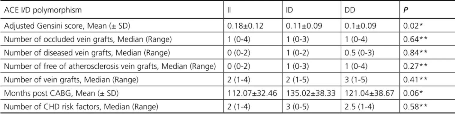
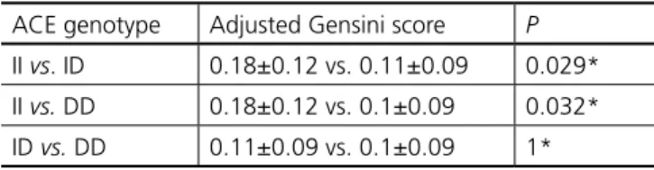
Relationship between ACE I/D polymorphism and severity of vein graft atherosclerosis and comparison of the number of occluded, diseased and free of atherosclerosis vein grafts, number of vein grafts, months after CABG and number of CHD risk factors according to genotype are reported in Table 3. According to Gensini score, ACE I/D polymorphism was associated with progression and development of atherosclerosis in venous bypass grafts (P=0.02). Bonferroni’s post hoc- test showed patients who were homozygous for I allele had higher adjusted Gensini score and rate of atherosclerosis (Table 4). Although there was no statistical difference in the number of diseased vein grafts per genotype, Gensini score showed bypass degeneration was more severe in II genotype. Without regarding adjusted Gensini score, ACE II genotype resulted in vein graft failure in earlier postoperative period (statistically marginal signiicant difference in months after CABG, P=0.06).
Table 1. Demographic characteristics and clinical features of
the studied patients with vein graft atherosclerosis.
Parameter N (%) / Mean±SD
n 102
Sex (Female/Male) 18 (17.6)/84 (82.4)
Age (year) 64.89±8.54
Body Mass Index (kg/m²) 26.52±3.68
Hypertension (positive) 62 (60.8)
Hypercholesterolemia (positive) 95 (93.1) Diabetes mellitus (positive) 30 (29.4) Cigarette smoking (positive) 17 (16.7)
Family history (positive) 68 (66.7)
Total Coronary Heart Disease risk factors 3 (1-5)*
Total number of vein grafts 254
Left anterior descending 12
Left circumlex 5
Diagonal 54
Optus margin 95
Right coronary artery 81
Ramous 7
Occluded vein grafts 107
Diseased vein grafts 66
Free of atherosclerosis 81
Number of vein grafts per patient 3 (1-5)* Months elapsed from Coronary Artery
Bypass Graft surgery
128.7±38.36
Vein graft Gensini score** 13.99±11.41
Adjusted vein graft Gensini score† 0.11±0.1
*Median (range)
** Vein graft Gensini score: the mean of Gensini score of total grafts in each patient
†Adjusted vein graft Gensini score: vein graft Gensini score was divided by months elapsed from Coronary Artery Bypass Graft surgery
Table 2. Genotype and allele frequencies of ACE I/D
polymorphism in studied patients with vein graft atherosclerosis.
ACE I/D
polymorphism Genotype N (%) Allele
N (Absolute frequency)
II 14 (13.7) D 112 (0.55)
ID 64 (62.7) I 92 (0.45)
DD 24 (23.6)
ACE=Angiotensin Converting Enzyme
Table 3. Effect of ACE I/D polymorphism on progression of atherosclerosis in vein grafts of the studied patients.
ACE I/D polymorphism II ID DD P
Adjusted Gensini score, Mean (± SD) 0.18±0.12 0.11±0.09 0.1±0.09 0.02*
Number of occluded vein grafts, Median (Range) 1 (0-4) 1 (0-3) 1 (0-4) 0.64**
Number of diseased vein grafts, Median (Range) 0 (0-2) 1 (0-2) 0.5 (0-3) 0.84**
Number of free of atherosclerosis vein grafts, Median (Range) 0 (0-2) 1 (0-3) 1 (0-4) 0.27**
Number of vein grafts, Median (Range) 2 (1-4) 2 (1-5) 3 (1-5) 0.41**
Months post CABG, Mean (± SD) 112.07±32.46 135.02±38.33 121.04±38.67 0.06*
Number of CHD risk factors, Median (Range) 2 (1-4) 3 (0-5) 2.5 (1-4) 0.58**
REFERENCES
1. Marui A, Kimura T, Nishiwaki N, Mitsudo K, Komiya T, Hanyu M, et al.; CREDO-Kyoto PCI/CABG Registry Cohort-2 Investigators. Com-parison of ive-year outcomes of coronary artery bypass grafting ver-sus percutaneous coronary intervention in patients with left ventric-ular ejection fractions≤50% versus >50% (from the CREDO-Kyoto PCI/CABG Registry Cohort-2). Am J Cardiol. 2014;114(7):988-96.
DISCUSSION
The results of our study show that ACE II genotype could be considered a risk factor for long-term graft failure after CABG. ACE II may have some roles in the progression of atheromatous plaque of vein grafts in earlier postoperative periods as compared with two other genotypic groups. Few studies about the inluence of ACE I/D polymorphism on venous bypass graft atherosclerosis are available. Studies carried out in Turkey and Germany demonstrated different results[25-27]. Ortlepp et al.[25]
reported ACE I/D polymorphism was not associated with venous bypass degeneration in the long term in their 101 studied patients. Dayi et al.[26] indicated DD genotype inluenced
vein graft occlusion in late postoperative period in their 87 consecutively selected patients. On the other hand, our study shows no statistical difference among ACE genotypic groups and the number of occluded vein grafts (P=0.64, Kruskal-Wallis test). In a study done by Völzke et al.[27] on 247 patients, they
demonstrated ACE DD genotype increased the rate of mortality and cardiovascular morbidity in the midterm after CABG.
Although ACE DD genotype was associated with the increasing risk of cardiovascular disease in many studies, some studies reported other type of genotypes as risk factors. Zee et al.[28] showed I allele was risk factor for essential hypertension.
Ismail et al.[29] found signiicantly higher frequency of ACE
II genotype in hypertensive patients aged 20-40 years. In Northern Indian population I allele was associated with essential hypertension[30]. ACE ID genotype was responsible for peripheral
vascular disease in Western Turkish patients[31].
The results of the study of ACE I/D polymorphism and cardiovascular disease in Iranian population seem conlicting. The presence of D allele exacerbated the risk of early onset coronary artery disease in west population of Iran[32]. Shaiee
et al.[33] showed different results. They reported no association
between ACE DD genotype and the risk of coronary artery disease. They collected study population from patients referred to Shahid Rajaei Cardiovascular Medical and Research Center, Tehran, Iran[33]. Despite the adverse effect of ACE DD genotype
on hypertension in type 2 diabetic population reported by Nakhjavani et al.[34], Nikzamir et al.[35] found no relation between
ACE I/D polymorphism and the presence of metabolic syndrome in patients with type 2 diabetes.
In our study the frequency distribution of II, ID and DD genotype are 13.7%, 62.7% and 23.6% respectively. These values are similar to the results found by Nikzamir et al.[35]
(16.5%, 58.2% and 25.3%), but are different from the data reported by Vaisi-Raygani et al.[32] (17.3%, 40% and 42.7%),
Shaiee et al.[33] (16.22%, 32.24% and 51.51%) and Nakhjavani
Authors’ roles & responsibilities
NZ Collected the data and helped in data analysis; final approval of the manuscript
MH Contributed in designing and conducting the study; final approval of the manuscript
MM Contributed in designing and conducting the study; final approval of the manuscript
HM Rechecked the statistical analysis and revised the
manuscript; final approval of the manuscript
NE Rechecked the statistical analysis and revised the
manuscript; final approval of the manuscript
AMS Proposed the idea; managed the research project; rechecked the statistical analysis; prepared the manuscript and final approval of the manuscript
et al.[34] (27.5%, 50% and 22.5%). Frequency distribution of
ACE I/D polymorphism observed in various parts of Iran is different and it may be responsible for the discrepancies in the reported results. It seems that different ACE I/D polymorphism interactions with cardiovascular disease could be attributed to the various ethnicities studied in previously mentioned Iranian studies.
We also think that the heterogeneity of our sample considering clinical proile, pharmacological treatment and lifestyle, as well as the surgical technique for myocardial revascularization, could inluence the follow-up as well as its relation with the genetic proile. Lack of serum ACE activity measurement and also the small sample size are main limitations of this study.
CONCLUSION
Although there are conlicting results about ACE I/D polymorphism and the degree of venous bypass graft degeneration, this study suggests an association between ACE genotype II and atherosclerosis of saphenous vein grafts, however, large samples considering clinical, demographic and ethnic proile are necessary to conirm these results.
ACKNOWLEDGMENTS
This study is result of a Doctor of Pharmacy thesis project which was inancially supported by the Vice-Chancellery for Research and Technology of the Isfahan University of Medical Sciences. Authors would like to thank all personnel of the poisoning emergency room of the Noor and Ali-Asghar [PBUH] University Hospital for their sincere help. Authors would like to thank Mr. Rory O’Conner for inal English editing.
Table 4. Comparison of angiographic severity of coronary
ar-tery disease in different ACE genotypes.
ACE genotype Adjusted Gensini score P
II vs. ID 0.18±0.12 vs. 0.11±0.09 0.029*
II vs. DD 0.18±0.12 vs. 0.1±0.09 0.032*
ID vs. DD 0.11±0.09 vs. 0.1±0.09 1*
2. Serruys PW, Morice MC, Kappetein AP, Colombo A, Holmes DR, Mack MJ, et al.; SYNTAX Investigators. Percutaneous coronary inter-vention versus coronary-artery bypass grafting for severe coronary artery disease. N Engl J Med. 2009;360(10):961-72
3. Daemen J, Boersma E, Flather M, Booth J, Stables R, Rodriguez A, et al. Long-term safety and eficacy of percutaneous coronary intervention with stenting and coronary artery bypass surgery for multivessel coronary artery disease: a meta-analysis with 5-year pa-tient-level data from the ARTS, ERACI-II, MASS-II, and SoS trials. Circulation. 2008;118(11):1146-54.
4. Hlatky MA, Boothroyd DB, Bravata DM, Boersma E, Booth J, Brooks MM, et al. Coronary artery bypass surgery compared with percuta-neous coronary interventions for multivessel disease: a collaborative analysis of individual patient data from ten randomised trials. Lan-cet. 2009;373(9670):1190-7.
5. Sá MPBO, Ferraz PE, Escobar RR, Nunes EO, Soares AMMN, Sá FBCA, et al. Five-year outcomes following PCI with DES versus CABG for unprotected LM coronary lesions: meta-analysis and meta-regres-sion of 2914 patients. Rev Bras Cir Cardiovasc. 2013;28(1):83-92. 6. Sen O, Gonca S, Solakoglu S, Dalcik H, Dalcik C, Ozkara A.
Compar-ison of conventional and no-touch techniques in harvesting saphe-nous vein for coronary artery bypass grafting in view of endothelial damage. Heart Surg Forum. 2013;16(4):E177-83.
7. Sabik JF 3rd. Understanding saphenous vein graft patency.
Circula-tion. 2011;124(3):273-5.
8. Harskamp RE, Lopes RD, Baisden CE, de Winter RJ, Alexander JH. Saphenous vein graft failure after coronary artery bypass surgery: pathophysiology, management, and future directions. Ann Surg. 2013;257(5):824-33.
9. Shukla N, Jeremy JY. Pathophysiology of saphenous vein graft failure: a brief overview of interventions. Curr Opin Pharmacol. 2012;12(2):114-20.
10. Parang P, Arora R. Coronary vein graft disease: pathogenesis and prevention. Can J Cardiol. 2009;25(2):e57-62.
11. Belczak CEQ, Tyszka AL, Godoy JMP, Ramos RN, Belczak SQ, Caffaro RA. Clinical complications of limb undergone harvesting of great saphenous vein for coronary artery bypass grafting using bridge technique. Rev Bras Cir Cardiovasc. 2009;24(1):68-72.
12. Roy H, Bhardwaj S, Yla-Herttuala S. Molecular genetics of athero-sclerosis. Hum Genet. 2009;125(5-6):467-91.
13. Arnett DK, Baird AE, Barkley RA, Basson CT, Boerwinkle E, Ga-nesh SK, et al.; American Heart Association Council on Epidemi-ology and Prevention; American Heart Association Stroke Council; Functional Genomics and Translational Biology Interdisciplinary Working Group. Relevance of genetics and genomics for preven-tion and treatment of cardiovascular disease: a scientiic statement from the American Heart Association Council on Epidemiology and Prevention, the Stroke Council, and the Functional Genomics and Translational Biology Interdisciplinary Working Group. Circulation. 2007;115(22):2878-901.
14. Radaelli G, Bodanese LC, Guaragna JCVC, Borges AP, Goldani MA, Petracco JB, et al. The use of inhibitors of angiotensin-converting enzyme and its relation to events in the postoperative period of CABG. Rev Bras Cir Cardiovasc. 2011;26(3):373-9.
15. Zintzaras E, Raman G, Kitsios G, Lau J. Angiotensin-converting enzyme insertion/deletion gene polymorphic variant as a mark-er of coronary artmark-ery disease: a meta-analysis. Arch Intmark-ern Med. 2008;168(10):1077-89.
16. Urhan Küçük M, Sucu N, Şahan Firat S, Aytaçoğlu BN, Vezir Ö, Bozali C, et al. Role of ACE I/D gene polymorphisms on the effect of ramipril in inlammatory response and myocardial injury in patients undergoing coronary artery bypass grafts. Eur J Clin Pharmacol. 2014;70(12):1443-51.
17. Teranishi J, Yamamoto R, Nagasawa Y, Shoji T, Iwatani H, Okada N, et al. ACE insertion/deletion polymorphism (rs1799752) modiies
the renoprotective effect of renin-angiotensin system blockade in patients with IgA nephropathy. J Renin Angiotensin Aldosterone Syst. 2015;16(3):633-41.
18. Pacurari M, Kafoury R, Tchounwou PB, Ndebele K. The Renin-An-giotensin-aldosterone system in vascular inlammation and remod-eling. Int J Inlam. 2014;2014:689360.
19. Fang H, Chen W, Gao Y, Shen Y, Luo M. Molecular mechanisms associated with Angiotensin-converting enzyme-inhibitory peptide activity on vascular extracellular matrix remodeling. Cardiology. 2014;127(4):247-55.
20. Zablocki D, Sadoshima J. Angiotensin II and oxidative stress in the failing heart. Antioxid Redox Signal. 2013;19(10):1095-109. 21. Dikalov SI, Nazarewicz RR, Bikineyeva A, Hilenski L, Lassègue B,
Griendling KK, et al. Nox2-induced production of mitochondrial su-peroxide in angiotensin II-mediated endothelial oxidative stress and hypertension. Antioxid Redox Signal. 2014;20(2):281-94.
22. Oldroyd KG, Phadke KV, Phillips R, Carson PH, Clarke M, Davis JA. Cardiac catheterisation by the Judkins technique as an outpatient procedure. BMJ. 1989;298(6677):875-6.
23. Gensini GG. A more meaningful scoring system for determining the severity of coronary heart disease. Am J Cardiol. 1983;51(3):606. 24. Shanmugam V, Sell KW, Saha BK. Mistyping ACE heterozygotes.
PCR Methods Appl. 1993;3(2):120-1.
25. Ortlepp JR, Janssens U, Bleckmann F, Lauscher J, Merkelbach-Bruse S, Hanrath P, et al. A chymase gene variant is associated with ath-erosclerosis in venous coronary artery bypass grafts. Coron Artery Dis. 2001;12(6):493-7.
26. Dayi SU, Tartan Z, Terzi S, Kasikcioglu H, Uyarel H, Orhan G, et al. Inluence of angiotensin converting enzyme insertion/deletion poly-morphism on long-term total graft occlusion after coronary artery bypass surgery. Heart Surg Forum. 2005;8(5):E373-7.
27. Völzke H, Engel J, Kleine V, Schwahn C, Dahm JB, Eckel L, et al. An-giotensin I-converting enzyme insertion/deletion polymorphism and cardiac mortality and morbidity after coronary artery bypass graft surgery. Chest. 2002;122(1):31-6.
28. Zee RY, Lou YK, Grifiths LR, Morris BJ. Association of a polymor-phism of the angiotensin I-converting enzyme gene with essential hypertension. Biochem Biophys Res Commun. 1992;184(1):9-15. 29. Ismail M, Akhtar N, Nasir M, Firasat S, Ayub Q, Khaliq S. Association
between the angiotensin-converting enzyme gene insertion/dele-tion polymorphism and essential hypertension in young Pakistani patients. J Biochem Mol Biol. 2004;37(5):552-5.
30. Srivastava K, Sundriyal R, Meena PC, Bhatia J, Narang R, Saluja D. Association of angiotensin converting enzyme (insertion/deletion) gene polymorphism with essential hypertension in northern Indian subjects. Genet Test Mol Biomarkers. 2012;16(3):174-7.
31. Başar Y, Salmayenli N, Aksoy M, Seçkin S, Aydin M, Ozkök E. ACE gene polymorphism in peripheral vascular disease. Horm Metab Res. 2007;39(7):534-7.
32. Vaisi-Raygani A, Ghaneialvar H, Rahimi Z, Nomani H, Saidi M, Bahrehmand F, et al. The angiotensin converting enzyme D allele is an independent risk factor for early onset coronary artery disease. Clin Biochem. 2010;43(15):1189-94.
33. Shaiee SM, Firoozrai M, Salimi S, Zand H, Hesabi B, Mohebbi A. Angiotensin converting enzyme DD genotype not associated with increased risk of coronary artery disease in the Iranian population. Pathophysiology. 2010;17(3):163-7.
34. Nakhjavani M, Esfahanian F, Jahanshahi A, Esteghamati A, Nikzamir AR, Rashidi A, et al. The relationship between the insertion/deletion polymorphism of the ACE gene and hypertension in Iranian patients with type 2 diabetes. Nephrol Dial Transplant. 2007;22(9):2549-53. 35. Nikzamir A, Nakhjavani M, Golmohamadi T, Dibai L. Association