Atrial Fibrillation, Neurocognitive Decline and
Gene Expression After Cardiopulmonary Bypass
Rahul S. Dalal
1, MD; Ashraf A. Sabe
1, MD; Nassrene Y. Elmadhun
1, MD; Basel Ramlawi
2, MD; Frank W. Sellke
1, MD
Abstract
Objective: Atrial fibrillation and neurocognitive decline are common complications after cardiopulmonary bypass. By utilizing genomic microarrays we investigate whether gene expression is associated with postoperative atrial fibrillation and neurocognitive decline.
Methods: Twenty one cardiac surgery patients were prospectively matched and underwent neurocognitive assessments pre-operatively and four days postpre-operatively. The whole blood collected in the pre-cardiopulmonary bypass, 6 hours after-cardiopulmonary bypass, and on the 4th postoperative day was
hybridized to Affymetrix Gene Chip U133 Plus 2.0 Microarrays. Gene expression in patients who developed postoperative atrial fibrillation and neurocognitive decline (n=6; POAF+NCD) was compared with gene expression in patients with postoperative atrial fibrillation and normal cognitive function (n=5; POAF+NORM) and patients with sinus rhythm and normal cognitive function (n=10; SR+NORM). Regulated genes were identified using JMP Genomics 4.0 with a false discovery rate of 0.05 and fold change of >1.5 or <-1.5.
Results: Eleven patients developed postoperative atrial fibrillation. Six of these also developed neurocognitive decline. Of the 12 patients with sinus rhythm, only 2 developed neurocognitive decline. POAF+NCD patients had unique regulation of 17 named genes preoperatively, 60 named genes six hours after cardiopulmonary bypass, and 34 named genes four days postoperatively (P<0.05) compared with normal patients. Pathway analysis demonstrated that these genes are involved in cell death, inflammation, cardiac remodeling and nervous system function.
Conclusion: Patients who developed postoperative atrial fibrillation and neurocognitive decline after cardiopulmonary bypass may have differential genomic responses compared to normal patients and patients with only postoperative atrial fibrillation, suggesting common pathophysiology for these conditions. Further exploration of these genes may provide insight into the etiology and improvements of these morbid outcomes.
Keywords: Atrial Fibrillation. Cardiopulmonary Bypass. Genes. Microarray Analysis.
INTRODUCTION
Surgical advancements have allowed an increasingly older population to undergo cardiac surgery and cardiopulmonary bypass (CPB) with a low mortality risk. Efforts have therefore focused on reducing postoperative morbidity. Neurocognitive decline (NCD, up to 80% incidence) and atrial ibrillation (AF, 20-45% incidence) remain two of the most common complications after CPB[1,2]. Coronary artery
bypass graft (CABG) guidelines by the American College of Cardiology/ American Heart Association describe two types of neurocognitive
deicits, with type 2 representing the vast majority[3]. Type 2 deicits
are global and may include confusion and intellectual and memory decline without a known focal lesion and may signiicantly impair patients’ quality of life. The etiology of these deicits is likely related to multiple factors including age, procedure, CPB time, hypoxia, and inlammation[4]. Up to 30% of type 2 deicits persist for at least one
year and early NCD appears to predict long-term deicits[5].
Like NCD, the high incidence of postoperative AF (POAF) has persisted. POAF generally occurs by postoperative day four and may precipitate heart failure and cerebrovascular emboli[6-8]. Because of
increased hospital stay and readmissions, it is estimated that healthcare costs for patients who develop POAF are $10,000 higher than for those who do not[7]. Though several factors have been correlated
with POAF after cardiac surgery, our inability to eliminate its incidence may be related to unknown pathophysiologic mechanisms. Studies have proposed that oxidation and inlammation after CPB induce cardiomyocyte damage and predispose to the development of atrial arrhythmias[9]. Experiments in a canine model of rapid atrial pacing
demonstrated that statins, which are known for their anti-inlammatory Abbreviations, acronyms & symbols
AF = Atrial fibrillation
CABG = Coronary artery bypass graft CPB = Cardiopulmonary bypass NCD = Neurocognitive decline POAF = Postoperative atrial fibrillation SR = Sinus rhythm
DOI: 10.5935/1678-9741.20150070
1Division of Cardiothoracic Surgery, Cardiovascular Research Center, Warren Alpert
Medical School of Brown University, Providence, RI, USA.
2Methodist DeBakey Heart & Vascular Center, Methodist Hospital, Houston, Texas,
USA.
This study was carried out in the Division of Cardiothoracic Surgery, Cardiovascular Research Center, Warren Alpert Medical School of Brown University, Providence, RI.
Financial Support: Funding for this research was provided by the National Heart, Lung, and Blood Institute (R01HL46716, Dr. Sellke; T35HL094308, Dr. Dalal) and NIH Training grant 5T32-HL094300-03 (Drs. Sabe and Elmadhun).
No conlicts of interest exist for any of the authors.
Correspondence Address: Frank W. Sellke
2 Dudley Street, MOC 360 Providence, RI, USA 02905 E-mail: [email protected]
Article received on May 2nd, 2015
and anti-oxidant properties, reduced shortening of the atrial effective refractory period and thus POAF susceptibility[10]. In a case-control
study, our group previously demonstrated that patients with POAF had elevated serum peroxide levels, excess myocardial oxidation, and an increased oxidative genomic response compared with patients in sinus rhythm (SR)[11].
While these complications have been studied independently, prior research suggests an association between POAF and neurologic abnormalities[12]. In a prospective observational study, Stanley et al.[13]
found signiicantly more cognitive deicits in patients who developed POAF, which was also associated with worse cognitive functioning six weeks after surgery. While it is thought that the paroxysmal nature of POAF, embolization, and decreased cardiac output increase risk for neurologic dysfunction, it remains unknown if there are common pathways by which both NCD and POAF arise.
High-throughput microarray provides a practical approach to investigate genomic changes and disease development. Microarrays can screen the entire human genome for regulated genes and bring light to the underlying pathways that may promote morbidities like NCD and POAF. We previously utilized microarray to demonstrate increased expression of genes involved with inlammation and neurologic dysfunction in patients who developed NCD after CPB compared to patients without NCD (NORM)[14]. We now examine gene
expression changes in patients who develop both POAF and NCD (POAF+NCD) compared to patients spared of these complications (SR+NORM) and those who develop POAF alone (POAF+NORM). To further investigate the underlying pathophysiology of these disease processes we utilize modern microarray and bioinformatics techniques to identify genes that may be associated with the combined incidence of these complications.
METHODS
Patient Enrollment and Matching
We performed a single-institution, prospective cohort study approved by the Beth Israel Deaconess Medical Center Institutional Review Board/Committee on Clinical Investigations in Boston, MA. Forty-two consecutive patients were scheduled for urgent or elective primary CABG, valve replacement (mitral or aortic), or a combination of both requiring CPB. All study participants were provided informed written consent for surgical procedures and blood collection for this investigation. Patients with pre-operative documented AF, high-grade carotid stenosis, known calciied aortas, recent cerebrovascular accident, severe neurologic deicits, serum creatinine>2.0 mg/dL, and hepatic cirrhosis were excluded. Subjects undergoing aortic root/arch procedures, on antiarrhythmic medications, or unable to complete neurocognitive assessments were also excluded.
POAF was deined as sustained AF conirmed by electrocardiogram before postoperative day ive that required anticoagulation or cardioversion. Of the 42 subjects enrolled, only the subset that developed both POAF and NCD was prospectively matched with selected SR+NORM and POAF+NORM patients based on pre-operative baseline characteristics (i.e. sex, age, hypercholesterolemia, hypertension, diabetes mellitus, white blood cell count, β-blocker use), intraoperative characteristics (i.e. CPB and aortic cross-clamp time, cardiotomy suction and antiibrinolytic use, procedure type), and postoperative characteristics (i.e. β-blocker use and time to extubation). Subsequent serologic and molecular studies were performed in a blinded fashion.
Surgical Technique
We followed our institution’s conventional operative approach regarding general anesthesia induction, midline sternotomy, systemic heparinization, CPB, and invasive monitoring as previously described[14].
Neurocognitive Assessment
Patients underwent neurocognitive assessments performed by trained, blinded psychometricians between 1 and 10 days pre-operatively, on postoperative day 4, and in the 3rd month of the postoperative
period. Patients were also evaluated for depression using the Geriatric Depression Scale. Memory, attention, language, global cognition, and executive functioning were assessed using 8 validated tools:
The Hopkins Verbal Learning Test measured verbal learning, recall, and retention by assessing the maximum number of items learned, the number of items recalled after 20 minutes divided by the maximum number learned, and the number of items correctly named from a list. Working memory and attention span were measured using Digit Span. Attention shifting ability was assessed by recording the time needed to complete Trailmaking A and B. Confrontational naming was measured using the Boston Naming Test. Fluency was evaluated by requiring patients to generate words beginning with a speciic letter (phonemic luency) or in a category (semantic luency). The Visual Search and Acuity Test and Stroop Color-Word Inference Test measured visuospatial abilities and executive function. Premorbid intelligence was measured using the Wechsler Test of Adult Reading. In accordance with the “Statement of consensus on assessment of neurobehavioral outcomes after cardiac surgery,” NCD was deined as a 1-standard deviation deicit from baseline on 25% of tasks[15].
Blood Collection and Microarray Processing
Blood samples were drawn from patients via central venous catheter pre-operatively immediately after anesthesia induction (pre-CPB), 6 hours postoperatively in the intensive care unit (post-(pre-CPB), and on postoperative day four (4D). Whole blood was drawn into PAXgene tubes (QIAGEN Inc, Valencia, Ca) for extraction and mRNA stabilization per the manufacturer’s instructions.
RNA extraction and puriication from whole blood, cDNA synthesis, and generation of biotin-labeled cRNA were performed by the Beth Israel Deaconess Medical Center Proteomics Core according to prior protocols[16,17]. All cRNA samples were hybridized to Affymetrix
GeneChip HG-U133 Plus 2.0 microarrays (Affymetric INc, Santa Clara, Ca). Chips were scanned using the HP G2500A ChipScanner (Affymetrix) and dChip software (Wong et al.[18], Boston, MA) was used
for quality control analysis and signal measurement. No outliers were identiied and all samples underwent subsequent pathway analysis.
Gene Expression and Pathway Analysis
Raw microarray data underwent gene expression analysis using JMP Genomics 4.0 (SAS, Cary, NC) for normalization, quality control, and statistical analysis. The Robust Multichip Average method normalized and compared composite chip data. Gene expression in Pre-CPB, Post-CPB, and 4D blood samples for POAF+NCD patients were compared to corresponding samples from SR+NORM and POAF+NORM using one-way ANOVA. A post-hoc false discovery rate algorithm with alpha of 0.05 minimized false positive results. Signiicantly, regulated genes met two criteria: 1) –log (P-value) exceeding the threshold calculated by JMP Genomics for each comparison and 2) fold change in gene expression >1.5 or <-1.5 between groups. A 1.5-fold change cutoff was chosen here and in a prior study of this patient population to reduce background noise while not limiting results to the most labile genes[14,19]. Signiicantly regulated genes were uploaded into Ingenuity
Pathway Analysis (IPA, Ingenuity Systems, Redwood City, CA) to generate top canonical pathways regulated by the selected genes.
Real-time PCR
RESULTS
Patient Characteristics
Patients with POAF+NCD (n=6) were prospectively matched with SR+NORM (n=10) and POAF+NORM (n=5). Table 1 lists well-matched baseline characteristics of these subjects and shows no signiicant differences in race, sex, age, and co-morbidities as calculated by one-way ANOVA. Patients underwent similar intraoperative courses with regard to anesthesia, CPB technique, temperature, and perioperative monitoring. There were no differences in other postoperative complications, such as focal neurologic deicits or cerebrovascular events in patients with POAF compared to SR during the study period. Of 11 total POAF patients, 6 developed NCD (54.5%), and of 12 SR
patients, only 2 developed NCD (16.7%). After three months, all but one patient returned regained normal cognitive function[20].
Gene Expression and Confirmation
We previously published comprehensive gene expression databases of patients with POAF or SR before and after CPB as well as patients with and without NCD after CPB, including unsupervised hierarchical sample clustering, and conirmation of microarray gene-expression data with real-time PCR[11,20]. Our described microarray GeneChip
identiied 54,675 transcripts. Complete lists of genes regulated in the comparisons of POAF+NCD vs. SR+NORM or POAF+NORM are provided in Tables 2 to 7.
Table 1. Characteristics for matching of patients who developed POAF and NCD with controls.
Characteristic A
POAF+NCD (n=6)
B SR+NORM (n=10)
C
POAF+NORM (n=5) P-value Pre-operative data
Age (y)a 66.5±7.4 69.2±7.1 73.4±5.8 0.28
Sex (% male) 83.3 (5/6) 100 (10/10) 80.0 (4/5) 0.40
Hypertension (% of group) 83.3 (5/6) 70.0 (7/10) 40.0 (2/5) 0.34
Hypercholesterolemia (% of group) 50.0 (3/6) 50.0 (5/10) 20.0 (1/5) 0.54
Diabetes mellitus (% of group) 50.0 (3/6) 30.0 (3/10) 40.0 (2/5) 0.76
Leukocytes (103 cells/μL)a 7.4±2.1 7.2±2.0 10.3±2.9 0.05
Hematocrit (%) 35.6±4.3 34.5±4.0 37.7±7.6 0.53
Glucose (mg/dL) 193±131 163±68 118±38 0.38
Intraoperative data
Procedure (% CABG) 83.3 (5/6) 70.0 (7/10) 80.0 (4/5) 0.84
CPB time (min)a 78.3±32.6 78.9±26.3 70.6±20.1 0.84
Cross-clamp time (min)a 57.7±23.9 63.0±21.0 46.4±21.3 0.40
aValues are mean ± SD
CABG=coronary artery bypass graft; CPB=cardiopulmonary bypass; POAF=post-operative atrial fibrillation; SR=sinus rhythm
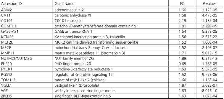
Table 2. Pre-CPB gene expression in patients with POAF+NCD compared with SR+NORM – complete list.
Accession ID Gene Name FC P-values
ADM2 adrenomedullin 2 1.66 1.12E-05
CA11 carbonic anhydrase XI 1.58 4.47E-05
CD101 CD101 molecule 2.19 1.15E-04
COMTD1 catechol-O-methyltransferase domain containing 1 1.81 2.29E-05
GAS6-AS1 GAS6 antisense RNA 1 1.54 5.37E-05
KCNIP3 Kv channel interacting protein 3, calsenilin 1.56 2.51E-22
MCF2L MCF.2 cell line derived transforming sequence-like 1.52 1.00E-04
MECR mitochondrial trans-2-enoyl-CoA reductase 1.52 2.19E-07
MMP11 matrix metallopeptidase 11 (stromelysin 3) 1.71 5.01E-15
NUTM2F/NUTM2G NUT family member 2G 1.89 6.31E-13
PHF20 PHD inger protein 20 0.65 1.78E-05
PYCR1 pyrroline-5-carboxylate reductase 1 1.53 5.37E-05
RGS12 regulator of G-protein signaling 12 1.52 9.77E-06
TOM1L2 target of myb1-like 2 (chicken) 1.60 1.15E-04
VGLL1 vestigial like 1 (Drosophila) 1.87 3.02E-07
WIZ widely interspaced zinc inger motifs 1.83 8.91E-10
ZBED5 zinc inger, BED-type containing 5 1.63 1.07E-04
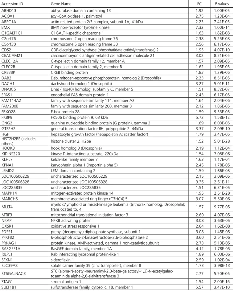
Table 3. Post-CBP gene expression in patients with POAF+NCD compared with SR+NORM – complete list.
Accession ID Gene Name FC P-values
ABHD13 abhydrolase domain containing 13 1.92 1.00E-05
ACOX1 acyl-CoA oxidase 1, palmitoyl 2.75 1.23E-04
ARPC1A actin related protein 2/3 complex, subunit 1A, 41kDa 2.23 7.41E-05
BMX BMX non-receptor tyrosine kinase 7.32 1.00E-14
C1GALT1C1 C1GALT1-speciic chaperone 1 1.63 1.82E-08
C2orf76 chromosome 2 open reading frame 76 2.38 5.25E-08
C5orf30 chromosome 5 open reading frame 30 2.56 6.17E-06
CDS2 CDP-diacylglycerol synthase (phosphatidate cytidylyltransferase) 2 1.95 4.07E-10 CEACAM21 carcinoembryonic antigen-related cell adhesion molecule 21 3.02 8.71E-05
CLEC12A C-type lectin domain family 12, member A 1.57 2.09E-05
CLEC2B C-type lectin domain family 2, member B 1.62 1.95E-05
CREBBP CREB binding protein 1.83 1.29E-06
DAB2 Dab, mitogen-responsive phosphoprotein, homolog 2 (Drosophila) 2.23 8.51E-05
DACH1 dachshund homolog 1 (Drosophila) 3.27 5.01E-11
DNAJC5 DnaJ (Hsp40) homolog, subfamily C, member 5 1.51 8.32E-07
EPAS1 endothelial PAS domain protein 1 2.43 6.17E-05
FAM114A2 family with sequence similarity 114, member A2 1.64 2.04E-06
FAM200B family with sequence similarity 200, member B 2.12 1.86E-05
FBXO28 F-box protein 28 1.59 9.33E-05
FKBP9 FK506 binding protein 9, 63 kDa 5.72 1.58E-12
GNG2 guanine nucleotide binding protein (G protein), gamma 2 1.69 6.03E-05
GTF2H2 general transcription factor IIH, polypeptide 2, 44kDa 3.37 2.09E-10
HGF hepatocyte growth factor (hepapoietin A; scatter factor) 1.79 3.47E-05
HIST2H2BE (includes
others) histone cluster 2, H2be 1.52 5.01E-28
HOOK3 hook homolog 3 (Drosophila) 2.19 1.12E-04
KIDINS220 kinase D-interacting substrate, 220kDa 1.54 7.08E-06
KLHL7 kelch-like family member 7 1.63 1.17E-04
KPNA1 karyopherin alpha 1 (importin alpha 5) 2.45 1.78E-05
LEMD2 LEM domain containing 2 1.59 1.66E-05
LOC100506229 uncharacterized LOC100506229 2.15 3.09E-05
LOC100506328 uncharacterized LOC100506328 9.36 2.51E-11
LOC285835 uncharacterized LOC285835 1.51 6.31E-05
MAPK14 mitogen-activated protein kinase 14 1.95 2.51E-28
MARCH5 membrane-associated ring inger (C3HC4) 5 3.07 5.50E-06
MLLT4 myeloid/lymphoid or mixed-lineage leukemia (trithorax homolog, Drosophila); translocated to, 4 1.57 9.77E-05
MTIF3 mitochondrial translational initiation factor 3 2.60 4.07E-05
NKAP NFKB activating protein 3.08 3.63E-05
OXSR1 oxidative stress responsive 1 2.84 1.62E-08
PDSS1 prenyl (decaprenyl) diphosphate synthase, subunit 1 3.08 1.45E-05
PFKFB2 6-phosphofructo-2-kinase/fructose-2,6-biphosphatase 2 3.60 2.51E-06
PRKAG1 protein kinase, AMP-activated, gamma 1 non-catalytic subunit 2.73 5.13E-05
RASGEF1A RasGEF domain family, member 1A 4.12 1.78E-05
RILPL1 Rab interacting lysosomal protein-like 1 1.89 6.03E-06
SFXN1 siderolexin 1 2.59 1.02E-04
SLC39A8 solute carrier family 39 (zinc transporter), member 8 1.73 3.98E-13
ST6GALNAC3 ST6
(alpha-N-acetyl-neuraminyl-2,3-beta-galactosyl-1,3)-N-acetylgalac-tosaminide alpha-2,6-sialyltransferase 3 2.77 5.50E-06
STAG1 stromal antigen 1 1.54 2.00E-16
SULT1B1 sulfotransferase family, cytosolic, 1B, member 1 5.57 3.47E-10
SYNE1 spectrin repeat containing, nuclear envelope 1 3.11 3.09E-06
TANK TRAF family member-associated NFKB activator 1.94 3.24E-05
TIMM23 translocase of inner mitochondrial membrane 23 homolog (yeast) 1.84 2.51E-16
TMLHE trimethyllysine hydroxylase, epsilon 1.95 3.47E-05
TOR1AIP2 torsin A interacting protein 2 1.55 6.31E-25
TRPS1 trichorhinophalangeal syndrome I 2.09 7.08E-05
UBE2H ubiquitin-conjugating enzyme E2H 3.47 2.19E-07
VAMP3 vesicle-associated membrane protein 3 1.74 8.51E-05
WDFY3 WD repeat and FYVE domain containing 3 2.69 1.02E-05
YIPF4 Yip1 domain family, member 4 1.79 1.23E-06
YKT6 YKT6 v-SNARE homolog (S. cerevisiae) 1.72 3.39E-05
ZNF350 zinc inger protein 350 1.57 5.13E-05
FC=fold change
Table 4. 4D gene expression in patients with POAF+NCD compared with SR+NORM – complete list.
Accession ID Gene Name FC P-values
AGPAT6 1-acylglycerol-3-phosphate O-acyltransferase 6 1.54 5.61E-04
ATP13A4 ATPase type 13A4 1.63 6.46E-06
BCL2L1 BCL2-like 1 3.17 1.58E-13
C20orf203 chromosome 20 open reading frame 203 0.45 4.37E-07
CASC7 cancer susceptibility candidate 7 (non-protein coding) 1.70 3.98E-11
CDC42BPA CDC42 binding protein kinase alpha (DMPK-like) 1.94 1.86E-08
CDCA7 cell division cycle associated 7 1.72 5.75E-05
CTSO cathepsin O 0.54 8.71E-05
DDX17 DEAD (Asp-Glu-Ala-Asp) box helicase 17 7.10 5.01E-27
DLD dihydrolipoamide dehydrogenase 2.13 3.16E-14
DOCK1 dedicator of cytokinesis 1 2.09 6.31E-50
DSC2 desmocollin 2 2.01 4.07E-06
FRMD8 FERM domain containing 8 2.42 2.45E-06
GLCCI1 glucocorticoid induced transcript 1 2.61 3.16E-16
GRB10 growth factor receptor-bound protein 10 1.79 3.16E-11
HNMT histamine N-methyltransferase 1.65 1.05E-04
IDE insulin-degrading enzyme 1.52 6.61E-06
LOC284080 uncharacterized LOC284080 1.51 8.13E-05
MMD monocyte to macrophage differentiation-associated 1.57 2.00E-15
NCR1 natural cytotoxicity triggering receptor 1 1.90 2.57E-05
NEDD4L neural precursor cell expressed, developmentally down-regulated 4-like, E3
ubiquitin protein ligase 1.60 6.31E-11
PLXNB1 plexin B1 1.53 1.00E-15
PRKAA2 protein kinase, AMP-activated, alpha 2 catalytic subunit 1.54 5.37E-05
PRRT1 proline-rich transmembrane protein 1 1.98 3.98E-05
REEP1 receptor accessory protein 1 1.52 2.63E-07
RHCE/RHD Rh blood group, D antigen 1.69 8.13E-05
RIOK3 RIO kinase 3 4.54 9.55E-05
RPL10 ribosomal protein L10 0.40 8.91E-05
RPL18 ribosomal protein L18 0.47 1.95E-05
SMC3 structural maintenance of chromosomes 3 1.53 4.47E-06
SRSF1 serine/arginine-rich splicing factor 1 2.04 4.37E-05
ST7 suppression of tumorigenicity 7 1.58 6.76E-05
TFAP2E transcription factor AP-2 epsilon (activating enhancer binding protein 2 epsilon) 1.93 5.89E-06
UBE2H ubiquitin-conjugating enzyme E2H 2.94 7.76E-06
Table 5. Pre-CPB gene expression in patients with POAF+NCD compared with POAF+NORM – complete list.
Accession ID Gene Name FC P-values
ACTR3BP5 ARP3 actin-related protein 3 homolog B (yeast) pseudogene 5 0.57 3.55E-09
AP5S1 adaptor-related protein complex 5, sigma 1 subunit 1.87 1.00E-04
C14orf166B chromosome 14 open reading frame 166B 1.54 1.58E-12
CA11 carbonic anhydrase XI 1.79 8.91E-06
CCDC36 coiled-coil domain containing 36 0.35 5.01E-21
CIZ1 CDKN1A interacting zinc inger protein 1 2.42 1.10E-04
FHAD1 forkhead-associated (FHA) phosphopeptide binding domain 1 1.51 6.31E-06
FKRP fukutin related protein 0.54 4.17E-05
GTPBP3 GTP binding protein 3 (mitochondrial) 0.49 2.75E-05
KCNIP3 Kv channel interacting protein 3, calsenilin 1.58 6.31E-19
KHSRP KH-type splicing regulatory protein 1.56 4.79E-10
LOC100507477 uncharacterized LOC100507477 1.99 3.98E-05
MCF2L MCF.2 cell line derived transforming sequence-like 1.66 4.17E-05
MMP11 matrix metallopeptidase 11 (stromelysin 3) 1.67 5.01E-12
NFATC1 nuclear factor of activated T-cells, cytoplasmic, calcineurin-dependent 1 1.61 6.46E-05
NUTM2F/NUTM2G NUT family member 2G 1.89 3.16E-11
PHAX phosphorylated adaptor for RNA export 0.59 2.45E-05
PYCR1 pyrroline-5-carboxylate reductase 1 1.75 5.89E-06
RIN1 Ras and Rab interactor 1 1.78 7.94E-05
SLC24A6 solute carrier family 24 (sodium/lithium/calcium exchanger), member 6 0.45 1.20E-04
SYT17 synaptotagmin XVII 0.57 3.98E-14
TACC2 transforming, acidic coiled-coil containing protein 2 0.57 3.16E-18
TMEM259 transmembrane protein 259 1.64 7.41E-06
TUBG1 tubulin, gamma 1 1.79 8.71E-05
VGLL1 vestigial like 1 (Drosophila) 1.90 1.74E-06
WIZ widely interspaced zinc inger motifs 1.74 1.48E-07
WNK2 WNK lysine deicient protein kinase 2 0.48 3.98E-31
XYLT2 xylosyltransferase II 0.65 1.05E-04
ZNF528 zinc inger protein 528 0.36 8.51E-05
FC=fold change
Table 6. Post-CPB gene expression in patients with POAF+NCD compared with POAF+NORM – complete list.
Accession ID Gene Name FC P-values
ANKMY2 ankyrin repeat and MYND domain containing 2 0.61 1.58E-12
ANKRD6 ankyrin repeat domain 6 0.54 1.38E-08
AP4E1 adaptor-related protein complex 4, epsilon 1 subunit 0.50 4.68E-05
BCS1L BC1 (ubiquinol-cytochrome c reductase) synthesis-like 0.53 7.76E-05
CDS2 CDP-diacylglycerol synthase (phosphatidate cytidylyltransferase) 2 2.03 3.09E-09
CEBPG CCAAT/enhancer binding protein (C/EBP), gamma 0.41 1.26E-05
CLEC2B C-type lectin domain family 2, member B 1.97 1.70E-06
DACH1 dachshund homolog 1 (Drosophila) 2.47 8.32E-07
FKBP9 FK506 binding protein 9, 63 kDa 2.38 8.32E-05
GOLT1B golgi transport 1B 0.42 3.98E-13
GTF2H2 general transcription factor IIH, polypeptide 2, 44kDa 2.54 1.35E-06
HIST2H2BE (includes others) histone cluster 2, H2be 1.52 3.98E-26
HIVEP2 human immunodeiciency virus type I enhancer binding protein 2 0.62 6.03E-05
KMO kynurenine 3-monooxygenase (kynurenine 3-hydroxylase) 0.33 1.00E-31
LOC100506328 uncharacterized LOC100506328 5.99 1.38E-07
LOC728613 programmed cell death 6 pseudogene 0.48 6.92E-05
MAPK14 mitogen-activated protein kinase 14 1.98 6.31E-24
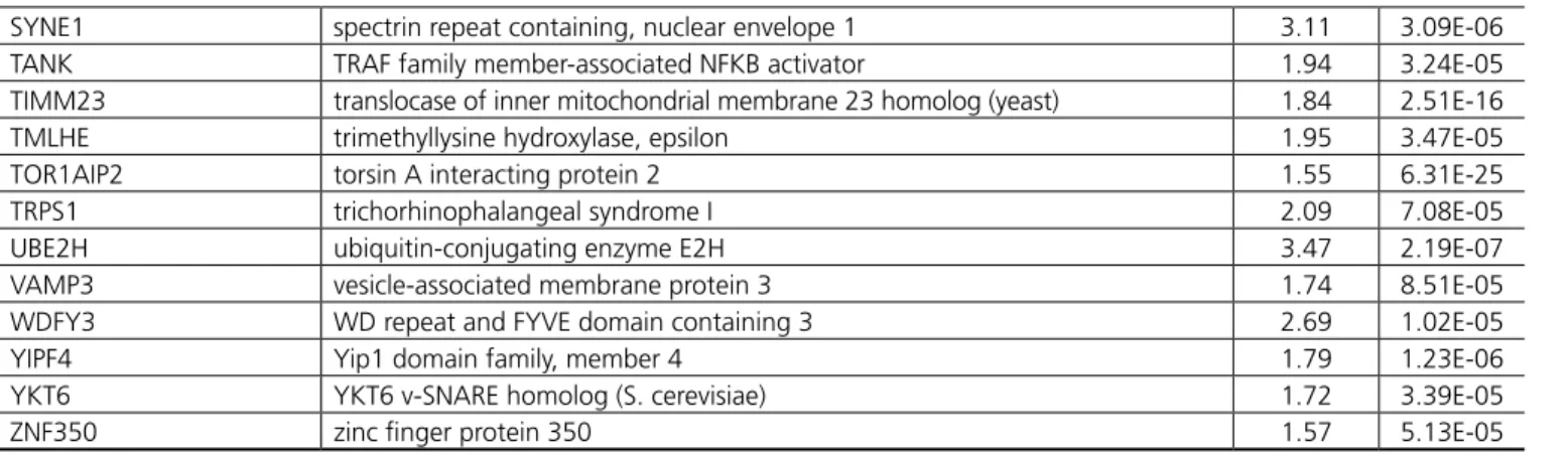
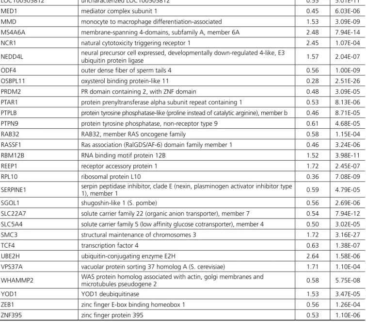
Table 7. 4D gene expression in patients with POAF+NCD compared with POAF+NORM – complete list.
Accession ID Gene Name FC P-values
ACSL6 acyl-CoA synthetase long-chain family member 6 1.62 8.13E-06
ADAMTS6 ADAM metallopeptidase with thrombospondin type 1 motif, 6 0.65 4.37E-05
ADRBK2 adrenergic, beta, receptor kinase 2 0.22 1.29E-09
AGPAT6 1-acylglycerol-3-phosphate O-acyltransferase 6 1.63 1.58E-12
BCL2L1 BCL2-like 1 2.75 3.09E-06
C20orf203 chromosome 20 open reading frame 203 0.31 3.16E-14
CASC7 cancer susceptibility candidate 7 (non-protein coding) 1.75 1.05E-10
CBL Cbl proto-oncogene, E3 ubiquitin protein ligase 0.64 3.02E-10
CDC42BPA CDC42 binding protein kinase alpha (DMPK-like) 2.24 2.24E-10
CDCA7 cell division cycle associated 7 1.88 1.70E-09
CHD2 chromodomain helicase DNA binding protein 2 0.48 2.40E-05
CHERP calcium homeostasis endoplasmic reticulum protein 0.46 8.91E-07
CLIC2 chloride intracellular channel 2 2.08 5.01E-27
DCAF15 DDB1 and CUL4 associated factor 15 0.48 5.89E-05
DDX17 DEAD (Asp-Glu-Ala-Asp) box helicase 17 6.55 2.29E-06
DLD dihydrolipoamide dehydrogenase 1.89 2.51E-25
DOCK1 dedicator of cytokinesis 1 2.10 1.00E-10
EPB41L4B erythrocyte membrane protein band 4.1 like 4B 0.65 7.94E-49
FRMD8 FERM domain containing 8 3.33 2.51E-17
GLCCI1 glucocorticoid induced transcript 1 2.43 1.29E-08
GRB10 growth factor receptor-bound protein 10 1.72 7.94E-14
HEMGN hemogen 2.75 1.45E-09
IDE insulin-degrading enzyme 1.56 2.82E-05
L1CAM L1 cell adhesion molecule 1.60 1.05E-05
continue
NEK6 NIMA-related kinase 6 0.59 1.58E-21
PDS5B PDS5, regulator of cohesion maintenance, homolog B (S. cerevisiae) 0.48 4.79E-05
PMM1 phosphomannomutase 1 0.63 1.66E-05
PPP2R1B protein phosphatase 2, regulatory subunit A, beta 1.50 1.10E-05
RIOK1 RIO kinase 1 0.54 1.00E-15
RNF144B ring inger protein 144B 0.60 2.88E-05
SIVA1 SIVA1, apoptosis-inducing factor 0.58 8.71E-08
SLC27A3 solute carrier family 27 (fatty acid transporter), member 3 0.60 1.58E-05
SLC39A8 solute carrier family 39 (zinc transporter), member 8 1.64 2.95E-10
SULT1B1 sulfotransferase family, cytosolic, 1B, member 1 3.68 3.09E-06
SYNE1 spectrin repeat containing, nuclear envelope 1 2.74 1.26E-04
TIMM23 translocase of inner mitochondrial membrane 23 homolog (yeast) 1.83 1.26E-13
TOR1AIP2 torsin A interacting protein 2 1.55 3.98E-22
TTLL12 tubulin tyrosine ligase-like family, member 12 0.54 3.55E-05
TUBB6 tubulin, beta 6 class V 0.61 8.13E-06
YIPF4 Yip1 domain family, member 4 1.68 8.32E-05
ZCCHC10 zinc inger, CCHC domain containing 10 0.61 2.09E-09
ZDHHC12 zinc inger, DHHC-type containing 12 0.47 4.37E-05
ZNF226 zinc inger protein 226 0.42 5.01E-29
ZNF350 zinc inger protein 350 1.83 7.94E-06
LOC100505812 uncharacterized LOC100505812 0.55 5.01E-11
MED1 mediator complex subunit 1 0.45 6.03E-06
MMD monocyte to macrophage differentiation-associated 1.53 3.09E-09
MS4A6A membrane-spanning 4-domains, subfamily A, member 6A 2.48 7.94E-14
NCR1 natural cytotoxicity triggering receptor 1 2.45 1.07E-04
NEDD4L neural precursor cell expressed, developmentally down-regulated 4-like, E3
ubiquitin protein ligase 1.57 2.04E-07
ODF4 outer dense iber of sperm tails 4 0.56 1.00E-09
OSBPL11 oxysterol binding protein-like 11 0.28 2.51E-26
PRDM2 PR domain containing 2, with ZNF domain 0.48 3.09E-05
PTAR1 protein prenyltransferase alpha subunit repeat containing 1 0.53 8.13E-06
PTPLB protein tyrosine phosphatase-like (proline instead of catalytic arginine), member b 0.46 8.71E-05
PTPN9 protein tyrosine phosphatase, non-receptor type 9 0.61 4.68E-05
RAB32 RAB32, member RAS oncogene family 0.58 1.15E-04
RASSF1 Ras association (RalGDS/AF-6) domain family member 1 0.46 3.24E-06
RBM12B RNA binding motif protein 12B 1.52 3.98E-11
REEP1 receptor accessory protein 1 1.72 2.45E-07
RPL10 ribosomal protein L10 0.36 7.08E-09
SERPINE1 serpin peptidase inhibitor, clade E (nexin, plasminogen activator inhibitor type 1), member 1 0.59 4.79E-05
SGOL1 shugoshin-like 1 (S. pombe) 0.56 2.69E-06
SLC22A7 solute carrier family 22 (organic anion transporter), member 7 0.54 7.94E-12
SLC5A4 solute carrier family 5 (low afinity glucose cotransporter), member 4 0.50 3.02E-05
SMC3 structural maintenance of chromosomes 3 1.72 3.16E-27
TCF4 transcription factor 4 0.63 1.38E-07
UBE2H ubiquitin-conjugating enzyme E2H 2.64 1.58E-06
VPS37A vacuolar protein sorting 37 homolog A (S. cerevisiae) 1.71 1.10E-04
WHAMMP2 WAS protein homolog associated with actin, golgi membranes and
microtubules pseudogene 2 0.58 5.75E-08
YOD1 YOD1 deubiquitinase 1.53 3.47E-05
ZEB1 zinc inger E-box binding homeobox 1 0.56 1.26E-04
ZNF395 zinc inger protein 395 0.53 1.10E-06
FC=fold change
Gene Expression and Pathway Analysis in POAF+NCD vs. SR+NORM
Figure 1 shows the distribution of regulated genes by fold-change for each time point in this comparison. Pre-CPB, 19 genes were signiicantly regulated in the POAF+NCD group compared to NORM+SR, of which 17 were named. Notably, 16 of these 17 genes were up-regulated, while 1 was down-regulated. Pathway analysis used to group genes by potential pathophysiologic functions demonstrated that these genes are related to cardiovascular disease, nervous system function, and cell death, as described in Table 8. Post-CPB, the number of genes increased to 65, of which 60 were named. All 60 were up-regulated, and while distinct from those regulated pre-operatively, pathway analysis demonstrated that many of these genes are associated with cardiovascular disease and remodeling, inlammation, and nervous system disorders, as seen in Table 9. At 4D, the number of genes decreased to 41, of which 34 were named. Of these, 30 were up-regulated while 4 were
down-regulated. Several genes, as listed in Table 10, are similarly involved with cardiovascular disease, nervous system function, inlammation, and protein degradation.
Gene Expression and Pathway Analysis in Patients with POAF+NCD vs. POAF+NORM.
were named. Twenty-seven of these were up-regulated, while 27 were down-regulated. IPA analysis again revealed that several genes affect cardiovascular disease, inlammation, and cell death. Selected genes grouped by pathophysiologic function for the
Fig. 1 - Distribution of genes regulated for POAF+NCD vs. SR+NORM.
POAF+NCD vs. POAF+NORM comparisons are found in Tables 11-13. While the majority of the genes identiied for these comparisons were distinct from that of POAF+NCD vs. SR+NORM across all time points, multiple genes overlap and are listed in Table 14.
Table 8. Pre-CPB Gene Expression in Patients with POAF and NCD compared with SR and NORM – selected genes grouped by poten-tial pathophysiologic function.
Accession ID Gene Name FC P-values
Cardiovascular disease
ADM2 adrenomedullin-2 1.66 1.00E-04
Nervous system function
KCNIP3 Kv channel interacting protein 3, calsenilin 1.56 2.51E-22
Cell death and survival
MMP11 matrix metallopeptidase 11 (stromelysin 3) 1.71 5.01E-15
FC=fold change
Table 9. Post-CPB gene expression in patients with POAF and NCD compared with SR and NORM – selected genes grouped by poten-tial pathophysiologic function.
Accession ID Gene Name FC P-values
Cardiovascular disease
BMX BMX non-receptor tyrosine kinase 7.32 1.00E-14
EPAS1 endothelial PAS domain protein 1 2.43 6.17E-05
HGF hepatocyte growth factor (hepapoietin A; scatter factor) 1.79 3.47E-05
MAPK14 mitogen-activated protein kinase 14 1.95 2.51E-28
Nervous system function
KIDINS220 kinase D-interacting substrate, 220kDa 1.54 7.08E-06
SYNE1 spectrin repeat containing, nuclear envelope 1 3.11 3.09E-06
YKT6 YKT6 v-SNARE homolog (S. cerevisiae) 1.72 3.39E-05
Inlammation
CREBBP CREB binding protein 1.83 1.29E-06
Pyschological disorders
TMLHE trimethyllysine hydroxylase, epsilon 1.95 3.47E-05
Table 10. 4D Gene expression in patients with AF and NCD compared with SR and NORM – selected genes grouped by potential pathophysiologic function.
Accession ID Gene Name FC P-values
Cardiovascular disease
BCL2L1 BCL2-like 1 3.17 1.58E-13
PRKAA2 protein kinase, AMP-activated, alpha 2 catalytic subunit 1.54 5.37E-05
Nervous system function
IDE insulin-degrading enzyme 1.52 6.61E-06
CDC42BPA CDC42 binding protein kinase alpha (DMPK-like) 1.94 1.86E-08
PLXNB1 plexin B1 1.53 1.00E-15
Inlammation
NCR1 natural cytotoxicity triggering receptor 1 1.90 2.57E-05
DOCK1 dedicator of cytokinesis 1 2.09 6.31E-50
SMC3 structural maintenance of chromosomes 3 1.53 4.47E-06
Protein degradation
DLD dihydrolipoamide dehydrogenase 2.13 3.16E-14
NEDD4L neural precursor cell expressed, developmentally down-regulated
4-like, E3 ubiquitin protein ligase 1.60 6.31E-11
UBE2H ubiquitin-conjugating enzyme E2H 2.94 7.76E-06
FC=fold change
Fig. 2 - Distribution of genes regulated for POAF+NCD vs. POAF+NORM.
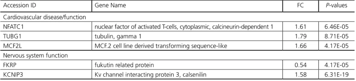
Table 11. Pre-CPB gene expression in patients with POAF+NCD compared with POAF+NORM – selected genes grouped by potential pathophysiologic function.
Accession ID Gene Name FC P-values
Cardiovascular disease/function
NFATC1 nuclear factor of activated T-cells, cytoplasmic, calcineurin-dependent 1 1.61 6.46E-05
TUBG1 tubulin, gamma 1 1.79 8.71E-05
MCF2L MCF.2 cell line derived transforming sequence-like 1.66 4.17E-05
Nervous system function
FKRP fukutin related protein 0.54 4.17E-05
KCNIP3 Kv channel interacting protein 3, calsenilin 1.58 6.31E-19
Table 12. Post-CPB gene expression in patients with POAF+NCD compared with POAF+NORM – selected genes grouped by poten-tial pathophysiologic function.
Accession ID Gene Name FC P-values
Cardiovascular disease/function
MAPK14 mitogen-activated protein kinase 14 1.98 6.31E-24
SYNE1 spectrin repeat containing, nuclear envelope 1 2.74 1.26E-04
CDS2 CDP-diacylglycerol synthase (phosphatidate cytidylyltransferase) 2 2.03 3.09E-09 Inlammation
HIVEP2 human immunodeiciency virus type I enhancer binding protein 2 0.62 6.03E-05
FC=fold change
Table 13. 4D Gene expression in patients with POAF+NCD compared with POAF+NORM – selected genes grouped by potential patho-physiologic function.
Accession ID Gene Name FC P-values
Cardiovascular disease
CBL Cbl proto-oncogene, E3 ubiquitin protein ligase 0.64 2.24E-10
SERPINE1 serpin peptidase inhibitor, clade E (nexin, plasminogen activator inhibitor type 1), member 1
0.59 2.69E-06
BCL2L1 BCL2-like 1 2.75 3.09E-06
MED1 mediator complex subunit 1 0.45 3.09E-09
RASSF1 Ras association (RalGDS/AF-6) domain family member 1 0.46 3.98E-11
Cell death/survival
IDE insulin-degrading enzyme 1.56 1.05E-05
RAB32 RAB32, member RAS oncogene family 0.58 3.24E-06
CDC42BPA CDC42 binding protein kinase alpha (DMPK-like) 2.24 1.70E-09
DOCK1 dedicator of cytokinesis 1 2.10 7.94E-49
L1CAM L1 cell adhesion molecule 1.60 5.01E-11
PTPN9 protein tyrosine phosphatase, non-receptor type 9 0.61 1.15E-04
SMC3 structural maintenance of chromosomes 3 1.72 1.38E-07
DDX17 DEAD (Asp-Glu-Ala-Asp) box helicase 17 6.55 2.51E-25
GRB10 growth factor receptor-bound protein 10 1.72 1.45E-09
PRDM2 PR domain containing 2, with ZNF domain 0.48 8.13E-06
TCF4 transcription factor 4 0.63 1.58E-06
ZEB1 zinc inger E-box binding homeobox 1 0.56 1.10E-06
Inlammation
ADRBK2 adrenergic, beta, receptor kinase 2 0.22 1.29E-09
NCR1 natural cytotoxicity triggering receptor 1 2.45 2.04E-07
FC=fold change
Table 14. Signiicantly regulated genes overlapping across multiple comparisons.
Comparisons Overlapping Regulated Genes
POAF+NCD vs. SR+NORM (Pre-CPB)
POAF+NCD vs. AF+NORM (Pre-CPB) ca11, kcnip3, mcf2l, mmp11, nutm2f/nutm2g, pycr1, vgll1, wiz POAF+NCD vs. SR+NORM (Post-CPB)
POAF+NCD vs. AF+NORM (Post-CPB)
cds2, clec2b, dach1, fkbp9, gtf2h2, hist2h2be, mapk14, slc39a8, sult1b1, syne1, timm23, tor1aip2, yipf4, znf350
POAF+NCD vs. SR+NORM (4D) POAF+NCD vs. AF+NORM (4D)
agpat6, bcl2l1, c20orf203, casc7, cdc42bpa, cdca7, ddx17, dld, dock1, frmd8, glcci1, grb10, ide, mmd, ncr1, nedd4l, reep1, rpl10, smc3, ube2h
POAF+NCD vs. SR+NORM (Post-CPB) POAF+NCD vs. SR+NORM (4D) POAF+NCD vs. AF+NORM (4D)
DISCUSSION
AF and NCD after cardiac surgery have each been extensively studied. Much of the literature for POAF has pointed to inlammation and oxidative stress as promoting factors. Indeed, prior work from our group demonstrated signiicantly elevated genomic markers of oxidative stress in the blood of patients who develop POAF after CPB[11]. We similarly used microarray to study NCD patients
and found increased expression of blood inlammatory mediators from those undergoing CPB[14]. Given that the genomic regulation
of systemic cytotoxic insults such as oxidation and inlammation appear to promote POAF and NCD when studied individually, we sought to determine if genomic responses differ in patients who develop both complications.
Our current microarray study shows that the expression proiles of patients who develop both POAF and NCD after CPB differ from those who develop neither complication nor POAF alone. The greatest amount of gene regulation occurred postoperatively, suggesting that CPB may induce a differential genomic response in susceptible patients. Furthermore, POAF+NCD vs. POAF+NORM had the most gene regulation at 4D, while POAF+NCD vs. SR+NORM had the most gene regulation post-CPB with a largely different set of genes identiied. This suggests that POAF and NCD after CPB may be linked pathophysiologically through mechanisms distinct from those inducing POAF alone, with more genomic changes occurring at an earlier stage.
Many genes regulated post-CPB in POAF+NCD vs. SR+NORM are associated with pathologic cardiac remodeling. One such gene includes BMX, a non-receptor tyrosine kinase. Mitchell-Jordan et al.[21] demonstrated that BMX-knockout mice were
resistant to massive cardiac hypertrophy following transverse aortic constriction relative to wild type, indicating a signiicant role for BMX in cardiac remodeling. If the impressive 7.32-fold up-regulation of BMX in the blood of our POAF+NCD patients also relects their myocardial expression, excess cardiac remodeling after CPB may be a predisposing factor for POAF and NCD. Additional up-regulated genes identiied in this group with reported roles in cardiac remodeling include EPAS1, HGF, and MAPK14[22-24]. While
there is much evidence for oxidative stress in cardiac remodeling and AF[25], our study found genes implicated in remodeling but not
oxidative stress, perhaps due to our limited sample size. However, while Ramlawi et al.[20] demonstrated genomic regulation of
oxidative stress in POAF patients, they did not report genes directly related to cardiac remodeling. This difference may lie in the fact that our patients developed NCD in addition to POAF, introducing a potential association of cardiac remodeling with secondary neurologic effects.
Several genes identiied in the POAF+NCD vs. SR+NORM comparison are also directly implicated in neurologic dysfunction. KIDINS220 was up-regulated post-CPB and has been shown to accumulate with tau protein in the brains of Alzheimer Disease patients[26]. At 4D, there was also increased expression of PLXNB1,
which controls the behavior of microtubule tips and dendrite morphology[27]. Given its critical role in regulating the cytoskeleton
and dendrite growth, it is postulated to be involved in the pathogenesis of several neurological disorders.
Genes related to inlammation and cell death were also identiied in POAF+NCD vs. SR+NORM. KIDINS200, discussed above, has a known role in T-cell receptor-mediated T-cell activation in addition to its neurologic functions[28]. At 4D, up-regulated pro-inlammatory
genes include NCR1 and DOCK. NCR1 encodes a natural killer cell receptor that triggers cytotoxicity, while DOCK1 is involved in
cytoskeletal rearrangements required for phagocytosis[29,30]. Genes
involved with protein degradation were also identiied at 4D, including NEDD4L and UBE2H. NEDD4L encodes an E3 ubiquitin ligase and UBEH2 encodes ubiquitin-conjugating enzyme E2H, both of which target proteins for lysosomal degradation[31,32]. These
genes have no established relationship to either POAF or NCD after CPB, but given that systemic inlammatory and catabolic processes are known contributors to both complications, the regulation of these proteins at the genomic level may be relevant[33-36].
Our study has limitations, the most signiicant of which is the size of our patient population. A larger study may allow for the identiication of more genes that may characterize complete pathways, such as the oxidative stress response, as opposed to our identiication of several isolated genes related to various pathways. While our patients were well matched, our sample size also precludes us from respecting Hardy-Weinberg Equilibrium. However, we hope that our indings stimulate interest in larger studies of this nature.
Another limitation is our proiling gene expression in blood rather than heart or brain tissues, both of which were not feasible in this study and would not be a practical option for future patient management strategies. It is unknown if the genes involved with cardiovascular and neurologic function identiied in blood relect pathways in the heart and brain. However, several genes we identiied may have systemic effects through inlammation and cell death that may secondarily damage both heart and brain tissue and predispose these individuals to POAF and NCD.
CONCLUSION
Our indings may expand what is known about the pathophysiology underlying POAF and NCD. While we cannot assert a true genetic association between POAF and NCD given our limited sample size, our results suggest that differential genomic responses existed in our study sample of patients who developed both complications after cardiac surgery. There may have been an inluence of pathologic cardiac remodeling and involvement of genes with known roles in inlammation, cell death, and nervous system function that may have promoted POAF and NCD in our patient population. We hope that the database of regulated genes provided by this work sparks further study of differentially expressed pathways that may deepen our understanding of these important and costly complications and potentially offer means of risk stratiication and improved patient management.
Authors’ roles & responsibilities
RSD Analysis or interpretation of data; statistical analysis; final approval of the manuscript; study design AAS Study design; final approval of the manuscript NYE Study design; final approval of the manuscript BR Study design; final approval of the manuscript
REFERENCES
1. Gao L, Taha R, Gauvin D, Othmen LB, Wang Y, Blaise G. Postoperative cognitive dysfunction after cardiac surgery. Chest. 2005;128(5):3664-70.
2. Auer J, Weber T, Berent R, Ng CK, Lamm G, Eber B. Risk factors of postoperative atrial ibrillation after cardiac surgery. J Card Surg. 2005;20(5):425-31.
3. Eagle KA, Guyton RA, Davidoff R, Edwards FH, Ewy GA, Gardner TJ, et al.; American College of Cardiology; American Heart Association. ACC/AHA 2004 guideline update for coronary artery bypass graft surgery: a report of the American College of Cardiology/American Heart Association Task Force on Practice Guidelines (Committee to Update the 1999 Guidelines for Coronary Artery Bypass Graft Surgery). Circulation. 2004;110(14):e340-437.
4. Murkin JM. Etiology and incidence of brain dysfunction after cardiac surgery. J Cardiothorac Vasc Anesth. 1999;13(suppl 1):12-7; discussion 36-7.
5. Newman MF, Kirchner JL, Phillips-Bute B, Gaver B, Grocott H, Jones RH, et al.; Neurological Outcome Research Group and the Cardiothoracic Anesthesiology Research Endeavors Investigators. Longitudinal assessment of neurocognitive function after coronary-artery bypass surgery. N Engl J Med. 2001;344(6):395-402. 6. Creswell LL, Schuessler RB, Rosenbloom M, Cox JL. Hazards of
postoperative atrial arrhythmias. Ann Thorac Surg. 1993;56(3):539-49. 7. Aranki SF, Shaw DP, Adams DH, Rizzo RJ, Couper GS, VanderVliet
M, et al. Predictors of atrial ibrillation after coronary artery surgery. Current trends and impact on hospital resources. Circulation. 1996;94(3):390-7.
8. Ommen SR, Odell JA, Stanton MS. Atrial arrhythmias after cardiothoracic surgery. N Engl J Med. 1997;336(20):1429-34. 9. Fontes ML, Mathew JP, Rinder HM, Zelterman D, Smith BR, Rinder
CS; Multicenter Study of Perioperative Ischemia (McSPI) Research Group. Atrial ibrillation after cardiac surgery/cardiopulmonary bypass is associated with monocyte activation. Anesth Analg. 2005;101(1):17-23.
10. Shiroshita-Takeshita A, Schram G, Lavoie J, Nattel S. Effect of simvastatin and antioxidant vitamins on atrial ibrillation promotion by atrial-tachycardia remodeling in dogs. Circulation. 2004;110(16):2313-9.
11. Ramlawi B, Out H, Mieno S, Boodhwani M, Sodha NR, Clements RT, et al. Oxidative stress and atrial ibrillation after cardiac surgery: a case-control study. Ann Thorac Surg. 2007;84(4):1166-73. 12. Mathew JP, Parks R, Savino JS, Friedman AS, Koch C, Mangano DT,
et al. Atrial ibrillation following coronary artery bypass graft surgery: predictors, outcomes, and resource utilization. MultiCenter Study of Perioperative Ischemia Research Group. JAMA. 1996;276(4):300-6. 13. Stanley TO, Mackensen GB, Grocott HP, White WD, Blumenthal JA,
Laskowitz DT, et al.; Neurological Outcome Research Group; CARE Investigators of the Duke Heart Center. The impact of postoperative atrial ibrillation on neurocognitive outcome after coronary artery bypass graft surgery. Anesth Analg. 2002;94(2):290-5.
14. Sabe AA, Dalal RS, Chu LM, Elmadhun NY, Ramlawi B, Bianchi C, et al. Preoperative gene expression may be associated with neurocognitive decline after cardiopulmonary bypass. J Thorac Cardiovasc Surg. 2015;149(2):613-22.
15. Murkin JM, Newman SP, Stump DA, Blumenthal JA. Statement of consensus on assessment of neurobehavioral outcomes after cardiac surgery. Ann Thorac Surg. 1995;59(5):1289-95.
16. Jones J, Otu H, Spentzos D, Kolia S, Inan M, Beecken WD, et al. Gene signatures of progression and metastasis in renal cell cancer. Clin Cancer Res. 2005;11(16):5730-9.
17. Ruel M, Bianchi C, Khan TA, Xu S, Liddicoat JR, Voisine P, et al. Gene expression proile after cardiopulmonary bypass and cardioplegic arrest. J Thorac Cardiovasc Surg. 2003;126(5):1521-30.
18. Li C, Wong WH. Model-based analysis of oligonucleotide arrays:
expression index computation and outlier detection. Proc Natl Acad Sci U S A. 2001;98(1):31-6.
19. Dalman MR, Deeter A, Nimishakavi G, Duan ZH. Fold change and p-value cutoffs signiicantly alter microarray interpretations. BMC Bioinformatics. 2012;13 Suppl 2:S11.
20. Ramlawi B, Otu H, Rudolph JL, Mieno S, Kohane IS, Can H, et al. Genomic expression pathways associated with brain injury after cardiopulmonary bypass. J Thorac Cardiovasc Surg. 2007;134(4):996-1005.
21. Mitchell-Jordan SA, Holopainen T, Ren S, Wang S, Warburton S, Zhang MJ, et al. Loss of Bmx nonreceptor tyrosine kinase prevents pressure overload-induced cardiac hypertrophy. Circ Res. 2008;103(12):1359-62.
22. Scortegagna M, Ding K, Oktay Y, Gaur A, Thurmond F, Yan LJ, et al. Multiple organ pathology, metabolic abnormalities and impaired homeostasis of reactive oxygen species in Epas1-/- mice. Nat Genet. 2003;35(4):331-40.
23. Chen AL, Ou CW, He ZC, Liu QC, Dong Q, Chen MS. Effect of hepatocyte growth factor and angiotensin II on rat cardiomyocyte hypertrophy. Braz J Med Biol Res. 2012;45(12):1150-6.
24. Ren J, Zhang S, Kovacs A, Wang Y, Muslin AJ. Role of p38alpha MAPK in cardiac apoptosis and remodeling after myocardial infarction. J Mol Cell Cardiol. 2005;38(4):617-23.
25. Korantzopoulos P, Kolettis T, Siogas K, Goudevenos J. Atrial ibrillation and electrical remodeling: the potential role of inlammation and oxidative stress. Med Sci Monit. 2003;9(9):RA225-9.
26. López-Menéndez C, Gamir-Morralla A, Jurado-Arjona J, Higuero AM, Campanero MR, Ferrer I, et al. Kidins220 accumulates with tau in human Alzheimer’s disease and related models: modulation of its calpain-processing by GSK3 /PP1 imbalance. Hum Mol Genet. 2013;22(3):466-82.
27. Laht P, Otsus M, Remm J, Veske A. B-plexins control microtubule dynamics and dendrite morphology of hippocampal neurons. Exp Cell Res. 2014;326(1):174-84.
28. Deswal S, Meyer A, Fiala GJ, Eisenhardt AE, Schmitt LC, Salek M, et al. Kidins220/ARMS associates with B-Raf and the TCR, promoting sustained Erk signaling in T cells. J Immunol. 2013;190(5):1927-35. 29. Pessino A, Sivori S, Bottino C, Malaspina A, Morelli L, Moretta
L, et al. Molecular cloning of NKp46: a novel member of the immunoglobulin superfamily involved in triggering of natural cytotoxicity. J Exp Med. 1998;188(5):953-60.
30. Wu YC, Horvitz HR. C. elegans phagocytosis and cell-migration protein CED-5 is similar to human DOCK180. Nature. 1998;392(6675):501-4.
31. Zhou R, Snyder PM. Nedd4-2 phosphorylation induces serum and glucocorticoid-regulated kinase (SGK) ubiquitination and degradation. J Biol Chem. 2005;280(6):4518-23.
32. Kaiser P, Seufert W, Höfferer L, Koler B, Sachsenmaier C, Herzog H, et al. A human ubiquitin-conjugating enzyme homologous to yeast UBC8. J Biol Chem. 1994;269(12):8797-802.
33. Ramlawi B, Rudolph JL, Mieno S, Feng J, Boodwhani M, Khabbaz K, et al. C-Reactive protein and inlammatory response associated to neurocognitive decline following cardiac surgery. Surgery. 2006;140(2):221-6.
34. Hogan AM, Shipolini A, Brown MM, Hurley R, Cormack F. Fixing hearts and protecting minds: a review of the multiple, interacting factors inluencing cognitive function after coronary artery bypass graft surgery. Circulation. 2013;128(2):162-71.
35. Fontes ML, Amar D, Kulak A, Koval K, Zhang H, Shi W, et al. Increased preoperative white blood cell count predicts postoperative atrial ibrillation after coronary artery bypass surgery. J Cardiothorac Vasc Anesth. 2009;23(4):484-7.