Infected Cardiac Myxoma: an Updated Review
Shi-Min Yuan
1, MMed, PhD
Abstract
Objective: This study aims to present an updated clinical picture of the infected cardiac myxoma. Revankar & Clark made a systematic review of infected cardiac myxoma based on the literature before 1998. Since then, there has not been any updated information describing its recent changing trends.
Methods: A comprehensive literature search of infected cardiac myxoma was conducted on MEDLINE, Highwire Press and Google between 1998 and 2014.
Results: In comparison with Revankar & Clark’s series, the present series disclosed a significantly decreased overall mortality. It is believed that refinement of the prompt diagnosis
and timely management (use of sensitive antibiotics and surgical resection of the infected myxoma) have resulted in better outcomes of such patients.
Conclusion: The present series of infected cardiac myxoma illustrated some aggravated clinical manifestations (relative more occasions of high-grade fever, multiple embolic events and the presence of refractory microorganisms), which should draw enough attention to careful diagnosis and treatment. In general, the prognosis of infected cardiac myxoma is relatively benign and the long-term survival is always promising.
Keywords: Embolism. Infection. Myxoma.
INTRODUCTION
Cardiac myxomas are rare. The classical clinical manifestations of cardiac myxomas can be a triad with constitutional, obstructive and embolic symptoms[1]. The infected cardiac myxoma is a very rare condition with only sporadic cases reported in the literature. In 1998, Revankar & Clark[2] presented a complete literature collection of infected cardiac myxoma with a total of 40 cases, with detailed descriptions of the clinical features of this rare condition. They found no clear distinction between infected and uninfected cardiac myxomas, however, the infected myxomas are associated with more febrile symptoms and a higher risk of embolic events. In order to highlight the recent trends of infected cardiac myxoma, an updated review is made based on a renewed literature collection.
METHODS
A comprehensive literature search was conducted on MEDLINE, Highwire Press and Google between 1998 and 2014. The search terms included “myxoma”, “heart”, “heart valve”, “endocarditis”, “infected”, “bacteremia”, “blood culture” and “sepsis”. Data were extracted from the text, figures and tables, with details of the study population, demographics, onset symptom, duration of disease, risk factor, complication, microorganism, antibiotic use and surgical treatment, timing of surgical operation, follow-up duration and main outcomes including survival, postoperative complication, requirement of further surgical procedure and mortality.
DOI: 10.5935/1678-9741.20140112
1The First Hospital of Putian, Teaching Hospital, Fujian Medical University, Putian,
China.
Work carried out at The First Hospital of Putian, Teaching Hospital, Fujian Medical University, Putian, Fujian Province, People’s Republic of China.
No financial support.
Correspondence address: Shi-Min Yuan
Longdejing Street, 389 - Chengxiang District, Putian, Fujian Province, People’s Republic of China
E-mail: [email protected]
Article received on July 14th, 2014
Article accepted on September 23rd, 2014
Reference values of white blood cell count, hemoglobin, erythrocyte sedimentation rate and C-reaction protein were 4.0-10.0×10⁹/L, >12 g/dL (>11 g/dL for females), <20 mm/h and 0-1 mg/dL, respectively.
Quantitative data were presented as mean±standard deviation with range and median values, and intergroup differences were compared using the unpaired t test. Frequencies were compared using Fisher’s exact test. Results with P<0.05 were considered statistically significant.
Patients’ information
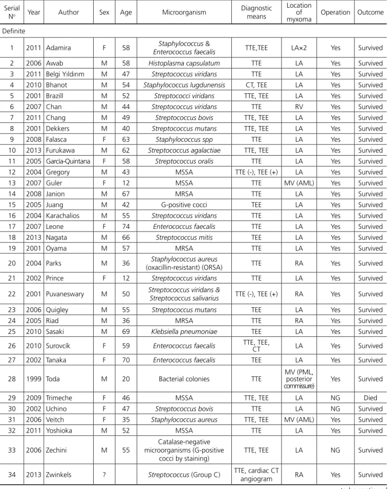
Table 1. Summary of 39 patients with infected cardiac myxoma. Serial
No Year Author Sex Age Microorganism
Diagnostic means
Location of
myxoma Operation Outcome
Definite
1 2011 Adamira F 58 Staphylococcus &
Enterococcus faecalis TTE,TEE LA×2 Yes Survived
2 2006 Awab M 58 Histoplasma capsulatum TTE LA Yes Survived 3 2011 Belgi Yıldırım M 47 Streptococcus viridans TTE LA Yes Survived 4 2010 Bhanot M 54 Staphylococcus lugdunensis CT, TEE LA Yes Survived 5 2001 Brazill M 52 Streptococci viridans TTE, TEE LA Yes Survived 6 2007 Chan M 44 Streptococcus viridans TTE RV Yes Survived 7 2011 Chang M 49 Streptococcus bovis TTE, TEE LA Yes Survived 8 2001 Dekkers M 40 Streptococcus mutans TTE, TEE LA Yes Survived 9 2008 Falasca F 63 Staphylococcus spp TTE LA Yes Survived 10 2013 Furukawa M 62 Streptococcus agalactiae TTE, TEE LA Yes Survived 11 2005 García-Quintana F 58 Streptococcus oralis TTE LA Yes Survived 12 2004 Gregory M 43 MSSA TTE (-), TEE (+) LA Yes Survived 13 2007 Guler F 12 MSSA TTE MV (AML) Yes Survived
14 2008 Janion M 67 MRSA TTE LA Yes Survived
15 2005 Juang M 42 G-positive cocci TEE LA Yes Survived 16 2004 Karachalios M 55 Streptococcus viridans TTE LA Yes Survived 17 2007 Leone F 74 Enterococcus faecalis TTE LA Yes Survived 18 2013 Nagata M 66 Streptococcus mitis TEE LA Yes Survived
19 2001 Oyama M 57 MRSA TTE LA Yes Survived
20 2004 Parks M 36 Staphylococcus aureus
(oxacillin-resistant) (ORSA) TTE RA Yes Survived 21 2002 Prince F 12 Streptococcus viridans TTE LA Yes Survived
22 2001 Puvaneswary M 50 Streptococcus viridans &
Streptococcus salivarius TTE (-), TEE (+) RA Yes Survived
23 2006 Quigley M 55 Streptococcus mutans TEE LA Yes Survived
24 2005 Riad M 36 MRSA TTE RA Yes Survived
25 2010 Sasaki M 69 Klebsiella pneumoniae TEE LA Yes Survived
26 2010 Surovcík F 59 Enterococcus faecalis TTE, TEE, CT LA Yes Survived
27 2002 Tanaka F 70 Enterococcus faecalis TEE LA Yes Survived
28 1999 Toda M 20 Bacterial colonies TTE
MV (PML, posterior
commissure) Yes Survived
29 2009 Trimeche F 46 MSSA TTE, TEE LA NG Died
30 2002 Uchino F 47 Streptococcus bovis TTE LA NG Survived 31 2006 Veitch F 35 Staphylococcus aureus TTE, TEE MV (AML) Yes Survived
32 2011 Yoshioka M 52 MSSA TTE LA Yes Survived
33 2006 Zechini M 55
Catalase-negative microorganisms (G-positive
cocci by staining)
TTE, TEE LA NG Survived
34 2013 Zwinkels ? Streptococcus (Group C) TTE, cardiac CT
Table 2. Risk factors.
Risk factor Present Revankar & Clark’s x2 P value
Recent dental problem 3 (7.7) 9 (22) 3.4 0.115
Dental surgery 1
Reconstructive dental procedures; coronary catheterization and
angioplasty 2 years earlier 1
Dental decay 1
Recent infection 4 (10.3) 4 (10) 0.0 1.000
Achilles tendon infection 2
Urinary tract infection 1
Cellulitis, web-space abscess of hand 1
Invasive procedure 4 (10.3) 2 (5) 0.8 0.432
Umbilical hernia repair 1
Amputation above knee for intractable osteomyelitis 1 Multiple surgery, closed trauma of the left knee 1 Acupuncture for weight reduction 1
Immunocompromised condition 5 (12.8) 3 (7.5) 0.6 0.481 Intravenous drug use, hepatitis C infection 2
Diabetes mellitus 1
Breast cancer (surgery, radiotherapy, chemotherapy), pharyngitis 1 Traveled to Mexico 3 months before, patent fossa ovalis 1
Total 16 (41.0) 18 (45) 0.1 0.821
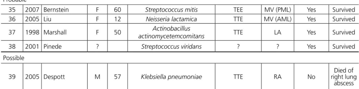
Probable
35 2007 Bernstein F 60 Streptococcus mitis TEE MV (PML) Yes Survived 36 2005 Liu F 12 Neisseria lactamica TTE MV (AML) Yes Survived
37 1998 Marshall F 50 Actinobacillus
actinomycetemcomitans TTE LA Yes Survived
38 2001 Pinede ? Streptococcus viridans ? ? Yes Survived
Possible
39 2005 Despott M 57 Klebsiella pneumoniae TTE RA No
Died of right lung
abscess
AML=anterior mitral leaflet; CT=computed tomography; F=female; LA=left atrium; M=male; MRSA=methicillin-resistant Staphylo-coccus aureus; MSSA=methicillin-sensitive StaphyloStaphylo-coccus aureus; MV=mitral valve; NG=not given; PML= posterior mitral leaflet; RA=right atrium; TEE=transesophageal echocardiography; TTE= transthoracic echocardiography
52) years (n=37). No age difference was found between male and female patients (50.7±11.4 years vs. 46.9±21.3 years, P=0.4786).
Risk factors
Recent dental procedures, recent infections, previous invasive procedures and immunocompromised conditions were the risk factors that led to a cardiac myxoma infected. No predominance was noted between the above risk factors. No significant difference was found in each risk factor between the present series and the series of Revankar & Clark[2] (Table 2).
Clinical features
Laboratory findings showed that leukocyte count was elevated in 20 patients (76.9%) and normal in 6 (23.1%) patients (x2=15.1, P=0.000) with a quantitative result of 15.7±7.3 (range, 4.9-33.6; median, 15.2) ×109/L (n=26). Hemoglobin was reduced indicating a mild anemia in 13 (92.9%) patients and normal in 1 (7.1%) patient (x2=20.6, P=0.000) with a quantitative result of
10.4±1.3 (range, 9-13.7; median, 10.8) g/dL (n=14). Erythrocyte sedimentation rate and C-reactive protein were elevated in all studied cases: erythrocyte sedimentation rate was 83.3±28.4 (range, 37-124; median, 83) mm/h (n=15) and C-reaction protein was 15.8±10.5 (range, 3-39.9; median, 13.2) mg/dL (n=17). Microscopic hematuria was present in 2 (5.4%) patients.
Table 3. Onset symptoms and signs of the patients with infected cardiac myxoma.
Feature Present Revankar & Clark’s x2 P value
Symptom
Fever 36 (97.3) 37 (92) 0.9 0.616
Embolic events 10 (27.0) 5 (12) 2.6 0.151
Weight loss 9 (24.3) 13 (32) 0.6 0.460
Dyspnea 8 (21.6) 3 (8) 3.1 0.106
Fatigue 8 (21.6) 11 (28) 0.4 0.605
Neurologic symptoms 7 (18.9) 8 (20) 0.0 1.000
Malaise 6 (16.2) 11 (28) 1.4 0.279
Rigor/shivers/chills 6 (16.2) 11 (28) 1.4 0.279
Night sweat 5 (13.5) 11 (28) 2.3 0.165
Weakness 3 (8.1) 5 (12) 0.4 0.713
Abdominal pain 3 (8.1) 3 (8) 0.0 1.000
Anorexia 3 (8.1) 3.4 0.106
Edemas 3 (8.1) 3.4 0.106
Chest pain/distress 2 (5.4) 2.2 0.228
Cough 2 (5.4) 4 (10) 0.6 0.676
Nausea/vomiting 2 (5.4) 1 (2) 0.4 0.605
Headache 1 (2.7) 4 (10) 1.7 0.360
Arm pain 1 (2.7) 1.1 0.481
Arthralgia 1 (2.7) 6 (15) 3.5 0.110
Diarrhea 1 (2.7) 3 (8) 0.9 0.616
Hemoptysis 1 (2.7) 1.1 0.481
Jaundice 1 (2.7) 1.1 0.481
Lethargy 1 (2.7) 1.1 0.481
Back pain 1 (2.7) 1.1 0.481
Myalgias 1 (2.7) 1.1 0.481
Palpitation 1 (2.7) 1.1 0.481
Septic shock 1 (2.7) 1.1 0.481
Sign
Temperature (ºC)
<37.8 5/28 (17.9) 3/32 (9) 0.9 0.454
37.8-38.9 9/28 (32.1) 19/32 (59) 4.5 0.042
>38.9 14/28 (50) 10/32 (31) 2.2 0.189
Heart murmur 18 (48.6) 26 (65) 2.1 0.172
Loud S1 3 (8.1) 14 (35) 8.1 0.006
Extra heart sound 9 (22) 9.4 0.002
“Tumor plop” 2 (5) 1.9 0.494
Splenomegaly 3 (8) 2.9 0.241
Skin lesions 3 (8.1) 6 (15) 0.9 0.484
Microorganisms
All 39 patients had microbiological evidence confirmed by one or more investigations including blood culture, culture of resected myxoma, histopathological inspection for bacteria or confirmation by polymerase chain reaction. Blood culture for microbiological isolation was positive in 37, negative in 1 and unstated in 1. Cultures of resected myxoma tissues were done in 15 patients: 9 (60%) were positive and 6 (40%) were negative. Histopathological studies of the resected myxomas were performed in 30 patients. They were histopathologically inspected for bacteria in 14 patients: 13 (92.9%) were positive and 1 (7.1%) was negative. The 41 bacteria of 39 patients were summarized in Table 4.
Histopathology showed inflammatory cell infiltrate in 11 (36.7%), necrosis in 4 (13.3%) and micro-/focal abscesses in 2 (6.7%) patients, respectively.
Complications
There were 12 patients in whom complications developed. Of these, embolic events in 10 (8 were multiple sites or multiple organs), sepsis in 4 (one was septic shock), disseminated intravascular coagulation in 3 and lung abscess in 1. The embolic events were shown in Table 5. In comparison, 18 patients of Revankar & Clark’s[2] series developed embolic events and only one of them were multisites (80% (8/10) vs. 5.6% (1/18), x2=16.3, P=0.000).
Treatment
Thirty-eight of 39 patients received a surgical treatment of infected cardiac myxoma and the reason that only patient did not undergo any surgical procedure was due to rapid deterioration leading to death. Of the 38 surgical operations, 30 were isolated cardiac myxoma resections, 5 were cardiac
Table 4. Pathogens of infective cardiac myxoma.
Pathogens Present Revankar & Clark’s x2 P value
Streptococci 17 (41.5) 20 (50) 0.6 0.507
Streptococcus viridans 7
Streptococcus mutans 2
Streptococcus bovis 2
Streptococcus mitis 2
Streptococcus (Group C) 1
Streptococcus agalactiae 1
Streptococcus oralis 1
Streptococcus salivarius 1
Staphylococci 12 (29.3) 7 (18) 1.6 0.295
MSSA 4
MRSA 3
Staphylococcus aureus 2 (1 was oxacillin-resistant (ORSA))
Staphylococcus lugdunensis 1
Staphylococcus spp 1
Staphylococcus (species not given) 1
Enterococcus faecalis 4 (9.8) 2 (5) 0.7 0.675
Gram-negative bacilli 2 (4.9) 3 (8) 0.2 0.675
Klebsiella pneumoniae 2
Gram-negative cocci 1 (2.4) 1.0 1.000
Neisseria lactamica 1
Fungus 1 (2.4) 3 (8) 1.1 0.359
Histoplasma capsulatum 1
Actinomyce 1 (2.4) 1.0 1.000
Actinobacillus actinomycetemcomitans 1
Unknown 3 (7.3) 3 (8) 0.0 1.000
Bacterial colonies 1
Table 5. Embolic events.
Location n (%)
Cerebral 1 (10)
Cerebral, peripheral (extremities) 1 (10) Cerebral, splenic 1 (10) Cerebral, splenic, renal 1 (10)
Coronary (left anterior descending coronary artery) 1 (10)
Multiple (location not indicated) 1 (10)
Multiple peripheral (left common + external iliac
arteries + right deep femoral artery) 1 (10)
Pulmonary 1 (10)
Splenic 1 (10)
Splenic, renal 1 (10)
myxoma resection with concurrent cardiac surgical procedures (mitral valve replacement in 2, and mitral valve replacement + coronary artery bypass grafting, mitral valve replacement + embolectomy of the left anterior descending coronary artery + coronary artery bypass grafting, and pulmonary valve replacement in 1 patient each) and 3 with staged peripheral operations (3rd toe of right foot amputation & right femoro-poplitereal bypass; right common iliac artery thrombectomy and embolectomy of the left common and external iliac and right deep femoral arteries in 1 patient each).
The timing for surgical operation was reported in 26 patients, 4 of them were operated on an urgent basis and 22 had a delay of 18±13.5 (range, 3-42; median, 14) days (n=21) after admission for the purpose of sufficient preoperative antibiotic treatment and stabilization of patients’ conditions. Preoperative antibiotic treatment was described in 26 patients. The frequently used antibiotics included vancomycin (7 patients), penicillin (4 patients) and ampicillin (3 patients). Sometimes, they were used along with gentamicin (80 mg every 8 h). Other antibiotics were imipenem, meropenem, peperacillin, trimethoprim/ sulfamethoxazole, oxicillin, ampicillin, nafcillin, teicoplanin, azitromicine, ceftriaxone, clindamycin and amoxicillin–clavulanic acid.
The duration of preoperative antibiotic use was 22.3±13.4 (range, 7-42; median, 18) days (n=12). Postoperative antibiotic regimens included vancomycin (500 mg every 6 h), ceftazidime (2 g every 8 h) and netilmicin (150 mg every 12 h) followed by cefepime (1 g every 12 h) plus teicoplanin (400 mg daily), nafcillin (2 g intravenously every 6 h) and gentamicin and antifungal drugs with a therapeutic course of 31.2±5.1 (range, 27-42; median, 28) days (n=9).
Outcomes
During a follow-up period of 11.1±14.5 (0.1-58; median, 8.5) months (n=16), 37 (92.6%) patients survived and 2 (7.4%) died. Of the 37 survived patients, 35 (94.6%) were event-free and 2 (5.4%) were complicated with ruptured saccular
abdominal aortic aneurysm with renal infarct and septic emboli requiring aortoiliac bypass in one patient, and increased cerebral hematoma requiring craniotomy in another. One patient died of rupture of a right lung abscess 3 weeks after admission and the other died of disseminated intravascular coagulation on postoperative day 10. The overall mortality was 5.1% and the operative mortality was 2.6%, with a significantly reduced overall mortality in comparison with that of the patient series of Revankar & Clark (overall mortality: 5.1% vs. 21%, x2=4.0, P=0.047; operative mortality: 3% vs. 2.6%, x2=1.4, P=0.239). The overall survival rate was 92.6% (Figure 1). The survival rates of the surgical and non-surgical patients were 96.2% and 0%, respectively (Figure 2).
Fig. 1 - The overall survival of the present series was 92.6%.
DISCUSSION
In 1998, Revankar & Clark[2] defined infected cardiac myxoma in three levels based on clinical and pathological findings of the myxoma:
Definite infected cardiac myxoma
1. Documented myxoma by pathology and 2a. Microorganisms seen on pathology or
2b. Positive blood cultures and inflammation on pathology. Probable infected cardiac myxoma
1. Documented myxoma by pathology and
2. Positive blood cultures or inflammation on pathology. Possible infected cardiac myxoma
1. Characteristic appearance by transthoracic or transesophageal echocardiography and
2. Positive blood cultures.
Using these criteria, the three-level infected cardiac myxoma accounted for 85%, 12.5% and 2.5% respectively as reported by Revankar & Clark[2], and 87.2%, 10.3% and 2.6% in the present series.
The diagnosis of infected cardiac myxoma can be a challenge. The time interval from symptom onset to establishment of the diagnosis varied from 0 to 126 (median, 4) months[40].Pathogens can be evidenced by blood culture, culture or staining of resected myxoma, and occasionally confirmation by polymerase chain reaction is helpful. The transthoracic or transesophageal echocardiography is a reliable means for the detection of a cardiac myxoma. Only in patients with rapid deterioration leading to sudden death was the diagnosis of a cardiac myxoma established by autopsy.
Comparisons between the two series revealed that the present series were characterized by few occasions of moderate-grade fever (while more occasions of high-moderate-grade fever in spite of lack of a statistical significance) and abnormal heart sound, but more uncommon microorganisms and more severe complications -- multiple embolic events. Other clinical features of the two patient series did not differ from each other. The somehow aggravated situations may contribute to the presence of refractory microorganisms, such as Gram-negative bacteria, fungus and actinomyce.
In comparison with Revankar & Clark’s[2] series, the present series disclosed a significantly decreased overall mortality. It is believed that refinement of the prompt diagnosis and timely management (use of sensitive antibiotics and surgical resection of the infected myxoma) have resulted in better outcomes of such patients.
CONCLUSION
In conclusion, the present series of infected cardiac myxoma illustrated somehow aggravated clinical manifestations (relative more occasions of high-grade fever, multiple embolic events and the presence of refractory microorganisms), which should draw sufficient attention to be careful in the diagnosis and treatment. In general, the prognosis of infected cardiac myxoma is relative benign and now the long term survival is always promising.
Authors’ roles & responsibilities
SMY Study conception and design; analysis and/or interpretation of data; manuscript writing, final approval of the manuscript.
REFERENCES
1. Nath MP, Singh B, Chakrabarty A. Left atrial myxoma presenting as stroke: case report & review of literature. Indian Anaesth Forum [Accessed March 23, 2014]. Available at: http://www.theiaforum.org 2. Revankar SG, Clark RA. Infected cardiac myxoma. Case report and
literature review. Medicine (Baltimore). 1998;77(5):337-44. 3. Adamira M, Justik P, Ulman J, Brezina A, Mirejovsky T, Trnkova M.
Two sporadic infected cardiac myxomas in 1 patient. Tex Heart Inst J. 2011;38(2):191-3.
4. Awab A, Hamadani M, Sud B, Voskuhl GW. Infected atrial myxoma. Med J Aust. 2006;185(6):332.
5. Belgi Yıldırım A, Er A, Küçük M, Ozbilim G. Infected giant left atrial myxoma: an unusual phenomenon. Anadolu Kardiyol Derg. 2011;11(1):83-5.
6. Bhanot N, Sahud AG, Bhat S, Lane S, Manyam H, Chan-Tompkins NH. Fever of unknown origin: a case of cardiac myxoma infected with Staphylococcus lugdunensis. South Med J. 2010;103(7):697-700. 7. Brazill PL. Infected cardiac myxoma: an unusual phenomenon. Can
Oper Room Nurs J. 2001;19(3):20-1.
8. Chan V, Veinot JP, Hynes M, Lapierre H, Ruel M. Infected right ventricular myxoma and pulmonary valve endocarditis. J Thorac Cardiovasc Surg. 2007;134(1):248-9.
9. Chang JH, Kim JY, Yoon JW, Seol MD, Won DJ, Cho WH, et al. A case of infected left atrial myxoma with concomitant mitral valve endocarditis. Korean Circ J. 2011;41(10):618-21.
10. Dekkers P, Elbers HR, Morshuis WJ, Jaarsma W. Infected left atrial myxoma. J Am Soc Echocardiogr. 2001;14(6):644-5.
11. Falasca K, Ucciferri C, Mancino P, Di Girolamo A, Vecchiet J. Infected atrial myxoma: a rare cause of fever. Infez Med. 2008;16(1):40-2. 12. Furukawa A, Kishi S, Aoki J. Large infected atrial myxoma with
vegetations. Rev Esp Cardiol. 2013;66(4):310.
13. García-Quintana A, Martín-Lorenzo P, Suárez de Lezo J, Díaz-Escofet M, Llorens R, Medina A. Infected left atrial myxoma. Rev Esp Cardiol. 2005;58(11):1358-60.
14. Gregory SA, O’Byrne WT 3rd, Fan P. Infected cardiac myxoma. Echocardiography. 2004;21(1):65-7.
15. Guler N, Ozkara C, Kaya Y, Saglam E. Ruptured abdominal aortic aneurysm after resection of an infected cardiac myxoma. Tex Heart Inst J. 2007;34(2):233-5.
16. Janion M, Sielski J, Ciuraszkiewicz K. Sepsis complicating giant cardiac myxoma. Am J Emerg Med. 2008;26(3):387.e3-4.
17. Juang SE, Lai HC, Lan YC, Liu TJ, Lai HC. Left atrial infective endocarditis with giant vegetation without involvement of the mitral valve: case report of transesophageal echocardiography in diagnosis. Acta Anaesthesiol Taiwan. 2005;43(3):165-7.
18. Karachalios G, Bablekos G, Karachaliou I, Zoganas L, Charalabopoulos A, Charalabopoulos K. Left atrial myxoma prolapsing into the left ventricle. Case report and review of the literature. Chemotherapy. 2004;50(6):297-301.
19. Leone S, dell’aquila G, Giglio S, Magliocca M, Maio P, Nigro FS, et al. Infected atrial myxoma: case report and literature review. Infez Med. 2007;15(4):256-61.
21. Oyama H, Nakayama M, Ikeda A, Maeda M, Miyahara T, Inoue S, et al. A case of cardiac myxoma with multiple brain hemorrhage. No Shinkei Geka. 2001;29(6):533-7.
22. Parks JD, Thangathurai D, Farhoomand L, Riad MG. Infected atrial myxoma presenting with septic shock. [Accessed March 30, 2014]. Available at: http://www.usc.edu/schools/medicine//departments/ anesthesiology/assets/WARC2004/parks1.pdf
23. Prince JM, Larsen R, Janner D. Infected cardiac myxoma. Pediatr Infect Dis J. 2002;21(2):177-8.
24. Puvaneswary M, Thomson D. Magnetic resonance imaging features of an infected right atrial myxoma. Australas Radiol. 2001;45(4):501-3.
25. Quigley RL, Meursing DF, Rossman MI. Left atrial myxoma and mitral valve endocarditis--a cause and effect: a case report. Heart Surg Forum. 2006;9(1):E486-7.
26. Riad MG, Parks JD, Murphy PB, Thangathurai D. Infected atrial myxoma presenting with septic shock. J Cardiothorac Vasc Anesth. 2005;19(4):508-11.
27. Sasaki A, Baba T. Infected left atrial myxoma: report of a case. Kyobu Geka. 2010;63(11):1009-11.
28. Surovcík R, Jebavý P, Feuereisl R, Frídl P, Pavlovic J, Stĕrba D. Infected myxoma as a cause of acute infective endocarditis. Vnitr Lek. 2010;56(2):154-6.
29. Tanaka M, Kawahito K, Adachi H, Yamaguchi A, Ino T. Infected left atrial myxoma with mitral valve endocarditis. Jpn J Thorac Cardiovasc Surg. 2002;50(3):137-9.
30. Toda R, Moriyama Y, Shiota K, Toyohira H, Taira A. Myxoma of mitral valve associated with infective endocarditis. Jpn J Thorac Cardiovasc Surg. 1999;47(6):285-7.
31. Trimeche B, Bouraoui H, Garbaa R, Mahdhaoui A, Ben Rhomdane M, Ernez-Hajri S, et al. Systemic embolism and septic shock complicated left atrial myxoma: case report. Case Rep Med. 2009.
[Accessed March 30, 2014]. Available at: http://www.hindawi.com/ journals/crim/2009/306375/abs/
32. Uchino K, Mochida Y, Ebina T, Tobe M, Kobayashi S, Yano Y, et al. Infected left atrial myxoma. Intern Med. 2002;41(11):957-60. 33. Veitch AM, Manghat NE, Kakani NK, Lewis CT, Ring NJ. Systemic
septic embolisation secondary to an atrial myxoma in a young woman. Emerg Radiol. 2006;12(3):137-9.
34. Yoshioka D, Takahashi T, Ishizaka T, Higuchi T. Successful surgical resection of infected left atrial myxoma in a case complicated with disseminated intravascular coagulation and multiple cerebral infarctions: case report. J Cardiothorac Surg. 2011;6:68.
35. Zechini B, Cipriani P, Papadopoulou S, Di Nucci G, Petrucca A, Teggi A. Endocarditis caused by Lactococcus lactis subsp. lactis in a patient with atrial myxoma: a case report. Diagn Microbiol Infect Dis. 2006;56(3):325-8.
36. Zwinkels RL, van der Sar-van der Brugge S, Sleeswijk Visser SJ. An unexpected cause of sepsis in a patient with dental decay. Rare example of an infected right atrial calcified myxoma, with extensive calcified pulmonary emboli. Acute Med. 2013;12(1):34, 59-61. 37. Bernstein JM, Leasure W, Buel A. Getting to the heart of the matter.
Skinmed. 2007;6(6):290-2.
38. Liu YL, Liu XH, Cai KH. Cardiac valve myxomas with infected endocarditis in the youngerly. Zhonghua Bing Li Xue Za Zhi. 2005;34(10):695-6.
39. Marshall C, McDonald M. Recurrent subacute bacterial endocarditis as a presentation of left atrial myxoma. Aust N Z J Med. 1998;28(3):350.
40. Pinede L, Duhaut P, Loire R. Clinical presentation of left atrial cardiac myxoma. A series of 112 consecutive cases. Medicine (Baltimore). 2001;80(3):159-72.