The host defense mechanisms are not well developed in neonates. The aim of this study was to evaluate the dynamics of microbial colonization of the oral cavity in newborns. Eighty-one samples of the oral microbiota were obtained from 51 healthy newborns 10 min to 53 h after birth by gently rubbing sterile swabs onto the surface of the tongue, cheek mucosa, alveolar process and palate. After microbiological processing, counting of the colony forming units of streptococci, staphylococci and Gram-negative aerobic bacilli was performed.Between 10 min and 8 h, Staphylococcus epidermidis was detected in 30.7% of the samples; between 8 and 16 h, S. epidermidis was detected in 69.5% of the samples and streptococci in 56.5% of the samples; between 16 and 24 h, S. epidermidis, streptococci and S. aureus were detected in 77.78%, 85.18% and 37.03% of the samples, respectively. Between 24 and 53 h, S. epidermidis was detected in 88.89%, streptococci in 94.4% and S. aureus in 33.3% of the samples. Mutans streptococci were not detected in any of the samples. The adoption of strict hygienic measures by the mother and the nursing staff should be emphasized to avoid or at least delay the occurrence of infections caused by microorganisms in newborns. In addition, hospital procedures must be aseptic and invasive interventions must be minimized.
Dynamics of Microbial Colonization
of the Oral Cavity in Newborns
Paulo Nelson-Filho, Izabela Gonçalves Borba, Késsia Suênia Fidelis de Mesquita, Raquel Assed Bezerra Silva, Alexandra Mussolino de Queiroz, Léa Assed Bezerra Silva
Department of Pediatric Dentistry, School of Dentistry of Ribeirão Preto, University of São Paulo, Ribeirão Preto, SP, Brazil
Correspondence: Prof. Dr. Paulo Nelson Filho, Av. do Café, s/n, Monte Alegre, 14040-904 Ribeirão Preto, SP, Brasil. Tel: +55-16-3602-4786. Fax: +55-16-3633-0999. e-mail: [email protected]
Key Words: newborns, defense, microbial colonization, microorganisms.
Introduction
Most bacterial infections in neonates are usually acquired during or immediately after delivery (1). The host defense mechanisms in neonates are not well developed at the moment of birth and some commensals may become opportunistic pathogens, particularly in compromised babies who stay in the hospital for treatment of congenital abnormalities (1-5). Premature infants in neonatal intensive care units (NICU) (6) and those undergoing invasive life-saving medical interventions are at greater risk of developing infection (7).
Vaginal delivery inevitably results in exposure of the newborn to the normal maternal genital tract microflora (8). Although the newborn’s oral cavity is usually free microorganisms (9,10), colonization begins within a few hours after birth with microorganisms from the mother, nurses and sometimes from the environment (10). At birth and during the first months of life, the oral mucosa surfaces of babies can provide niches for bacterial colonization (3). It has been demonstrated that 8 days after birth, there is considerable variability in oral bacteria, and both aerobes (9) and anaerobes (11) can be detected in the oral cavity. The Gram-positive bacterial commensal Streptococcus salivarius is a pioneer colonizer of the human oral cavity (1,10), being detected from 8 h after birth (1). Large populations persist at this site for the host’s lifetime (9,12), and this bacterial species represents up to 98% of the total oral microbiota until the eruption of teeth (13).
The metabolic activity of pioneer species alters the
oral environment, providing acceptable conditions for colonization by other microbial species (10). Before tooth eruption, aerobic microorganisms (streptococci and staphylococci) are prevalent in the oral microbiota, with predominance of facultative microorganisms (14).
Interventions sustaining those who are hospitalized can result serious infections due to common nosocomial pathogens, particularly coagulase-negative staphylococci bacteria (CoNS) (7). Staphylococcus aureus (2,4), a coagulase-positive staphylococci bacterium, and S. epidermidis (5), a coagulase-negative staphylococci bacterium, are the most common cause of nosocomial infections in newborns, resulting in considerable morbidity and mortality (15).
Different mutans streptococci species may cause of several pathologies. Streptococcus mutans and S. sobrinus, for example, are important species associated with dental caries (16) and colonize the oral cavity after tooth eruption. However, S. salivarius is present naturally on the tongue and cheek mucosa because it has a unique ability to adhere to these surfaces (17). S. sanguinis colonization in infants occurs during a small “window of infectivity” around the age of 9 months. This colonization is tooth-dependent and is associated with the time of tooth emergence, but the levels of S. sanguinis decrease after mutans streptococci colonization (18).
416
P
. Nelson-Filho et al.
at a nursery in a university hospital.
Material and Methods
Eighty-one samples of the oral microbiota were obtained from 51 newborns (32 males and 19 females) after granting written parental consent and authorization from the Head of the Department of Pediatrics, in a university hospital.
Newborns of mothers who had no complications during pregnancy were selected, regardless of the form of delivery (vaginal or caesarean section). Only healthy newborns delivered at term, with appropriate weight for the gestational age and who were not under medication participated in this study. All newborns were under continued exclusive breast-feeding.
Bacterial samples for microbiological analysis were collected from the neonates in the birthing room within 10 min to 53 h after birth by gently rubbing the surface of tongue, cheek mucosa, alveolar process and palate with sterile swabs. Immediately after sample collection, the swabs were stored in sterile test tubes containing 1 mL of phosphate buffered saline (PBS). Samples were grown on Mitis Salivarius (MS) (Difco, Detroit, MI, USA) agar in 10x60 mm Petri dishes and on modified SB-20 (SB-20M) (19), Agar Ni and Mac Conkey (MC) (Difco) culture media in 20x100 mm Petri dishes.
Using a sterile pipette, 0.05 mL of each sample was
collected and spread onto the MS agar surface using a sterile bent glass rod as the spreading device. Then, 0.50 mL of each sample was subjected to serial decimal dilutions of up to 10-4 in PBS, and 0.05 mL of the resulting dilutions was sown on the MS agar surface. After seeding, the Petri dishes were incubated in anaerobic jars using the candle flame method at 37 °C for 48 h.
In the SB-20M medium, the samples were directly inoculated with cotton swabs, providing 5 samples per dish and the Petri dishes were incubated in anaerobic jars using the candle flame method at 37 °C for 48 h. Also, other 5 samples per dish were seeded in the MC and Ni media. The Ni dishes were incubated at 37 °C for 48 h and the MC dishes were incubated at 37°C for 24 h.
After the incubation period, the number of colony
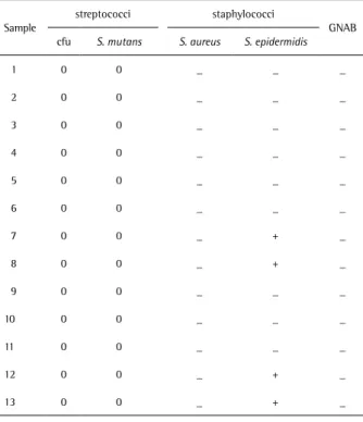
Table 2. Microbiological analysis of samples (n=13) between 8 h and 16 h after birth
Sample
streptococci staphylococci
GNAB cfu S. mutans S. aureus S. epidermidis
14 200 0 _ _ _
15 0 0 _ _ _
16 40 0 _ _ _
17 0 0 _ _ _
18 0 0 _ + _
19 60 0 _ + _
20 0 0 _ + +
21 0 0 _ + _
22 1,050 0 _ + _ 23 670,000 0 _ + _
24 80 0 _ + _
25 0 0 _ + _
26 18,700 0 _ + _
27 0 0 _ _ _
28 0 0 _ + _
29 550,000 0 _ + _ 30 5,400 0 _ + _
31 0 0 _ _ _
32 4,600 0 _ + _ 33 3,800,000 0 _ + _ 34 158,000 0 _ + _
35 0 0 _ _ _
36 3,400 0 _ + _ +: Positive bacterial growth. -: Negative bacterial growth. GNAB: Gram-negative aerobic bacilli.
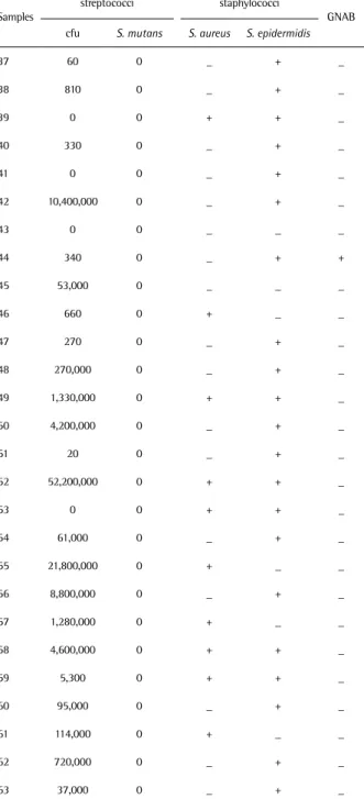
Table 1. Microbiological analysis of samples (n=13) between 10 min and 8 h after birth
Sample
streptococci staphylococci
GNAB cfu S. mutans S. aureus S. epidermidis
1 0 0 _ _ _
2 0 0 _ _ _
3 0 0 _ _ _
4 0 0 _ _ _
5 0 0 _ _ _
6 0 0 _ _ _
7 0 0 _ + _
8 0 0 _ + _
9 0 0 _ _ _
10 0 0 _ _ _
11 0 0 _ _ _
12 0 0 _ + _
13 0 0 _ + _
417
Colonization of the oral cavity of newborns
forming units (cfu) of total streptococci (viridans) in the MS medium, mutans streptococci in the SB-20M medium, S. aureus and S. epidermidis in the Ni medium, and Gram-negative aerobes in the MC medium were counted with a stereoscopic microscope.
Results
Between 10 min and 8 h after birth, S. epidermidis was detected in 4 of the 13 samples. In the other 9 samples, there was no bacterial growth until 8 h (Table 1).
Between 8 h and 16 h after birth, 13 of the 23 samples showed growth of streptococci, ranging from 40 to 3,800,000 cfu. S. epidermidis was detected in 16 samples, and only 1 sample showed growth of Gram-negative aerobic bacilli. Five samples did not show bacterial growth (Table 2).
Between 16 h and 24 h after birth, 23 of the 27 samples showed growth of streptococci, ranging from 20 to 52,200,000 cfu. Ten samples showed growth of S. aureus, and 21 samples showed growth of S. epidermidis. Gram-negative aerobic bacilli were detected in only 1 sample. Only 1 sample showed absence of bacterial growth (Table 3).
Between 24 h and 53 h after birth, all samples presented bacterial growth. Seventeen of the 18 samples showed growth of streptococci, ranging from 100 to 70,200,000 cfu. Sixteen samples showed growth of S. epidermidis and 6 samples showed growth of S. aureus. Gram-negative aerobic bacilli were found in 2 samples (Table 4).
Mutans streptococci were not detected in any of the 81 samples analyzed, regardless of time after birth.
Table 3. Microbiological analysis of samples (n=27) between 16 h and 24 h after birth
Samples
streptococci staphylococci
GNAB cfu S. mutans S. aureus S. epidermidis
37 60 0 _ + _
38 810 0 _ + _
39 0 0 + + _
40 330 0 _ + _
41 0 0 _ + _
42 10,400,000 0 _ + _
43 0 0 _ _ _
44 340 0 _ + +
45 53,000 0 _ _ _
46 660 0 + _ _
47 270 0 _ + _
48 270,000 0 _ + _ 49 1,330,000 0 + + _ 50 4,200,000 0 _ + _
51 20 0 _ + _
52 52,200,000 0 + + _
53 0 0 + + _
54 61,000 0 _ + _ 55 21,800,000 0 + _ _ 56 8,800,000 0 _ + _ 57 1,280,000 0 + _ _ 58 4,600,000 0 + + _ 59 5,300 0 + + _ 60 95,000 0 _ + _ 61 114,000 0 + _ _ 62 720,000 0 _ + _ 63 37,000 0 _ + _ +: Positive bacterial growth. -: Negative bacterial growth. GNAB: Gram-negative aerobic bacilli.
Table 4. Microbiological analysis of samples (n=17) between 24 h and 53 h after birth
Samples
streptococci staphylococci
GNAB cfu S. mutans S. aureus S. epidermidis
64 37,800,000 0 _ + _ 65 19,000,000 0 _ + _ 66 4,200,000 0 _ + _ 67 9,400,000 0 + + _
68 0 0 _ + _
69 4,600,000 0 _ + _ 70 15,600,000 0 + + _ 71 6,200,000 0 _ + + 72 28,800,000 0 _ + _ 73 54,600,000 0 + + _ 74 7,200,000 0 + + _ 75 56,600,000 0 _ + + 76 16,400,000 0 _ + _ 77 2,810,000 0 + + _ 78 20,200,000 0 _ _ _
79 100 0 _ _ _
418
P
. Nelson-Filho et al.
Discussion
Identification of the indigenous oral microbiota is complicated because the individual may have had contact with different types of microorganisms. The presence of these microorganisms may be transient in the oral cavity, depending on the conditions encountered during their metabolism and growth (9).
Previous studies have reported that the oral cavity of children is sterile until 10 h (14) and 24 h (9) after birth and other findings point to an increase in the number of viable microorganisms between 6 and 10 h after birth (14). In the present study, only 30,7% of the samples showed bacterial growth between 10 min and 8 h after birth.
Silva et al. (4) identified S. aureus in the nostrils, mouth and anus of 49% of newborns. In this study, between 16 and 24 h after birth, S. aureus were present in 37.03% of the samples, and after 53 h of life, S. aureus were present in 33.3% of the samples, thereby showing a considerable risk of early infection. According to Zdrazílek et al. (2),S. aureus frequently colonize nipples and are regularly found in the umbilical stump and in eye and mouth corners of babies. Therefore, mothers should be instructed to wash their hands and then the nipples prior to breast-feeding to delay the colonization of S. aureus in newborns, who have immature immune systems. Although it is likely that the transmission of microorganisms may occur from the mother to the newborn, the microorganisms may also be transmitted via the hands of the nursing staff (20) and use of indwelling vascular catheters, high-calorie hyperalimentation infusions, and assisted ventilation (21).
The analysis of oral samples from 51 newborns at 1 and 8 days after birth revealed that approximately half of the children presented very low staphylococci levels (9). In this study, S. aureus, a pathogenic species, was detected in approximately 19.8% of the samples from newborns, possibly due to the contamination of the oral cavity by extrinsic sources, such as the attendants (8).
In the present study, S. epidermidis was the first microorganism to colonize newborn’s oral cavity between 10 min and 8 h (30.7%) after birth, and the counts of this microorganism increased by approximately 90% after 53 h of neonatal life. According to Van Mellaert et al. (5),S. epidermidis, previously considered a human commensal, are now regarded as a frequent cause of nosocomial infections and the most common cause of device-related infections. Thus, early and intense infection caused by this microorganism in the first hours of life can be dangerous. Preventive hygienic measures should be strengthened and encouraged.
During the first year of life, streptococci, staphylococci, neisseria, lactobacilli, veillonella (9), actinomyces and fusobacteria (10) are the most prevalent microorganisms
in the oral cavity of the infant (9). In this study, the most prevalent genera in the samples tested were streptococci in approximately 65.4% of the samples and staphylococci in approximately 75.3% of the samples. The present results agree with those of McCarthy et al. (9),demonstrating that streptococci make up 98% of the total bacteria in the first and second day of life. Streptococcus mitis biovar 1 and Streptococcus oralis were also detected in the oral cavity of newborns until 3 days after birth (22). S. mitis is considered as the major component of the oral streptococcal flora in infants at the end of the first year of life (12).
There is controversy regarding the colonization of the oral cavity in newborns by S. mutans. Some authors report that newborns (12,23) do not harbor S. mutans on their mucous membranes, whereas there is a report demonstrating that S. mutans can be detected in children as young as 2 months old (24) and at 3 months of age (25), prior to tooth eruption (25). In the present study, S. mutans were not detected in the samples, and this result is in agreement with the several reports in the literature (12,23).
The extent to which perinatal events influence the acquisition of indigenous microorganisms in the oral cavity remains unclear. Additional studies on the natural history of these indigenous microorganisms may provide general information on the indigenous biota. However, early colonization of newborn’s oral cavity by many bacterial species in those who have immature immune systems can predispose them to a disease.
In conclusion, the number of bacterial species in the oral cavity of neonates increased over time. Streptococci and S. epidermidis were detected in nearly all samples in the evaluated time spam. S. aureus was detected in one-third of the samples, and the number of samples without any evidence of bacteria decreased from 69.3% in the first period of evaluation (between 10 min and 8 h after birth) to 0% in the last period of evaluation (between 24 and 53 h after birth). The adoption of strict hygienic measures by the mother and the nursing staff should be emphasized to delay the occurrence of infections caused by microorganisms in newborns. In addition, hospital procedures must be aseptic and invasive interventions must be minimized as much as possible.
Resumo
Os mecanismos de defesa do hospedeiro não estão bem desenvolvidos em neonatos. O objetivo deste estudo foi avaliar a dinâmica da colonização microbiana da cavidade oral em recém-nascidos. Oitenta e uma amostras da microbiota oral foram obtidas a partir de 51 recém-nascidos saudáveis, entre 10 min e 53 h após o nascimento, esfregando-se, suavemente, cotonetes esterilizados sobre a superfície da língua, mucosa bucal, processo alveolar e palato. Após o processamento microbiológico foi realizada a contagem de unidades formadoras de colônias de estreptococos, estafilococos e bacilos aeróbios Gram-negativos. Entre 10 min e 8 h,
419
Colonization of the oral cavity of newborns
entre 8 e 16 h, S. epidermidis foram detectados em 69,5% das amostras e streptococci foram detectados em 56,5% das amostras. Entre 16 e 24 h, S. epidermidis, streptococci e S. aureus foram detectados em 77,78%, 85,18% e 37,03% das amostras, respectivamente. Entre 24 e 53 h, S. epidermidis
foram detectados em 88,89% das amostras, streptococci em 94,4% e S. aureus em 33,3% das amostras. S. mutans não foram encontrados em nenhuma amostra. A adoção de medidas de higiene rigorosas por parte da mãe e da equipe de enfermagem deve ser enfatizada para evitar ou pelo menos retardar a ocorrência de infecções causadas por microorganismos em recém-nascidos. Adicionalmente, procedimentos hospitalares devem ser assépticos e intervenções invasivas devem ser minimizadas.
Acknowledgements
The authors are indebted to Dr. Izabel Yoko Ito (in memoriam) of the Department of Clinical Analyses, Toxicology and Bromatology, School of Pharmaceutical Sciences of Ribeirão Preto, University of São Paulo, for the invaluable collaboration during microbiological processing and analysis. The authors also acknowledge Jaqueline E. C. V. Menezes for her assistance during sample collection.
References
1. Rotimi VO, Duerden BI. The development of the bacterial flora in normal neonates. J Med Microbiol 1981;14:51-62.
2. Zdrazílek J, Petrás P, Srámová H, Subertová V, Masková L.
Staphylococcusaureus at a maternity ward. I. Colonization of mothers
and neonates and survival of various S. aureus types in colonized individuals. J Hyg Epidemiol Microbiol Immunol 1988;32:49-57. 3. Könönen E. Development of oral bacterial flora in young children. Ann
Med 2000; 32:107-112.
4. Silva HA, Abdallah VOS, Carneiro CL, Gontijo Filho PP. Infection and colonization by Staphylococcus aureus in a high risk nursery of a Brazilian teaching hospital. Braz J Infect Dis 2003;7:381-386. 5. Van Mellaert L, Shahrooei M, Hofmans D, Eldere JV. Immunoprophylaxis
and immunotherapy of Staphylococcus epidermidis infections: challenges and prospects. Expert Rev Vaccines 2012;3:319-334. 6. Bokulich NA, Mills DA, Underwood M. Surface microbes in the neonatal
intensive care unit: changes with routine cleaning and over time. J Clin Microbiol 2013;51:2617-2624.
7. Marchant EA, Boyce GK, Sadarangani M, Lavoie PM. Neonatal sepsis due to coagulase-negative staphylococci. Clin Dev Immunol 2013 [Epub ahead of print. DOI: 10.1155/2013/586076].
8. Ross JM, Needham JR. Genital flora during pregnancy and colonization of the newborn. J R Soc Med 1980;73:105-110.
9. McCarthy C, Snyder ML, Parker RB. The indigenous oral flora of man. The newborn to the 1 year-old infant. Arch Oral Biol 1965;10:61-70. 10. MacFarlane TW, Samaranayake LP. Oral ecosystem and dental plaque.
In: Clinical Oral Microbiology. Editor: Sayer L. London: Wright, 1989. 11. Brook I. Anaerobic infections in children. Adv Exp Med Biol
2011;697:117-152.
12. Smith DJ, Anderson JM, King WF, van Houte J, Tauhman MA. Oral streptococcal colonization of infants. Oral Microbiol Immunol 1993;8:1-4.
13. Cortelli JR, Aquino DR, Cortelli SC, Franco GCN, Fernandes CB, Roman-Torres CVG et al. Detection of periodontal pathogens in oral mucous membranes of edentulous individuals. J Periodontol 2008;79:1962-1965.
14. Kosteka F. Relation of the teeth to the normal development of microbial flora in the oral cavity. The Dental Cosmos 1924; 66:927-935. 15. Tapia-Rombo CA, Ugarte-Torres RG, Alvarez-Vázquez E, Salazar-Acuña
AH. Risk factors for intrahospital infection in newborns. Arch Med Res 2001;32:304-311.
16. Coykendall AL. Classification and identification of the viridans streptococci. Clin Microbiol Rev 1989;2:315-328.
17. Gibbons RJ, Houte JV. Selective bacterial adherence to oral epithelial surfaces and its role as an ecological determinant. Infect Immun 1971;3:567-573.
18. Caufield PW, Dasanayake AP, Li Y, Pan Y, Hsu J, Hardin JM. Natural History of Streptococcus sanguinis in the oral cavity of infants: evidence for a discrete window of infectivity. Infect Immun 2000;68:4018-4023.
19. Saravia ME, Nelson-Filho P, Ito IY, da Silva LA, Emilson CG. Morphological differentiation between S. mutans and S. sobrinus on modified SB-20 culture medium. Res Microbiol 2011;166:63-67. 20. Safdar N, Maki DG. The commonality of risk factors for nosocomial
colonization and infection with antimicrobial-resistant Staphylococcus
aureus, enterococcus, Gram-negative bacilli, Clostridium difficile, and
Candida. Ann Intern Med 2002;136:834-844.
21. Baltimore RS. Neonatal nosocomial infections. Seminars in perinatology 1998;22:25-32.
22. Pearce C, Bowden GH, Evans M, Fitzsimmons SP, Johnson J, Sheridan MJ, et al.. Identification of pioneer viridians streptococci in the oral cavity of human neonates. J Med Microbiol 1995;42:67-72. 23. Straetemans MM, van Loveren C, de Soet JJ, de Graaff J, ten Cate JM.
Colonization with mutans streptococci and lactobacilli and the caries experience of children after the age of five. J Dent Res 1998;77:1851-1855.
24. Tankkunnasombut S, Youcharoen K, Wisuttisak W, Vichayanrat S, Tiranathanagul S. Early colonization of mutans streptococci in 2- to 36-month-old Thai children. Pediatr Dent 2009;31:47-51.
25. Wan AK, Seow WK, Walsh LJ, Bird P, Tudehope DL, Purdie DM. Association of Streptococcusmutans infection and oral developmental nodules in pre-dentate infants. J Dent Res 2001;80:1945-1948.