This study evaluated in vitro the influence of thermocycling and water storage (WS) on
the shear bond strength (SBS) of composite resin in cavities prepared in primary tooth enamel with conventional bur or Er:YAG laser. The test surfaces were obtained from 48 primary molars and randomly assigned to 2 groups (n=24), according to cavity preparation: A: bur-preparation and B: Er:YAG laser irradiation. The specimens were restored with an etch-and-rinse adhesive system and composite resin. Each group was divided into 4 subgroups (n=6) according to WS duration and number of thermal cycles (TCs): I: 24 h WS/no thermocycling; II: 7 days WS/500 TCs; III: 1 month WS/2,000 TCs; IV: 6 months WS/12,000 TCs. The specimens were tested to failure in shear strength at a crosshead speed of 0.5 mm/min. Data were analyzed statistically by two-way ANOVA and Tukey’s test. SBS means (S.D.) in MPa were: AI: 17.45 (2.03), AII:16.38 (1.49), AIII: 6.88 (0.66), AIV: 7.77 (1.53), BI: 12.32 (0.99), BII: 15.37 (2.24), BIII: 15.05 (2.01) and BIV-5.51 (1.01). WS duration and number of TCs influenced significantly the SBS values only for BIV (p<0.05). AI presented the highest SBS value, which was statistically similar to those of AII, BII and BIII. In conclusion, the adhesion of an etch-and-rinse adhesive to Er:YAG laser-irradiated primary tooth enamel was affected by the methods used to simulate degradation of the adhesive interface only when 6 months WS/12,000 TCs were employed.
Bond Durability of Er:YAG
Laser-Prepared Primary Tooth Enamel
Maria Cristina Borsatto1, Mayara Garcia Martinelli1, Marta Maria Martins Giamatei Contente1, Talitha de Siqueira Mellara1, Jesus Djalma Pécora2, Rodrigo Galo3
1Department of Pediatric Dentistry; 2Department of Restorative Dentistry, 3Department of Dental Materials
and Prosthodontics, Ribeirão Preto School of Dentistry, University of São Paulo, Ribeirão Preto, SP, Brazil
Correspondence: Profa. Dra. Maria Cristina Borsatto, Avenida do Café, S/N, Monte Alegre, 14040-904 Ribeirão Preto, SP, Brasil. Tel: +55-16-3602-4114. Fax: +55-16-3633-0999. e-mail: [email protected]
Ke y Wo rd s : E r : YA G l a s e r, thermocycling, water storage, shear bond strength, primary teeth.
Introduction
Contemporary dental principles recommend, as often as possible, noninvasive rather than invasive strategies.In this context, cavity preparation with high-level lasers, such as Er:YAG laser, has gained popularity in the last years (1). This technology has been presented as a viable option for replacing the conventional high-speed air turbine and low-speed burs, as it is claimed to offer greater patient comfort by reducing vibration, pressure and noise associated with rotary dental instruments (1). These characteristics are of special interest for pediatric dentistry.
As a rule, adhesive dental materials are developed to be applied on tooth substrate prepared with rotary instruments and treated with conventional techniques (2). It is yet to be determined how the morphological changes produced by laser ablation could affect the behavior of these materials. Protocols that mimic the natural aging process of dental restoration should be used to test the bond strength (BS) of contemporary adhesive systems to Er:YAG-prepared teeth. The structural and morphological differences between primary and permanent enamel substrates may interfere with the adhesion mechanism (3). The presence of a thicker “prismless” layer and a less mineralized substrate in primary teeth is an example of these differences (4), and reinforce the need of caution when extrapolating results obtained with permanent teeth to primary teeth. To the best of our knowledge, the adhesion of composite resin to a
laser-prepared primary enamel surface after a long-term degradation process has not yet been assessed.
In an attempt to reproduce the natural aging process of a dental restoration, thermocycling protocols (5) and water storage (WS) of bonded specimens (6) have been suggested as efficient methods to provide in vitro simulation of in vivo conditions. Thermocycling has been the most used method to stress the adhesive interface (7), while WS has been shown to reduce BS, even after short periods of storage, indicating the degradation of bonds over time (8).
This study assessed the shear bond strength (SBS) of a composite resin to bur- and Er:YAG-prepared primary tooth enamel after different thermocycling regimens and WS periods. The tested null hypothesis was that adhesion to Er:YAG laser-prepared primary tooth enamel is not affected by methods used to simulate adhesive interface degradation.
Material and Methods
331
E
r:
Y
A
G laser-prepared primary bond
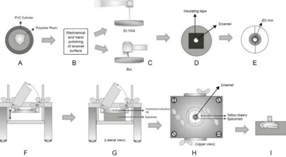
2 mm below the cementoenamel junction and crowns were embedded in polyester resin using polyvinyl chloride rings (2.1 cm diameter and 1.1 cm height) (Fig. 1A) (9). After resin polymerization, the rings were discarded and the enamel was polished with wet silicon carbide papers (Buehler Ltd., Lake Bluff, IL, USA) in a low-speed polishing machine (Politriz DP-9U2; Struers, A/S) (Fig. 1B) followed by hand polishing to obtain flat, smooth test surfaces, which were cleaned by rubber cup/pumice prophylaxis for 10 s. A 3-mm-diameter bond site was demarcated in each surface, and the test surfaces were randomly assigned to 2 groups (n=48), according to the cavity preparation: A: bur preparation and B: Er:YAG laser preparation (Fig. 1C). The A group was prepared with a #245 carbide bur (KG Soresen, Barueri, SP, Brazil) and a high-speed handpiece (Dabi-Atlante, Ribeirão Preto, SP, Brazil) with water/air spray for 10 s at 250 r.p.m. The high-speed handpiece was fixed in a cavity preparation machine (Marcelo Nucci ME, São Carlos, SP, Brazil) in which axle movement was monitored by digital comparison clocks, giving a precision of 0.01 mm in cavity dimensions. New burs were replaced after every five preparations.
In B group, the Er:YAG laser device used was the Kavo Key Laser 2 model (Kavo Dental GmbH & Co, Biberach, Germany), emitting at 2.94-mm wavelength Er:YAG laser, with 250 mJ pulse energy and a 2 Hz repetition rate. The laser beam (spot size=0.63 mm) was delivered in non-contact and focused mode with a fine water mist for 20 s. The device's 2051 handpiece was attached to the flexible fiber delivery system. The irradiation distances were standardized using a custom-made apparatus consisting of a holder
that positioned the handpiece in such a way that the laser beam was delivered perpendicular to enamel surface at a fixed working distance of 12 mm from the target site. This was used in conjunction with a semi-adjustable base, to which the specimen was fixed with wax. A previously trained operator handled the base's micrometer screws to alternately move it in right-to-left and forward-to-backward directions, thereby allowing the laser beam to provide a precise irradiation of the demarcated site.
After cavity preparation, bonding was performed according to the manufacturer’s instructions: surfaces were etched with a 35% phosphoric acid gel (Scotchbond etchant; 3M ESPE, St. Paul, MN, USA) for 15 s, rinsed thoroughly for 15 s, and dried with a mild, oil-free air stream to obtain a uniformly white, dull, chalk-like appearance. Single Bond 2 Adper adhesive system (3M ESPE) was applied in two consecutive coats with disposable tips (Microbrush Corporation, Grafton, WI, USA). The adhesive system was slightly thinned with a mild oil-free air stream and photoactivated for 10 s with a visible light-curing unit with a 450-mW/cm2 intensity (XL 3000; 3M/ESPE), as measured with a curing radiometer (Demetron Research Corp., Danbury, CT, USA).
After completion of the bonding protocol, each specimen was fixed in a clamping device to maintain the test enamel surface parallel to a flat base (Fig. 1F). A split bisected polytetrafluoroethylene jig was positioned on the tooth/resin block, providing a cylindrical cavity (4 mm height x 3 mm diameter), which coincided with the demarcated bonding site (Fig. 1E). A resin cylinder with the same dimensions of the jig adhered to the dentin site was
332
M.C. Borsatto et al.
built incrementally by filling a split polytetrafluoroethylene jig with Filtek Z250 light-cured composite resin (3M/ESPE) (Fig. 1G-I).
The restored specimens were maintained in distilled water at 37°C and randomly assigned to 4 subgroups (n=6), according to the duration of water storage (WS) and number of thermal cycles (TCs) employed for adhesive interface aging: I: 24 h WS/no TCs, II: 7 days WS/500 TCs, III: 1 month WS/2,000 TCs and IV: 6 months WS/12,000 TCs. This way, we had in the bur-prepared group (A): AI, AII, AIII and AIV; and in the laser-prepared group (B): BI, BII, BIII and BIV. Each cycle consisted of water baths between 5 °C and 55 °C in a thermocycling machine (Ética Equipamentos Científicos S/A, São Paulo, SP, Brazil) with a dwell time of 30 s and transfer time of 7 s each. The specimens were subjected to 500 thermal cycles per week and were kept in distilled water at 37 °C in the intervals.
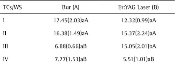
After the pre-determined time of WS/thermocylcing of each subgroup, shear bond strength (SBS) was determined using a knife-edge blade in a universal testing machine (Mod. MEM 2000; EMIC Ltda, São José dos Pinhais, PR, Brazil) at a crosshead speed of 0.5 mm/min and with a 50 kgf load cell. The means (in MPa) and standard deviations were calculated and data were analyzed statistically by two-way ANOVA. Tukey's test was used for multiple comparisons at a 5% significance level (Table 1).
A single examiner blinded to the groups examined the debonded specimens with a stereomicroscope at 40× magnification to assess the failure modes (adhesive, cohesive or mixed).
Results
SBS means and standard deviations are presented in Table 1. The analysis of the data revealed that the different periods of water storage and thermocycling had influenced the shear bond values in the Er:YAG laser-prepared groups only for the specimens that were stored 6 months associated with 12,000 TC (BIV group).
The bur-prepared group of specimens that were stored for 24 h in water and that were not submitted to
thermocycling (AI) showed the highest SBS, which were statistically similar to those obtained for the bur-prepared group submitted to 500 TCs/1 week WS (AII) and to the Er:YAG laser-prepared specimens that were submitted to 500 TCs/1 week WS (BII) and 2,000 TCs/1 month WS (BIII).
For each WS period and thermocycling regimen, the data revealed that cavity preparation with both Er:YAG and bur yielded SBS values that were statistically similar in the groups subjected to 500 TCs/1 week WS (II) and 12,000 TCs/6 months WS (IV).
Failure mode distribution is presented in Table 2. The failure pattern on the debonded surfaces showed that most specimens in the laser-irradiated enamel group fractured more adhesively and there was an increase of the number of mixed failures when the WS periods and numbers of thermal cycles were increased. No cohesive fracture was found in both groups.
Discussion
The durability of adhesion between resin composite and dental substrate is an important issue because it is directly related to the clinical performance of dental restorations. The degradation of bonds and morphological changes in the bond structure of the tooth-restoration interface aged in the oral environment for long periods have been reported in vivo (10,11). We failed to reject the null hypothesis because bond was degraded by long-term WS and thermocycling
in vitro methods. Immersion in water has been the most
common artificial technique to predict the behavior of resin-based restorative materials (12) because the presence of water is crucial for their deterioration (13) and its effect is very important when an etch-and-rinse adhesive system is used. In addition, thermocycling is a thermal fatigue method that evaluates bond durability by inducing repetitive contraction/expansion stresses at the tooth-material interface (14) and by simulating thermal changes that occur in the oral environment by eating, drinking and breathing (15). Simulation of thermal stress and long-term degradation in the oral environment adversely affected the adhesion to Er:YAG laser-irradiated enamel.
An increasing popularity of laser technology in dental practice has been observed because of the possibility of removing hard dental tissues safely and effectively (16) with preservation of health structures (17) and less noise or Table 1. Shear bond strength (MPa) means and standard deviations
in each group
TCs/WS Bur (A) Er:YAG Laser (B)
I 17.45(2.03)aA 12.32(0.99)aA
II 16.38(1.49)aA 15.37(2.24)aA
III 6.88(0.66)aB 15.05(2.01)bA
IV 7.77(1.53)aB 5.51(1.01)aB
Different letters indicate statistically significant difference (p=0.004).
Table 2. Failure mode distribution in each group
Failure
mode AI AII AIII AIV BI BII BIII BIV
Adhesive 75 65 65 50 100 75 65 50
333
Er:Y
A
G laser-prepared primary bond
vibration than with dental burs. Patients perceive laser as a more comfortable system, with significantly less need for local anesthesia (1,14). Therefore, there is a major interest in investigating the interaction pattern and the durability of the bond between the currently available adhesive systems and laser-irradiated teeth. It is important to point out that all adhesive materials have been developed to be applied to permanent, sound tooth substrate prepared with rotary instruments and treated with conventional techniques. There is a need for assessing the longevity of bond interface degradation of primary enamel prepared with laser and bur.
Data revealed that the SBS values decreased in the bur-prepared group subjected to 2000TCs/1 month WS and only in the 12,000TCs/6 months WS Er:YAG laser prepared group. Similar results were reached in an earlier investigation that tested the bond durability of laser-irradiated enamel on permanent teeth (18). The explanation for this result may be the Er:YAG laser’s micro-ablative process, which causes vaporization of water and dental organic components and subsequent micro-explosive destruction of inorganic substrates, resulting in a rough (16) and irregular surface (19). The morphology of laser-prepared cavities leads to an increased area of adhesive interface, which likely takes more time to be degraded.
Also, the Er:YAG laser-treated surface presents fusion and sealing of the enamel micropores (20)and, consequently, a reduction in solubility (21,22). This is despite the action of acid etching that dissolves the hydroxyapatite (23), which may leave the adhesive interface vulnerable to hydrolytic degradation (10).In addition, chemical reactions have been responsible for the degradation of resin-enamel bonds over time and, consequently, a decrease in bond strength, including a loss of stability in the adhesive systems (10) and the extraction of resin-material from the hybrid layer (24). In the present study, laser irradiation followed by phosphoric acid etching produced similar SBS values to those recorded in subgroups II and IV of the bur-prepared group. It has been reported that enamel surfaces of primary teeth prepared with Er:YAG laser exhibited a favorable pattern for adhesive procedures (25). This is likely because the laser-treated surface presents a surface microroughness with an irregular topography with extensive fused areas that could reduce enamel solubility (25). Phosphoric acid application after Er:YAG laser irradiation reduces superficial irregularities, creating a more homogeneous pattern (24), which could explain why some authors suggest their combination for increasing adhesion when treating primary teeth enamel (25).
Regarding the failure in the fractured specimens, the mixed failures in the irradiated groups could be attributed to the fact that the Er:YAG laser beam does not provide
a uniform, homogeneous etching pattern (24) and leaves non-lased areas between pulses (24). It may be speculated that failure occurs first in laser-ablated areas during the SBS test, more likely producing cohesive failure in dentin for the reasons mentioned above. Next, adhesive or cohesive failure in the resin occurs in the areas not reached by the laser beam, in which bond to the dentin substrate is expected to be stronger, producing a mixed failure mode. Also, it was observed in the present study, that the mixed failure increased with the increase of thermal cycles and storage periods, which lead to the weakening of the dental substrate. This could be explained by thedeleterious effect of WS and thermocycling.
Even when the limitations of an in vitro study are considered, the results of this research open perspectives for new researches that can evaluate the bond durability of Er:YAG laser-treated primary tooth structure. It is possible that testing other adhesive systems may produce different results. Researchers should explore how other adhesive systems interact with Er:YAG-irradiated primary teeth. The lack of studies testing the same methodology, technology, and materials on primary teeth was an obstacle to making reliable comparisons with this study’s outcomes. Although the findings obtained from permanent teeth have been assumed to apply to primary teeth, the existence of differences between primary and permanent substrates must be considered.
Based on the findings of this study, it may be concluded that the adhesion of an etch-and-rinse adhesive to Er:YAG laser-irradiated primary enamel was affected by the methods used to simulate degradation of the adhesive interface only when 12,000TCs/6 months WS were employed.
Resumo
Este estudo avaliou in vitro a influência da termociclagem (TC) e do
armazenamento em água (AA) na resistência ao cisalhamento de resina composta ao esmalte de dentes decíduos preparados com broca convencional e laser Er:YAG. As superfícies de trabalho foram obtidas de 48 molares decíduos e divididas aleatoriamente em dois grupos (n=24), de acordo com o tipo de preparo cavitário: A - preparo com broca; B - irradiação com laser Er:YAG. Os espécimes foram restaurados com um sistema adesivo etch-and-rinse e resina composta. Cada grupo foi dividido
em 4 subgrupos (n=6) de acordo com o tempo de armazenamento em água (AA) e o número de termociclagens (TCs): I - 24 h AA/0 TCs, II - 7 dias AA/500 TCs; III - 1 mês AA/2000 TCs; IV - 6 meses AA/12000 TCs. O teste de cisalhamento foi realizado em máquina de ensaio universal a uma velocidade de 0,5 mm/min. Os dados foram analisados estatisticamente pelo teste ANOVA a dois critérios e teste de Tukey. As médias de resistência ao cisalhamento (D.P.), em MPa, foram: AI: 17,45 (2,03), AII: 16,38 (1,49), AIII: 6,88 (0,66), AIV: 7,77 (1,53), BI: 12,32 (0,99), BII: 15,37 (2,24) , BIII: 15,05 (2,01) e BIV-5,51 (1,01). O tempo de armazenamento em água quanto a termociclagem influenciou significativamente os valores de resistência ao cisalhamento só para o grupo BIV (p<0,05). AI apresentou o maior valor de SBS, que foi estatisticamente semelhantes aos de AII, BII e BIII. Em conclusão, a adesão de um sistema adesivo etch-and-rinse
334
M.C. Borsatto et al.
métodos empregados para simulação da degradação da interface adesiva somente quando armazenamento em água por 6 meses e 12.000 ciclos de termociclagem foram empregados.
References
1. Aoki A, Ishikawa T, Yamada M, Otsuki M, Watanabe H, Tagami J, et al. Comparison between Er:YAG laser and conventional technique for root
caries treatment in vitro. J Dent Res 1998;77:1404-1014.
2. Torres CP, Gomes-Silva JM, Borsatto MC, Pécora JD, Palma-Dibb RG. Shear bond strength of self-etching and total-etch adhesive systems to Er:YAG laser-irradiated primary dentin. J Dent Child 2009;76, 67-73. 3. Low IM, Duraman N, Mahmood U. Mapping the structure, composition and mechanical properties of human teeth. Mater Sci Eng C. 2008;28:243-247.
4. Oliveira MAHM, Torres CP, Gomes-Silva JM, Chinelatti MA, Menezes FCH, Palma-Dibb RG, Borsatto MC. Microstructure and mineral composition of dental enamel of permanent and deciduous teeth. Microsc Res Tech 2009;73:572-577.
5. Li HP, Burrow MF, Tyas MJ. The effect of thermocycling regimens on the nanoleakage of dentin bonding systems. Dent Mater 2002;18:189-196. 6. Okuda M, Pereira PNR, Nakajima M, Tagami J, Pashley DH. Long-term
durability of resin dentin interface: Nanoleakage vs microtensile bond strength. Oper Dent 2002;27:289-296.
7. Bedran de Castro AKB, Pereira PN, Pimenta LAF. Long-term bond strength of restorations subjected to thermal-mechanical stresses over time. Am J Dent 2004;17:337-341.
8. Carrilho MRO, Carvalho RM, Tay FR, Pashley DH. Effects of storage media on mechanical properties of adhesive systems. Am J Dent 2004;17:104-108.
9. Torres CP, Ciccone JC, Ramos RP, Corona SA, Palma-Dibb RG, Borsatto MC. Tensile bond strength of self-etching adhesive systems to primary dentin. Am J Dent 2005;18:327-332.
10. Hashimoto M, Ohno H, Kaga M, Endo K, Sano H, Oguchi H. In vivo
degradation of resin-dentin bonds in humans over 1 to 3 years. J Dent Res 2000;79:1385-1391.
11. Yung FY, Gutknecht N, Franzen R, Fischer H. Shear strength of composite bonded to Er:YAG laser-prepared enamel: an in vitro comparative study. Lasers Med Sci 2013;28:879-889.
12. Amaral FLB, Colucci V, Palma-Dibb RG, Corona SAM. Assessment of
in vitro methods used to promote adhesive interface degradation: a
critical review. J Esthet Restor Dent 2007;19:340-354.
13. Örtengren U, Anderson F, Elgh U, Terselius B, Karlsson S. Influence of pH and storage time on the sorption and solubility behaviour of three composite resin materials. J Dent 2001;29:35-41.
14. De Munck J, Van Landuyt K, Peumans M, Poitevin A, Lambrechts P, Braem M, et al. A critical review of the durability of adhesion to tooth tissue: methods and results. J Dent Res 2005;84:118-132.
15. Gale MS, Darvell BW. Thermal cycling procedures for laboratory testing of dental materials restorations. J Dent 1999;27:89-99.
16. Hossain M, Nakamura Y, Yamada Y, Kimura Y, Nakamura G, Matsumoto K. Ablation depths and morphological changes in human and dentin after Er:YAG laser irradiation with and without water mist. J Clin Laser Med Surg 1999;17:105-109.
17. Hibst R, Keller U. Experimental studies of the application of the Er:YAG laser on dental hard substances: I. Measurement of the ablation rate. Lasers Surg Med 1989;9:338-344.
18. Amaral FLB, Colucci V, Souza-Gabriel AE, Chinelatti MA, Palma-Dibb RG, Corona SAM. Bond durability in erbium:yttrium-aluminum-garnet laser-irradiated enamel. Lasers Med Sci 2010;25:155-163.
19. Delfino CS, Souza-Zaroni WC, Corona SAM, Pécora JD, Palma- Dibb RG. Effect of Er:YAG laser energy on the morphology of enamel/adhesive system interface. Appl Surf Sci 2006;252:8476-8481.
20. Ying D, Chuah GK, Hsu CS. Effect of Er:YAG laser and organic matrix on porosity changes in human enamel. J Dent 2004;32:41-46. 21. Apel C, Schäfer C, Gutknecht N. Demineralization of Er:YAG and
Er,Cr:YSGG laser-prepared enamel cavities in vitro. Caries Res
2003;37:34-37.
22. Chimello-Sousa DT, Souza AE, Chinelatti MA, Pécora JD, Palma-Dibb RG, Corona SAM. Influence of Er:YAG laser irradiation distance on the bond strength of a restorative system to enamel. J Dent 2006;34:245-251. 23. Van Meerbeek B, Yoshida Y, Lambrechts P, Vanherle G, Duke ES, Eick JD,
et al. A TEM study of two water-based adhesive systems bonded to dry and wet dentin. J Dent Res 1998;77:50-59.
24. Souza-Gabriel AE, Chinelatti MA, Borsatto MC, Pécora JD, Palma-Dibb RG, Corona SA. SEM analysis of enamel surface treated by Er:YAG laser: influence of irradiation distance. Microsc Res Tech 2008;71:536-541. 25. Wanderley R, Monghini EM, Pécora JD, Palma-Dibb RG, Borsatto MC.
Shear bond strength to enamel of primary teeth irradiated with varying Er:YAG laser. Photomed Laser Surg 2005;23:260-267.