Constitutional and somatic methylation status of DMRH19 and KvDMR
in Wilms tumor patients
Leila C.A. Cardoso
1,3, Jair A. Tenorio Castaño
2, Hanna S. Pereira
3, Maria Angélica de F.D. Lima
3,7,
Anna Cláudia E. dos Santos
1, Paulo S. de Faria
4, Sima Ferman
5, Héctor N. Seuánez
1,3,Julián B. Nevado
2,
José Carlos Cabral de Almeida
6, Pablo Lapunzina
2and Fernando R. Vargas
1,3,71
Departamento de Genética, Universidade Federal do Rio de Janeiro, Rio de Janeiro, RJ, Brazil.
2Instituto de Genética Médica y Molecular, Hospital Universitario La Paz,
Centro de Investigación Biomédica en Red de Enfermedades Raras, Madrid, Spain.
3
Programa de Genética, Instituto Nacional de Câncer, Rio de Janeiro, RJ, Brazil.
4
Divisão de Patologia, Instituto Nacional de Câncer, Rio de Janeiro, RJ, Brazil.
5
Serviço de Oncologia Pediátrica, Instituto Nacional de Câncer, Rio de Janeiro, RJ, Brazil.
6Departamento de Genética Médica, Instituto Nacional de Saúde da Mulher, da Criança e do Adolescente
Fernandes Figueira, Fundação Oswaldo Cruz, Rio de Janeiro, RJ, Brazil.
7
Departamento de Genética e Biologia Molecular, Universidade Federal do Estado do Rio de Janeiro,
Rio de Janeiro, RJ, Brazil.
Abstract
The most frequent epigenetic alterations in Wilms tumor (WT) occur at WT2, assigned to 11p15. WT2 consists of two domains: telomeric domain 1 (DMRH19) that contains theIGF2 gene and an imprinted maternally expressed tran-script (H19) and centromeric domain 2 (KvDMR) that contains the genes KCNQ1, KCNQ1OT1 and CDKN1C. In this work, we used pyrosequencing and MS-MLPA to compare the methylation patterns of DMRH19/KvDMR in blood and tumor samples from 40 WT patients. Normal constitutional KvDMR methylation indicated that most of the epigenetic alterations in WT occur at DMRH19. Constitutional DMRH19 hypermethylation (HM DMRH19) was ob-served in two patients with Beckwith-Wiedemann syndrome. Pyrosequencing and MS-MLPA showed HM DMRH19 in 28/34 tumor samples: 16/34 with isolated HM DMRH19 and 12/34 with concomitant HM DMRH19 and KvDMR hypomethylation, indicating paternal uniparental disomy. With the exception of one blood sample, the MS-MLPA and pyrosequencing findings were concordant. Diffuse or focal anaplasia was present in five tumor samples and was as-sociated with isolated somatic HM DMRH19 in four of them. Constitutional 11p15 methylation abnormalities were present in 5% of the samples and somatic abnormalities in the majority of tumors. Combined analysis of DMRH19/KvDMR by pyrosequencing and MS-MLPA is beneficial for characterizing epigenetic anomalies in WT, and MS-MLPA is useful and reliable for estimation of DNA methylation in a clinical setting.
Keywords: epigenetic, histopathology, methylation, MS-MLPA, pyrosequencing.
Received: May 30, 2012; Accepted: July 24, 2012.
Introduction
Wilms tumor (WT) is the most frequent extra-cranial solid cancer in childhood, accounting for 6-7% of all child-hood neoplasias (Milleret al., 1995). In 80% of cases, WT is diagnosed in children less than five years old. In Brazil, the age-adjusted incidence for WT in children up to 14 years old varies from five to 18 cases per million (De Camargoet al., 2011). Bilateral presentation is observed in
5-10% of affected individuals (Dome and Huff, 2011). In most cases, WT is present as a sporadic, isolated disorder, although it may sometimes be a component of complex, congenital clinical entities such as WAGR (Wilms, Aniridia, Genitourinary congenital anomalies, Retarded development) syndrome, Denys-Drash syndrome (DDS: male pseudohermaphroditism, renal mesangial sclerosis) and Beckwith-Wiedemann syndrome (BWS: overgrowth, omphalocele and hemihypertrophy), among others (Scottet al., 2008a; Nakamura and Ritchey, 2010).
The most frequent epigenetic alterations in WT occur at the WT2 region in 11p15, secondary to either imprinting www.sbg.org.br
Send correspondence to Fernando Regla Vargas. Programa de Genética, Instituto Nacional de Câncer, Rua André Cavalcanti 37, 4º andar, 20231-050 Rio de Janeiro, RJ, Brazil. E-mail address: [email protected].
center mutations that result in isolated epigenetic defects or paternal uniparental disomies (UPD) (Scottet al., 2008b). WT2 consists of two domains that contain several genes: the telomeric domain 1 contains the insulin-like growth factor 2 (IGF2) gene and an imprinted maternally ex-pressed transcript (H19), while the centromeric domain 2 contains the potassium voltage-gated channel gene (KCNQ1), theKCNQ1opposite strand/antisense transcript 1 gene (KCNQ1OT1) and the dependent kinase inhibitor 1C gene (CDKN1C) (Smith et al., 2007; Scott et al., 2008a).
In domain 1,IGF2is normally expressed only by the paternal allele, in contrast toH19that transcribes an un-translated messenger RNA that is only maternally ex-pressed. The expression of these genes is regulated by the differentially methylated region H19 (DMRH19) located upstream of the H19 promoter; this region is normally methylated only in the paternal allele. Abnormal methyl-ation of DMRH19 leads to loss ofIGF2imprinting control and abnormalIGF2expression by the maternal allele; ex-pression of the latter gene is normally inhibited by ligation to the zinc-finger CCCTC-binding factor known as CTCF. The centromeric (domain 2) differentially methylated re-gion (KvDMR) is located in exon 10 ofKCNQ1OT1, along with the promotor region of this gene that is expressed in a tissue-specific manner normally only from the paternal al-lele. The KCNQ1OT1 transcript apparently cis-regulates the expression of other genes in domain 2. Loss of methyl-ation (LOM) inKCNQ10T1results in abnormal expression of the maternal allele, observed in 50-60% of BWS cases (Smithet al., 2007; Nativioet al., 2011; Romanelliet al., 2011).
Previous studies on the methylation status of DMRH19 and KvDMR in WT patients were based on time-consuming protocols (von Kanelet al., 2010) such as restriction fragment length polymorphism (RFLP) (Brown
et al., 2008), combined bisulfite restriction analysis (COBRA) (Satohet al., 2006; Brownet al., 2008; Cerrato
et al., 2008), blotting analysis with a methylation-sensitive restriction enzyme assay (Schneidet al., 1993; Moultonet al., 1994; Taniguchi et al., 1995; Squire et al., 2000), real-time PCR coupled with a methylation-sensitive restric-tion assay (Bruceet al., 2008; Gomeset al., 2009) and re-verse transcriptase-PCR coupled to a methylation-sensitive restriction enzyme (Okamotoet al., 1997; Squire et al., 2000).
In this work, we used a novel, rapid method of pyro-sequencing, a technique that enables high resolution quan-titative analysis of DNA methylation, and methylation sensitive-multiplex ligation-dependent probe amplification (MS-MLPA), a technique that allows the identification of changes in methylation patterns, to compare the
constitu-tional and somatic methylation patterns of DMRH19 and KvDMR in 40 WT patients.
Material and Methods
Subjects
Forty patients (24 males, 16 females) with docu-mented WT, treated at the Instituto Nacional de Câncer (Rio de Janeiro, Brazil), were included in this study. Pe-ripheral blood DNA samples from 31/40 patients and 34 fresh tumor DNA samples (from 29/40 patients) were used. All tumor samples were derived from primary tumors. For five patients more than one fresh tumor sample per patient (obtained at different times) was included: in two cases the first tumor sample was obtained from a biopsy while in three patients the samples were obtained from partial nephrectomies. All of the patients received with the same neoadjuvant chemotherapy treatment based on the Interna-tional Society of Paediatric Oncology (SIOP) WT 2001 trial protocol (Bhatnagar, 2009). The patients were exam-ined by a medical geneticist traexam-ined in dysmorphology. This study was approved by the Instituto Nacional de Cân-cer Ethics Committee (protocol number 87/08). All of the patients provided informed consent that was signed by par-ents or tutors.
Methods
DNA was extracted from peripheral blood and fresh tumor samples, essentially as described by Miller et al.
(1988) and Sambrooket al.(1989). Pyrosequencing reac-tions were done with 2mg of DNA previously modified with bisulfite (EZ DNA Methylation kit, Zymo Research, CA, USA) according to the manufacturer’s instructions. Primers complementary to the bisulfite-modified CpG is-lands in DMRH19 and KvDMR domains were designed with PSQ Assay Design Software v. 1.0.6 (Pyrosequencing AB, Uppsala, Sweden). Fragments spanning both domains were amplified with the following primers: DMRH19-F:
GGGGTTATTTGGGAATAGGATAT; DMRH19-R:
TAACTTAAATCCCAAACCATAACA; KvDMR-F:
TGTTTAGGTTAGGTTGTATTGTTG; KvDMR-R:
and pyrosequencing were prepared and run according to the Biotage AB®protocol (Uppsala, Sweden) in a PSQHS96A system (Pyrosequencing AB®, Uppsala, Sweden). Pyro-sequencing was done with Pyro-sequencing primers for each PCR product: GAATAGGATATTTATGGGAG for DMRH19-SF and GGGTATATAGTTTATTTTAGTA for KvDMR-SF (Roche Applied Science®). The CpG peaks were analyzed and the final methylation index (MI) was calculated from the average of the percentage for each CpG peak in the pyrogram. The data were analyzed with Pyro Q-CpG software v. 1.0.9 (Pyrosequencing AB). Standard deviations (SD) were calculated based on the average MI and the distribution of MI values for the patients and healthy controls.
The MS-MLPA 11p15 region kit SALSA ME030-B2 BWS/SRS (MRC-Holland, Amsterdam, Holland) contains 45 probes, 27 of which are specific for the BWS/SRS (Sil-ver Russel Syndrome) region in 11p15. This kit was used with 300 ng of genomic DNA and the methylation-sensitive restriction enzymeHhaI (Promega Corporation, Spain) according to the manufacturer’s protocol. The MS-MLPA PCR products were analyzed in an ABI 3130 auto-mated platform (Applied Biosystems®, Foster City, CA, USA). The raw data were analyzed with Excel-based
in-house software (Meth-HULP v. 1.1) developed by INGEMM (Instituto de Genética Médica y Molecular, Hos-pital Universitario La Paz, Madrid, Spain) (Romanelli et al., 2011). Normalization of the peak areas of undigested and digested samples from patients and controls (blood and kidney DNA samples) allowed visualization of the MI for each locus and sample. Methylation analysis was done by comparing restriction digested aliquots with paired undi-gested aliquots. Standard deviations were calculated based on the average MI and the distribution of MI values for the patients and healthy controls for each region (DMRH19 and KvDMR) analyzed.
Results
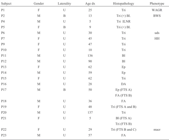
In this study, we investigated the constitutional and somatic 11p15 methylation patterns in 24 males and 16 fe-males with WT. There were four cases of bilateral tumor. The age at diagnosis ranged from five months to 137 months, with an average age at diagnosis of 43 months for all 40 patients and 44 months for patients with unilateral tu-mor. Diffuse or focal anaplasia was observed in five tumors (Table 1) and four of them also presented DMRH19 hyper-methylation (Table 2). Perilobar nephrogenic rests (PLNR)
Table 1- Clinical and histopathological data for 40 patients with Wilms tumor.
Subject Gender Laterality Age dx Histopathology Phenotype
P1 F U 25 Tri WAGR
P2 M B 13 Tri (+) Bl. BWS
P4 M U 5 Tri ILNR
P5 F B 9 Tri (+) Bl.
P6 M U 30 Tri uds
P7 F U 45 Tri HH
P9 F U 47 Tri
P10 F U 10 Tri
P11 M U 136 Bl
P12 M U 90 Bl
P13 F U 62 Ep
P14 M U 59 Ep
P15 F U 62 Tri
P16 M U 28 DA
P17 M B 50 Ep (FTS A)
FA (FTS B)
P18 M U 36 FA
P19 F U 48 Tri (FTS A and B)
P20 M U 137 Tri
P21 F U 5 Bl (FTS A)
Tri (FTS B)
P22 F U 29 Tri (FTS B and C) macr
Subject Gender Laterality Age dx Histopathology Phenotype
P24 F U 13 Tri
P25 M U 42 Tri
P26 M U 57 Tri HH Moe
P27 M B 61 Ep PLNR (FTS B)
Tri PLNR (FTS C)
P28 M U 67 Tri
P29 F U 49 Ep
P31 F U 34 Tri
P32 F U 6 Bl
P33 M U 54 Bl
P34 F U 35 Tri
P35 M U 46 Tri BWS
P36 M U 32 Bl; DA
P37 M U 48 Tri
P38 F U 19 Bl/Ep PLNR macr
P39 M U 48 Tri
P40 M U 28 Tri
P41 M U 26 Bl
P42 M U 56 St
P43 M U 13 Tri
Total (P): 40 43*
(A), (B) and (C) – tumor samples A, B and C, respectively. Age dx – age at diagnosis (months), B – bilateral, Bl – blastemal, Ep – epithelial, DA – diffuse anaplasia, F – female, FA – focal anaplasia, FTS – fresh tumor sample, HH – hemihypertrophy, ILNR – intralobar nephrogenic rests, M – male, macr – macrosomia, P – patient, PLNR – perilobar nephrogenic rests, Tri – triphasic, St – stromal, U – unilateral, uds – undiagnosed dysmorphic syndrome. *Av-erage age at diagnosis.
Table 1 (cont.)
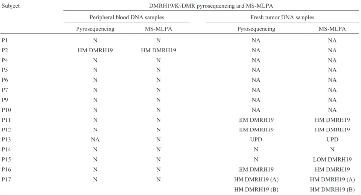
Table 2- Pyrosequencing and MS-MLPA for 11p15.
Subject DMRH19/KvDMR pyrosequencing and MS-MLPA
Peripheral blood DNA samples Fresh tumor DNA samples
Pyrosequencing MS-MLPA Pyrosequencing MS-MLPA
P1 N N NA NA
P2 HM DMRH19 HM DMRH19 NA NA
P4 N N NA NA
P5 N N NA NA
P6 N N NA NA
P7 N N NA NA
P9 N N NA NA
P10 N N NA NA
P11 N N HM DMRH19 HM DMRH19
P12 N N HM DMRH19 HM DMRH19
P13 NA N UPD UPD
P14 N N N N
P15 N N N LOM DMRH19
P16 N N HM DMRH19 HM DMRH19
P17 N N HM DMRH19 (A) HM DMRH19 (A)
were observed in two patients and intralobar rests (ILNR) in one patient. One patient with PLNR (P38) had macro-somia since birth and mild indentations on the ear lobes, but no other findings compatible with the clinical features of BWS. Eight patients had phenotypic abnormalities: one had WAGR syndrome (P1), two had BWS (P2 and P35), two had macrosomia defined by stature and weight above the 97thcentile for age and sex (P22 and P38), two had hemihypertrophy (P7 and P26), one of whom (P26) also presented with Moebius sequence (bilateral VII cranial nerve paralysis), and the eighth patient (P6) had an undiagnosed dysmorphic syndrome (Table 1).
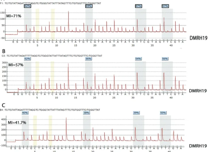
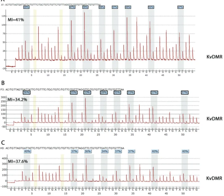
Pyrosequencing of DMRH19 in 31 blood DNA sam-ples detected hypermethylation in the two BWS patients (Table 2), with MI of 71% and 57%, respectively; these val-ues were higher than in healthy blood controls (MI = 45.4± 6.2%) (Figure 1). Pyrosequencing of KvDMR in 31 blood samples yielded normal MI estimates (Table 2, Figure 2), with the corresponding MI for healthy blood (control) be-ing 38.9±3.1%.
Pyrosequencing of DMRH19 in 34 fresh tumor sam-ples detected hypermethylation in 28 samsam-ples (Table 2). In five patients with more than one tumor sample, analysis of the two samples obtained at different times yielded similar Subject DMRH19/KvDMR pyrosequencing and MS-MLPA
Peripheral blood DNA samples Fresh tumor DNA samples
Pyrosequencing MS-MLPA Pyrosequencing MS-MLPA
P18 N N HM DMRH19 HM DMRH19
P19 N N UPD (A) UPD (A)
UPD (B) UPD (B)
P20 N N N N
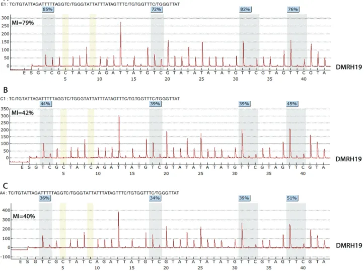
P21 NA NA UPD (FTS A) DMRH19 MI = 79%* UPD (A)
N (FTS B) DMRH19 MI = 42%* N (B)
P22 N N HM DMRH19 (B) HM DMRH19 (B)
HM DMRH19 (C) HM DMRH19 (C)
P23 NA NA HM DMRH19 HM DMRH19
P24 NA NA UPD UPD
P25 N N HM DMRH19 HM DMRH19
P26 N N NA NA
P27 N N HM DMRH19 (B) HM DMRH19 (B)
HM DMRH19 (C) HM DMRH19 (C)
P28 N N UPD UPD
P29 NA NA UPD UPD
P31 N N UPD UPD
P32 NA NA UPD UPD
P33 N N NA NA
P34 N N HM DMRH19 HM DMRH19
P35 HM DMRH19 NA UPD UPD
P36 NA NA N N
P37 NA NA HM DMRH19 HM DMRH19
P38 N N HM DMRH19 HM DMRH19
P39 N N NA NA
P40 N N UPD UPD
P41 N N N N
P42 N N UPD UPD
P43 NA NA HM DMRH19 HM DMRH19
Total (P): 40 31/40 31/40 34/40 34/40
(A), (B) and (C), tumor samples A, B and C, respectively. FTS – fresh tumor sample, HM – hypermethylation, LOM – loss of methylation, N – normal, NA – not available, P – patient, UPD – uniparental disomy. *P21 MI: DMRH19 methylation index for tumor samples from patient 21.
estimates of MI, except for one patient (p21) with an MI of 79% in sample A and 42% in sample B (Table 2, Figure 3). The corresponding MI for healthy renal tissue (control) was 42.6±15.3%.
KvDMR pyrosequencing in 34 fresh tumor DNA samples detected LOM in 12 samples (Table 2). The MI in healthy renal tissue (control) was 39.2± 10.8%. Pyrose-quencing showed that these 12 tumor samples with KvDMR LOM also had DMRH19 hypermethylation that was characteristic of paternal uniparental disomy (UPD) (Table 2).
MS-MLPA was done in 31 peripheral blood samples and in all 34 tumor samples (Table 2). DMRH19 hyper-methylation was observed in only one peripheral blood sample (P2); the remaining blood samples were normal (Figure 4A,B). MS-MLPA analysis of tumor samples showed isolated DMRH19 hypermethylation in 16/34 cases and in 12/34 samples a characteristic pattern of pater-nal UPD was observed (Table 2, Figure 4C). Isolated
DMRH19 LOM was detected in one tumor sample (P15) while a normal methylation pattern was observed in the re-maining 5/34 samples (Table 2).
Discussion
In this study, we used pyrosequencing and MS-MLPA to analyze the constitutional and somatic methyl-ation patterns in the DMRH19 and KvDMR regions of 40 WT patients. Data from tumor histopathology and phys-ical examination were compared with the methylation sta-tus in 11p15. The comparison of peripheral blood and fresh tumor samples confirmed that the epigenetic changes asso-ciated with WT were mostly somatic. Phenotypic abnor-malities characteristic of a syndromic form of WT were observed in 20% of patients (8/40). The average age at di-agnosis in our cohort was 43 months, which was similar to the 38 months reported by Scottet al. (2012).
Four patients had bilateral WT and tumor samples were available from two of them (P17 and P27). Two tumor samples from each of these patients were studied, one from each kidney (left and right). Sample A from patient P17 had a predominantly epithelial histology while sample B, which was collected from a subsequent nephrectomy on the contralateral kidney, showed focal anaplasia that probably reflected evolution of the WT. Both samples had DMRH19 hypermethylation and the DNA methylation indices were similar in the two samples from each of these patients.
Constitutional DMRH19 hypermethylation was ob-served in only two patients (P2 and P35), both with the BWS phenotype. These patients fit into a small group (5-7%) of BWS patients with isolated DMRH19 hyper-methylation reported by Blieket al.(2001). Interestingly,
these authors found that two of the four BWS patients who showed isolated DMRH19 hypermethylation also devel-oped WT (Blieket al., 2001). The normal constitutional methylation profiles shown by KvDMR pyrosequencing in the present study confirmed that most epigenetic alterations associated with WT involve DMRH19 but not KvDMR, as also reported by Prioloet al. (2008).
currently described in association with WT, indicating the heterogeneous etiology of this neoplasia (Dome and Huff, 2011). As already described in a proportion of these pa-tients, constitutive DMRH19 hypermethylation was ob-served in both BWS patients and somatic UPD was present in patient P35. Perilobar nephrogenic rests were observed in one of the patients with macrosomia (P38), as described above, and her tumor sample showed DMRH19 hyper-methylation.
DMRH19 pyrosequencing showed isolated hyper-methylation in 47% (16/34) of tumor samples, which was lower than the 71% previously reported with the COBRA assay in 28 WT samples (Brownet al., 2008). However, an-other study based on COBRA analysis (Cerrato et al., 2008) detected isolated somatic DMRH19 hypermethyl-ation in 10/40 WT patients (25%). Contrary to our study, in which 12 patients showed somatic LOM of KvDMR, Brownet al.(2008) observed no differences in the extent of KvDMR methylation between normal and tumor samples. Scottet al.(2012) reported that the proportion of isolated
DMRH19 hypermethylation observed with MS-MLPA was 31%, a value similar to our results.
One of our patients (P21) showed a difference in the extent of methylation between tumor samples A (79%) and B (42%). This discrepancy may reflect the fact that sample B was collected after successful chemotherapy with 90% tumor regression. DNA methylation levels are known to be influenced by various chemotherapeutic regimens. Among our patients, with exception of two biopsy samples, the re-maining tumor samples were obtained from patients who had been submitted to the same chemotherapeutic SIOP protocol (Bhatnagar, 2009). Variations in the rate of tumor regression in response to chemotherapy may have an im-pact on DNA methylation levels, as observed in patient P21.
Figure 4- MS-MLPA methylation indices. (A) Peripheral blood DNA from patient P2 showing isolated hypermethylation of DMRH19 (average MI = 84
cating paternal UPD in 11 out of 29 patients (38%). This proportion was similar to a previous UPD estimate (45%) in 40 WT patients (Cerratoet al., 2008).
We used MS-MLPA as a complementary procedure to analyze DMRH19 and KvDMR methylation. This assay yielded findings consistent with those obtained by pyrose-quencing, except for one tumor sample (P15) that showed DMRH19 LOM, probably because of a technical artefact. In addition, MS-MLPA confirmed the somatic, paternal UPD in 12 tumor samples.
Few studies have compared constitutional and so-matic 11p15 epigenetic alterations in WT patients. One of these used MS-MLPA to analyze blood DNA samples from 437 children with non-syndromic WT and identified con-stitutional defects in 13 patients (3%), including six pa-tients with DMRH19 hypermethylation and six with pater-nal UPD (Scottet al., 2008b). Constitutional 11p15 defects were observed in 5% (2/40) of our patients. The concomi-tant finding of macrosomia and somatic isolated DMRH19 hypermethylation in two patients (P22 and P38; Tables 1 and 2) indicated that these patients may represent somatic mosaics for this epigenetic disturbance. Additionally, pa-tient P38 also had PLNR, a condition previously reported to be associated with this phenotype (Dome and Huff, 2011). To the best of our knowledge, this is the first study to compare constitutional and somatic DMRH19 and KvDMR epigenetic alterations in WT patients by pyrose-quencing and MS-MLPA. Our findings highlight the bene-fits of the combined analysis of the DMRH19 and KvDMR regions. The MS-MLPA and pyrosequencing findings were highly consistent. MS-MLPA is a rapid, reliable technique that is less expensive than pyrosequencing and is adequate for accurate quantitative estimation of DNA methylation in a clinical setting.
Acknowledgments
This work was supported by Conselho Nacional de Desenvolvimento Científico (CNPq; grants 401966/2010-0, 476808/2010-3, 573806/2008-0) and Fun-dação de Amparo à Pesquisa do Estado do Rio de Janeiro (FAPERJ; E26/170.026/2008). Leila C.A. Cardoso was supported by a CNPq visiting fellowship for her training at the Instituto de Genética Médica y Molecular, Hospital Universitario La Paz, Spain, as part of her studies for a PhD degree from the Post-Graduate Program in Genetics, Uni-versidade Federal do Rio de Janeiro (UFRJ).
References
Beckwith JB (1998) Nephrogenic rests and the pathogenesis of Wilms tumor: Developmental and clinical considerations. Am J Med Genet 79:268-273.
Bhatnagar S (2009) Management of Wilms’ tumor: NWTSvs. SIOP. J Indian Assoc Pediatr Surg 14:6-14.
Bliek J, Maas SM, Ruijter JM, Hennekam RCM, Alders M, Westerveld A and Mannens MMAM (2001) Increased tu-mour risk for BWS patients correlates with aberrantH19and
not KCNQ1OT1methylation: Occurrence ofKCNQ1OT1
hypomethylation in familial cases of BWS. Hum Mol Genet 10:467-476.
Brown KW, Power F, Moore B, Charles AK and Malik KTA (2008) Frequency and timing of loss of imprinting at 11p13 and 11p15 in Wilms’ tumour development. Mol Cancer Res 6:1114-1123.
Bruce S, Hannula-Jouppi K, Lindgren CM, Lipsanen-Nyman M and Kere J (2008) Restriction site-specific methylation stud-ies of imprinted genes with quantitative real-time PCR. Clin Chem 54:491-499.
Cerrato F, Sparago A, Verde G, De Crescenzo, Citro V, Cubellis MV, Rinaldi MM, Boccuto L, Neri G, Magnani C, et al. (2008) Different mechanisms cause imprinting defects at the IGF2/H19 locus in Beckwith-Wiedemann syndrome and Wilm’s tumour. Hum Mol Genet 17:1427-1435.
De Camargo B, de Oliveira Ferreira JM, de Souza Reis R, Ferman S, Santos MO and Pombo-de-Oliveira MS (2011) Socioeco-nomic status and the incidence of non-central nervous sys-tem childhood embryonic tumours in Brazil. BMC Cancer 11:160-166.
Gomes MV, Huber J, Ferriani RA, Amaral Neto AM and Ramos ES (2009) Abnormal methylation at theKvDMR1 imprint-ing control region in clinically normal children conceived by assisted reproductive technologies. Mol Hum Reprod 15:471-477.
Miller RW, Young Jr JL and Novakovic B (1995) Childhood can-cer. Cancer 75:395-405.
Miller SA, Dykes DD and Polesky HF (1988) A simple salting out procedure for extracting DNA from human nucleated cells. Nucleic Acids Res 16:1215.
Moulton T, Crenshaw T, Hao Y, Moosikasuwan J, Lin N, Dem-bitzer F, Hensle T, Weiss L, McMorrow L, Loew T,et al. (1994) Epigenetic lesions at theH19locus in Wilms1 tu-mour patients. Nat Genet 7:440-447.
Nakamura L and Ritchey M (2010) Current management of Wilms’ tumour. Curr Urol Rep 11:58-65.
Nativio R, Sparago A, Ito Y, Weksberg R, Riccio A and Murrell A (2011) Disruption of genomic neighbourhood at the im-printedIGF2-H19locus in Beckwith-Wiedemann syndrome and Silver-Russell syndrome. Hum Mol Genet 20:1363-1374.
Okamoto K, Morison IM, Taniguchi T and Reeve AE (1997) Epigenetic changes at the insulin-like growth factor II/H19 locus in developing kidney is an early event in Wilms tumourigenesis. Proc Natl Acad Sci USA 94:5367-5371. Priolo M, Sparago A, Mammì C, Cerrato F, Laganà C and Riccio
A (2008) MS-MLPA is a specific and sensitive technique for detecting all chromosome 11p15.5 imprinting defects of BWS and SRS in a single-tube experiment. Eur J Hum Genet 16:565-571.
Sambrook J, Fritsch EF and Maniatis T (1989) Molecular Clon-ing: A Laboratory Manual. 2nd edition. Cold Spring Harbor Laboratory Press, New York, 545 pp.
Satoh Y, Nakadate H, Nakagawachi T, Higashimoto K, Joh K, Masaki Z, Uozumi J, Kaneko Y, Mukai T and Soejima H (2006) Genetic and epigenetic alterations on the short arm of chromosome 11 are involved in a majority of sporadic Wilm’s tumours. Br J Cancer 95:541-547.
Schneid H, Seurin D, Vazquez MP, Gourmelen M, Cabrol S and Le Boue Y (1993) Parental allele specific methylation of the human insulin-like growth factor II gene and Beckwith-Wiedemann syndrome. J Med Genet 30:353-362.
Scott RH, Douglas J, Baskcomb L, Nygren AO, Birch JM, Cole TR, Cormier-Daire V, Eastwood DM, Garcia-Minaur S, Lupunzina P,et al.(2008a) Methylation-specific multiplex ligation-dependent probe amplification (MS-MLPA) ro-bustly detects and distinguishes 11p15 abnormalities associ-ated with overgrowth and growth retardation. J Med Genet 45:106-113.
Scott RH, Douglas J, Baskcomb L, Huxter N, Barker K, Hanks S, Craft A, Gerrard M, Kohler JA, Levitt GA,et al.(2008b) Constitutional 11p15 abnormalities, including heritable im-printing center mutations, cause nonsyndromic Wilms tu-mour. Nat Genet 40:1329-1334.
Scott RH, Murray A, Baskcomb L, Turnbull C, Loveday C, Al-Saadi R, Williams R, Breatnach F, Gerrard M, Hale J,et al. (2012) Stratification of Wilms tumor by genetic and epigenetic analysis. Oncotarget 3:327-335.
Smith AC, Choufani S, Ferreira JC and Weksberg R (2007) Growth regulation, imprinted genes, and chromosome 11p15.5. Pediatr Res 61:43-47.
Squire JA, Li M, Perlikowski S, Fei YL, Bayani J, Zhang ZM and Weksberg R (2000) Alterations ofH19imprinting andIGF2 replication timing are infrequent in Beckwith-Wiedemann syndrome. Genomics 65:234-242.
Taniguchi T, Sullivan MJ, Ogawa O and Reeve AE (1995) Epi-genetic changes encompassing theIGF2/H19locus associ-ated with relaxation of IGF2 imprinting and silencing of H19in Wilms tumour. Proc Natl Acad Sci USA 92:2159-2163.
von Kanel T, Gerber D, Schaller A, Baumer A, Wey E, Jackson CB, Gisler FM, Heinimann K and Gallati S (2010) Quantita-tive 1-step DNA methylation analysis with naQuantita-tive genomic DNA as template. Clin Chem 56:1098-1106.
Internet Resources
Dome JS and Huff V (2011) GeneReviews: Wilms
Tumor Overview.
http://www.ncbi.nim.nih.gov/books/NBK1294 (October 12, 2011).
Editor: Marcio C. Silva-Filho