Hoon-Sang CHANG(a)
Sung-Ok HONG(b)
(a)Department of Conservative Dentistry, School of Dentistry, Chonnam National University, Gwangju, Korea.
(b)Department of Conservative Dentistry, School of Dentistry, Wonkwang University, Iksan, Korea.
Effects of layering technique on
the shade of resin overlays and the
microhardness of dual cure resin cement
Abstract: The purpose of this study was to assess the color of layered resin overlays and to test the early microhardness of dual cure resin cement (DCRC) light cured through the layered resin overlays. Resin overlays of 1.5 mm thickness were fabricated with the A3 shade of Z350 (Group 1L), the A3B and A3E shades of Supreme XT (Group 2L), and the A3, E3, and T1 shades of Sinfony (Group 3L) using one, two, and three layers, respectively (n = 7). Each layer of the resin overlays was set in equal thickness. The color of the resin overlays was measured with a colorimeter and compared with an A3 shade resin denture tooth. DCRC was light cured through the resin overlays, and the early
mi-crohardness of the DCRC was measured. The ΔE value between the
denture tooth and the resin overlays and the Vickers hardness number (VHN) of the DCRC were analyzed with one-way ANOVA and Tukey’s HSD test. The color differences were 8.9 ± 0.5, 5.3 ± 1.0, and 7.3 ± 0.5 and the VHNs were 19.4 ± 1.1, 21.1 ± 0.9, and 29.3 ± 0.6 for Groups 1L, 2L, and 3L, respectively. Therefore, to match the designated tooth color of resin inlays and to increase the early microhardness of DCRC, layered resin inlays are more appropriate than single-dentin-layer resin inlays. How-ever, the translucent layer should be used cautiously because the color difference of resin inlays with a translucent layer was affected more than those without a translucent layer.
Keywords: Color; Composite Resins; Hardness Tests.
Introduction
Dual cure resin cements (DCRCs) are intended to polymerize when they are exposed to curing light and when the base and catalyst compo-nents are mixed together.1 However, most DCRCs still require light
acti-vation and have demonstrated insuficient hardness when light curing
was omitted or attenuated by the tooth structure or the restoration. 2,3,4
The hardness of DCRCs was investigated, and it was reported that the thickness as well as the shade of the resin or ceramic inlays could affect the hardness of the resin cement.5,6
Esthetics in the posterior region are not as critical as in the anterior region, and the shade guides of many direct resin composite systems rec-ommend using a single dentin layer or dentin and enamel layers for pos-terior restorations. Some indirect resin composite systems are composed of dentin, enamel, and translucent layers, and manufacturers recommend
Declaration of Interests: The authors certify that they have no commercial or associative interest that represents a conflict of interest in connection with the manuscript.
Corresponding Author:
Hoon-Sang Chang E-mail: [email protected]
DOI: 10.1590/1807-3107BOR-2014.vol28.0016 Epub Jun 02, 2014
Submitted: Nov 28, 2013
using a layering technique for posterior restorations. The layering technique attempts to replace dentin and enamel by placing a more translucent layer over a more opaque layer of resin composite to create a perception of depth from within the restoration.7
However, studies have reported poor compatibility of resin composites of identical shade designation between pairs of shades,8,9 and the color of the
resto-ration was inluenced by the optical properties of the
enamel layer as well as those of the dentin layer.7,10
On the other hand, incorporation of enamel and translucent layers on resin overlays increased curing light penetration through layered resin overlays.11
Higher curing light penetration could enhance the light curing of DCRC beneath the resin overlays. Polymerization of resin cement is usually assessed by hardness tests,12,13 and many studies on
hard-ness measurements of DCRCs that were light cured through various indirect restorations were performed with one single shade.14,15 However, in clinical
situa-tions, resin inlays are usually fabricated with a lay-ering technique. Studies on the hardness of DCRCs light cured through resin overlays with a layering
technique are dificult to ind. Therefore, the pur
-pose of this study was to assess the inal shade of
resin overlays fabricated with a layering technique and to test the early microhardness of DCRC light cured through the layered resin overlays. The tested null hypothesis was that the layering technique had
no effect on the inal shade of the resin overlays and
on the early microhardness of DCRC light cured through the resin overlays.
Methodology
Resin overlay fabrication
Resin overlays of 15 mm diameter and 1.5 mm thickness were prepared in a custom-made
cylin-drical aluminum mold with a movable Telon plate
of 15 mm diameter inserted into the mold. A bolt
was attached to the opposite side of the Telon plate,
so that the space inside the hole could be adjusted by the rotation of the bolt, which was marked in 10 steps such that one step corresponded to a downward
movement of the Telon plate by 0.1 mm.12
Single-layer resin overlays (Group 1L) were
fab-ricated by lowering the Telon plate by 1.5 mm and
illing the empty space with Filtek Z350 (3M ESPE,
St Paul, USA), shade A3. The upper surface of the
aluminum mold was covered with a polyester ilm
and a glass slab to press the surface for removal of the excess resin composite. The resin composite was light cured with a light-emitting diode (LED) light
curing unit (LCU) (Elipar FreeLight 2, 3M ESPE, St
Paul, USA) for 5 seconds. After removing the glass
slab and the polyester ilm, the resin overlay was
light cured for 20 seconds using an overlapping cur-ing procedure to ensure that every part of the resin overlay was light cured.
Resin overlays with two layers (Group 2L) were
fabricated by lowering the Telon plate by 0.75 mm and illing the empty space with Filtek Supreme XT (3M ESPE, St Paul, USA), body shade A3B. After light curing as described previously, the Telon plate was lowered by another 0.75 mm, and Filtek Supreme XT, enamel shade A3E, was illed and light cured.
Resin overlays with three layers (Group 3L) were
fabricated by sequential lowering of the Telon plate
by 0.5 mm for layers of Sinfony indirect lab
com-posite (3M ESPE, Seefeld, Germany) dentin shade
A3, followed by enamel shade E3, and translucent shade T1. Seven resin overlays were fabricated in each group (n = 7).
To analyze the color of each layer constituting the resin overlays, resin overlay specimens were fabri-cated each in a single layer of 1.5 mm thickness as described above (n = 7).
Color measurement of resin overlays
Commission International de l’Eclairage (CIE) L*a*b* values of the resin overlays were measured with a colorimeter (ShadeEye NCC, Shofu Inc., Kyoto, Japan) by contacting the measuring tip to the middle of the resin overlays. The colorimeter was set in ‘Ana-lyze mode’, and before each color measurement, the instrument was calibrated with the white working standard provided by the manufacturer. The color measurement was processed in the lighting booth with 65K illumination (Spectralight III Booth,
Gre-tagMacbeth, Newburgh, USA) over a standard white
Table 2. Means and standard deviations of L*, a*, and b* values of each layer constituting resin overlays.
Resin Z350 Supreme XT Sinfony
Shade A3 A3B A3E A3 E3 T1
L* 75.8 ± 0.5a 79.3 ± 0.6c 78.5 ± 0.4b 79.1 ± 0.5bc 82.2 ± 0.4d 83.6 ± 0.4e
a* 2.2 ± 0.1a 1.3 ± 0.1b 0.6 ± 0.2c -0.3 ± 0.1e 0.1 ± 0.2d -0.4 ± 0.2e
b* 24.5 ± 0.3b 24.9 ± 0.7ab 19.6 ± 0.3c 25.5 ± 0.6a 7.7 ± 0.1d 2.7 ± 0.2e
Values followed by different superscript letters are significantly different (p < 0.05).
Table 1. Means and standard deviations of L*, a*, and b* values of layered resin overlays.
1L 2L 3L
L* 75.8 ± 0.5a 75.3 ± 1.1a 78.9 ± 0.8b a* 2.2 ± 0.1a 1.7 ± 0.2b -0.9 ± 0.4c b* 24.5 ± 0.3a 20.4 ± 0.6b 13.4 ± 0.4c
Values followed by different superscript letters are significantly different (p < 0.05). 1L = Z350 A3; 2L = Supreme XT A3B & A3E; 3L = Sinfony A3, E3 & T1.
was measured as a control. The color of the resin denture tooth was measured on the middle of the
lat labial surface of the central incisor.16 The color
difference (ΔE) between shade A3 of the resin den-ture tooth (L*1, a*1, b*1) and that of the resin overlays
(L*2, a*2, b*2) was calculated as follows:17 ΔE = [(L*1 – L*2)2 + (a*1 – a*2)2 + (b*1 – b*2)2]1/2
Early microhardness measurements of dual cure resin cement
A custom-made aluminum mold with an inner diameter of 3 mm and a Teflon plate insert was used.12 To control the light curing time of the DCRC,
the diameter of the DCRC specimen was controlled to be smaller than the diameter of the light guide tip of the LCU. The Teflon plate in the mold was
lowered by 1 mm, and DCRC (RelyX Unicem, 3M
ESPE, Seefeld, Germany) was mixed according to
the manufacturer’s instructions and illed into the
empty space. The upper surface of the aluminum mold including the DCRC was covered with a
poly-ester ilm and a previously fabricated resin overlay.
To control the DCRC thickness to 1 mm, a glass slab was used to put pressure on the upper surface of the mold. After removing the glass slab, the DCRC was light cured through the resin overlay with the LCU for 20 seconds according to the manufacturer’s instructions.12 The power density of the LCU was
measured at 1040 mW/cm2 with a handheld dental
radiometer (Cure Rite, Kerr, Milford, USA). After 10
minutes from the start of light curing, the Vickers microhardness of the DCRC was measured with a
microhardness tester (MHT-10, Anton Paar, Graz,
Austria) with a 100 g load for 10 seconds dwell time at 3 points in the center of the upper surface form-ing a small triangle, and the mean Vickers hardness number (VHN) was calculated.18 Seven DCRC
spec-imens that were light cured through layered resin overlays were used for the microhardness tests (n = 7).
The CIE L*a*b* values of the resin overlays and their constituents, the ΔE values between the denture tooth and the resin overlays, and the VHN of the DCRC were
analyzed with one-way ANOVA at a 5% signiicance
level followed by post-hoc comparisons with Tukey’s HSD test (SPSS 19.0, SPSS Inc., Chicago, USA).
Results
The CIE L*a*b* values of layered resin overlays and their constituents of 1.5 mm thickness are shown in Table 1 and Table 2, respectively. The L* value was higher in Group 3L compared to Groups 1L and 2L. The a* and b* values were higher in Group 1L com-pared to Groups 2L and 3L. In Table 2, the L* value was highest in Sinfony T1 followed by Sinfony E3, and Z350 A3 had the lowest L* value. The a* value of Z350 A3 was highest, followed by the layer con-stituents of Supreme XT and Sinfony. The b* values were highest in Sinfony A3, Supreme XT A3B, and Z350 A3, followed by Supreme XT A3E, Sinfony E3, and Sinfony T1 (p < 0.05).
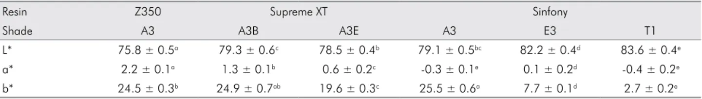
The ΔE values between the shade A3 resin denture
tooth and the resin overlays are shown in Figure 1.
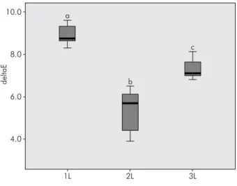
The VHN of DCRC light cured through resin
overlays is shown in Figure 2. The VHN of DCRC
light cured through Group 3L was 29.3 ± 0.6, through Group 2L was 21.1 ± 0.9, and through Group 1L was 19.4 ± 1.1 (p < 0.05).
Discussion
Generally, resin inlays are fabricated with resin composites exclusively used by dental laboratories.
However, some studies reported that the lexural
strength and surface hardness of direct resin com-posite materials were superior to those of indirect resin composites.19,20 Therefore, direct resin
compos-ite systems were used for resin overlay fabrication
with either a dentin layer or dentin and enamel layers and compared with indirect resin composite systems with dentin, enamel, and translucent layers. Because the incorporation of highly translucent enamel and
translucent layers is reported to affect the inal color
of the resin overlays,7 the color of the resin overlays
was measured and compared with a shade A3 resin denture tooth. In a previous study, a VITA Classi-cal Shade tab was used as a control with the metal tab holder removed.16 However, the removal of the
lingual part of the tab holder could affect the thick-ness of the shade tab, and thereby affect the color of the shade tab. In this study, a resin denture tooth was selected for the control because the lingual part of the denture tooth is similar in shape to that of a
human tooth without any modiications that could
affect the color measurement procedure.
The L* value of Group 3L was higher than that of the other groups, and this could be explained by the L* values of Sinfony T1 and Sinfony E3 being
sig-niicantly higher than those of other constituents of
resin overlays. The a* value was highest in Group 1L, followed by Group 2L and Group 3L. The a* values of Groups 1L and 2L were slightly positive, whereas that of Group 3L was slightly negative. In general, the a* values of the experimental groups were nearly neutral. The b* value was highest in Group 1L, fol-lowed by Group 2L and Group 3L. The b* value of the shades corresponding to dentin or body shades (Z350 A3, Supreme XT A3B, and Sinfony A3) were
signiicantly higher than the shades corresponding
to enamel shades (Supreme XT A3E, Sinfony E3 and Sinfony T1). Therefore, the resin overlays fabricated with enamel shades or enamel and translucent shades
showed lower b* values. Furthermore, the thickness
of the enamel plus translucent shades of Group 3L was 1.0 mm, compared to Group 2L with an enamel thickness of 0.75 mm, resulting in lower b* values.
When analyzing the CIE L*a*b* values of the individ-ual shades, the range of b* values (2.7 ± 0.2 to 25.5 ± 0.6) was greater than those of L* values (75.8 ± 0.5 to 83.6 ± 0.4) and a* values (-0.4 ± 0.2 to 2.2 ± 0.1). This implies that the dentin, enamel, and translucent shades of A3 are primarily dependent on the b* value rather than on L* or a* values. Even the CIE L*a*b* values were different among dentin shades and enamel shades. These shade
Figure 1. Color difference (ΔE) between a shade A3 resin denture tooth and layered resin overlays. ΔE value of the three groups was statistically significant (p < 0.05). 1L = Z350 A3; 2L = Supreme XT A3B & A3E; 3L = Sinfony A3, E3 & T1.
a
b
c
1L 4.0
6.0 8.0 10.0
deltaE
2L 3L
a b
c
1L 19.4
0 10 20 30
40 VHN
2L 21.1
3L 29.3
variations might be due to various factors such as the
amount, size, and shape of the iller.21 The iller con
-tent of Z350 and Supreme XT is 78.5% by weight, and
the iller is composed of zirconia/silica of 0.6 - 1.4 μm. However, Z350 has added silica nanoparticles of 20 nm,
whereas Supreme XT has silica nanoiller of 5 - 75 nm
in addition to the zirconia/silica. On the other hand,
Sinfony is illed with aluminum glass and silica of
0.6 μm, and the iller content is 50% by weight.22ΔE
values between the resin denture tooth and Groups 1L, 2L, and 3L were 8.9 ± 0.5, 5.3 ± 1.0, and 7.3 ± 0.5, respectively. ΔE = 0 ~ 2 is considered imperceptible,
ΔE = 2 ~ 3 just perceptible, ΔE = 3 ~ 8 moderately per-ceptible, and ΔE > 8 markedly perceptible.21 Therefore,
the ΔE of Group 1L was markedly perceptible, and those of Groups 2L and 3L were moderately percep-tible. These results are in accord with those of other studies on color measurements of resin composites. One study compared the enamel and dentin shades of resin composites with the VITA Classical Shade tabs and found that the range of ΔE values was 0.9 to 12.8.8
Costa et al.16 compared the inal shade of resin com
-posites prepared using the layering technique (enamel layer over dentin layer) with the corresponding VITA Classical Shade tabs and reported that only 28% (n = 72) of layered resin composites resulted in ΔE < 3.3, below the clinically perceptible limit.
To simulate the clinical situation of resin inlay cementation, the microhardness of DCRC was tested 10 minutes after the start of light curing the DCRC through resin overlays because resin inlays are
adjusted, inished, and polished immediately fol -lowing light curing DCRC, and these procedures can create stress affecting the adhesive cementation of resin inlays to tooth structure.1 The VHN increased
when the enamel layer was added to the dentin layer and increased further when the translucent layer was added to enamel and dentin layers for resin overlay fabrication. This result is supported by our previous study using the same type of resin overlays and LCU. The power density of the LCU measured through Groups 1L, 2L, and 3L was 163 ± 4 mW/cm2, 211 ±
5 mW/cm2, and 332 ± 6 mW/cm2, respectively.22 The
power density of the LCU through resin overlays could be enhanced using enamel and translucent layers because these layers were less effective than
the dentin layer in attenuation of the curing light.11,22
Therefore, the relatively higher translucency of the enamel and translucent layers, compared to that of the dentin layer, enhanced the curing light penetra-tion through the resin overlays and thus increased the photopolymerization of the DCRC. Consequently, the null hypothesis could be rejected.
The limitation of this study was that the colorim-eters, although commonly used in color measurement of teeth and tooth-colored restorations, have small apertures, and therefore have a tendency for edge-loss effects, which could lead to errors.23 Further studies
might explore the combination of various shade lay-ers with various thicknesses of individual laylay-ers for the fabrication of resin inlays to resemble the natural tooth color and simultaneously to increase the photo-polymerization of the resin cements used for luting.
Conclusion
Resin overlays fabricated with a single dentin layer showed markedly perceptible color differences com-pared to a shade A3 resin denture tooth. Addition-ally, the early microhardness of DCRC light cured through resin overlays with a single dentin layer had the lowest VHN values. On the other hand, resin over-lays with dentin and enamel layers and those with dentin, enamel, and translucent layers showed mod-erately perceptible color differences. However, the early microhardness of the DCRC was higher when it was light cured through resin overlays with den-tin, enamel, and translucent layers compared to resin overlays with dentin and enamel layers. Therefore, to match the designated tooth color of resin inlays and to increase the early microhardness of DCRC through resin inlays, multilayered resin inlays seem to be more appropriate than single-dentin-layer resin inlays. However, translucent layers should be used cautiously because the color difference of resin inlays with translucent layers was more affected than those without a translucent layer.
Acknowledgements
This study was inancially supported by the Chon -nam National University, 2012. The authors thank
3M Korea for their generous donation of materials
1. Arrais CAG, Giannini M, Rueggeberg FA. Kinetic analy -sis of monomer conversion in auto- and dual-polymerizing modes of commercial resin luting cements. J Prosthet Dent.
2009 Feb;101(2):128-36.
2. Hasegawa EA, Boyer DB, Chan DC. Hardening of dual-cured cements under composite resin inlays. J Prosthet Dent. 1991 Aug;66(2):187-92.
3. Sigemori RM, Reis AF, Giannini M, Paulillo LA. Curing depth of a resin-modified glass ionomer and two resin-based luting
agents. Oper Dent. 2005 Mar-Apr;30(2):185-9.
4. Braga RR, Cesar PF, Gonzaga CC. Mechanical properties of resin cements with different activation modes. J Oral Rehabil.
2002 Mar;29(3):257-62.
5. Calgaro PA, Furuse AY, Correr GM, Ornaghi BP, Gonzaga CC. Influence of the interposition of ceramic spacers on the degree of conversion and the hardness of resin cements. Braz Oral Res. 2013 Sep-Oct;27(5):403-9.
6. Park SH, Kim SS, Cho YS, Lee CK, Noh BD. Curing units’ ability to cure restorative composites and dual-cured composite cements under composite overlay. Oper Dent. 2004 Nov-Dec;29(6):627-35. 7. Kamishima N, Ikeda T, Sano H. Color and translucency
of resin composites for layering techniques. Dent Mater J.
2005 Sep;24(3):428-32.
8. Park SK, Lee YK. Shade distribution of commercial resin composites and color difference with shade guide tabs. Am J Dent. 2007 Oct;20(5):335-9.
9. Paravina RD, Kimura M, Powers JM. Color compatibility of resin composites of identical shade designation. Quintes-sence Int. 2006 Oct;37(9):713-9.
10. Lee YK, Powers JM. Calculation of colour resulting from composite/compomer layering techniques. J Oral Rehabil. 2004 Nov;31(11):1102-8.
11. Hong S-O, Oh Y, Min J-B, Kim J-W, Lee B-N, Hwang Y-C, et al. Power density of various light curing units through resin inlays with modified layer thickness. Restor Dent Endod. 2012 Aug;37(3):130-5.
12. Chang HS, Kim JW. Early hardness and shear bond strength of dual cure resin cement light cured through resin over-lays with different dentin layer thicknesses. Oper Dent. 2013 Nov 5. Epub ahead of print.
13. Hwang IN, Hong SO, Lee BN, Hwang YC, Oh WM, Chang HS. Effect of amulti-layer infection control barrier on the micro-hardness of a composite resin. J Appl Oral Sci. 2012 Sep-Oct;20(5):576-80.
14. Santos GB, Alto RV, Sampaio Filho HR, Silva EM, Fellows CE.
Light transmission on dental resin composites. Dent Mater. 2008 May;24(5):571-6.
15. Bueno ALN, Arrais CAG, Jorge ACT, Reis AF, Amaral CM. Light-activation through indirect ceramic restorations: does the overexposure compensate for the attenuation in light intensity during resin cement polymerization? J Appl Oral
Sci. 2011 Jan-Feb;19(1):22-7.
16. Costa J, Fox P, Ferracane J. Comparison of various resin com -posite shades and layering technique with a shade guide. J Esthet Restor Dent. 2010 Apr;22(2):114-24.
17. Nam SH, Lee HW, Cho SH, Lee JK, Jeon YC, Kim GC. High-efficiency tooth bleaching using non-thermal atmospheric pressure plasma with low concentration of hydrogen
per-oxide. J Appl Oral Sci. 2013 May-Jun;21(3):265-70.
18. Erdemir U, Yildiz E, Eren MM, Ozel S. Surface hardness evaluation of different composite resin materials: influence of sports and energy drinks immersion after a short-term
period. J Appl Oral Sci. 2013 Mar-Apr;21(2):124-31.
19. Soares CJ, Pizi EC, Fonseca RB, Martins LR. Mechani -cal properties of light-cured composites polymerized with several additional post-curing methods. Oper Dent.
2005 May-Jun;30(3):389-94.
20. Borba M, Della Bona A, Cecchetti D. Flexural strength and hardness of direct and indirect composites. Braz Oral Res.
2009 Jan-Mar;23(1):5-10.
21. Yamanel K, Caglar A, Ozcan M, Gulsah K, Bagis B. Assess -ment of color parameters of composite resin shade guides using digital imaging versus colorimeter. J Esthet Restor Dent. 2010 Dec;22(6):379-88.
22. Chang H-S, Lim Y-J, Kim J-M, Hong S-O. [Power density of light curing units through resin inlays fabricated with di-rect and indidi-rect composites]. J Korean Acad Conserv Dent. 2010 Sep;35(5):353-8. Korean.
23. Joiner A. Tooth colour: a review of the literature. J Dent. 2004 Jan;32 Suppl 1:3-12.