Adílis Kalina ALEXANDRIA Nicolli de Araujo MECKELBURG Ursula Tavares PUETTER Jordan Trugilho SALLES
Ivete Pomarico Ribeiro de SOUZA Lucianne Cople MAIA
(Universidade Federal do Rio de Janeiro – UFRJ, School of Dentistry, Department of Pediatric Dentistry and Orthodontics, Rio de Janeiro, RJ, Brazil.
Do pediatric medicines induce
topographic changes in dental enamel?
Abstract: The purpose of the present study was to evaluate the effect of common pediatric liquid medicines on surface roughness and tooth structure loss and to evaluate the pH values of these medicines at room and cold temperatures in vitro. Eighty-four bovine enamel blocks
were divided into seven groups (n = 12): G1-Alivium®, G2-Novalgina®,
G3-Betamox®, G4-Clavulin®, G5-Claritin®, G6-Polaramine® and
G7-Milli-Q water (negative control). The pH was determined and the samples were immersed in each treatment 3x/day for 5 min. 3D non-contact proilometry was used to determine surface roughness (linear Ra, volumetric Sa) and the Gap formed between treated and control areas in each block. Scanning electron microscopy (SEM) and energy dispersive spectrometry (EDS) were also performed. The majority of liquid medicines had pH ≤ 5.50. G1, G4, and G5 showed alterations in Ra when compared with G7 (p < 0.05). According to Sa and Gap results, only G5 was different from G7 (p < 0.05). Alteration in surface was more evident in G5 SEM images. EDS revealed high concentrations of carbon, oxygen, phosphorus, and calcium in all tested groups. Despite the low pH values of all evaluated medicines, only Alivium®, Clavulin®, and Claritin®
increased linear surface roughness, and only Claritin® demonstrated the
in vitro capacity to produce signiicant tooth structure loss.
Keywords: Dental Enamel; Administration, Oral; Pharmaceutical Preparations; Acidity; Topography.
Introduction
Medicines in liquid form are widely used for children because they facilitate intake.1,2,3 However, some of the inactive agents used in pediatric
liquid medications can cause damage to dental tissues because of their
low pH.3 Some medicines have acid in their compositions in order to
preserve their chemical stability and control their tonicity.4
Certain properties of acidic products in general may be related to the loss of surface structure of dental enamel: low endogenous pH, high titratable acidity, and minimal quantities of minerals such as calcium or phosphate, in their compositions.3,5,6,7,8 Acidic medications may cause
dental erosion with loss of dental tissue.1,4 Besides the presence of acids
in some children’s medications, other factors may also be related to changes in the surface morphology of dental enamel: high frequency of medication intake, bedtime consumption, high viscosity, and reduction in salivary low.3,5
Declaration of Interests: The authors certify that they have no commercial or associative interest that represents a conflict of interest in connection with the manuscript.
Corresponding Author: Lucianne Cople Maia E-mail: [email protected]
DOI: 10.1590/1807-3107BOR-2016.vol30.0011
Submitted: Oct 28, 2014
Ma ny ora l l iqu id med ici nes a re usua l ly recommended for sick children for long periods, and in cases of chronic diseases, they are administered daily. In vitro studies have shown that an acidic medication may reduce enamel hardness,9,10,11 but, to the best of our knowledge, the inluence of pediatric oral liquid medicines on enamel topography has not been studied yet. For this reason, the purpose of this in vitro study was to evaluate the effect of common pediatric liquid medicines on surface roughness and tooth structure loss and to evaluate the pH values of these medicines at room and cold temperatures.
Methodology
Pediatric medicines and pH analysis
The pediatric medicines used in this study
were two analgesics – Alivium® (Mantecorp, São
Paulo, Brazil) and Novalgina® (Sanofi-Aventis,
Paris, France); two antibiotics – Betamox® (Atral,
Castanheira do Ribatejo, Portugal) and Clavulin®
(GlaxoSmithKline, Brentford, United Kingdom); and
two antihistamines – Claritin® (Schering-Plough,
USA) and Polaramine® (Mantecorp, São Paulo,
Brazil) (Table 1).
The pH values of the selected medicines at cold and room temperatures were determined using a pH meter (Orion 261S, Thermo Fisher Scientiic Inc., Waltham, USA). After equipment calibration, three samples of each pediatric medicine were analyzed with pH electrodes. The average of the three measurements was used as the pH value (Table 1).
Specimen preparation
Sound bovine incisor crowns were cut using a water-cooled diamond saw (Bühler, Uzwil, Switzerland) to obtain enamel blocks (4 x 4 x 2 mm). These blocks were ixed with wax in an acrylic device to polish the enamel surface: 600- and 1200-grit silicon carbide papers (Extec Corp., Enield, USA), followed by a 1-µm diamond abrasive slurry (Extec Corp., Enield, USA) and washed ultrasonically in Milli-Q water (Merck Millipore, Darmstadt, Germany). Baseline surface microhardness (SMH) was measured using a microhardness tester (HVS-1000, Time Group Inc., Beijing, China) with a Knoop diamond under a 50-g load for 5 s, and ive indentations spaced 100 µm from each other were made at the center of the enamel surface to select the sample. A total of 84 blocks (mean 341.59 ± 34.15 kg/mm2) were selected for the present
study and randomly divided into seven groups (n = 12): G1 = Alivium®, G2 = Novalgina®, G3 = Betamox®,
G4 = Clavulin®, G5 = Claritin®, G6 = Polaramine®,
and G7 = Milli-Q water (experimental control). An acid-resistant nail varnish was used to divide the enamel surface into two distinct areas: 1) a sound window (unexposed area) – the right side of the enamel surface was covered with acid-resistant nail varnish (self-positive control); and 2) an experimental window (exposed area) – the left side was not covered.
Experimental protocols
Twelve blocks from each group were immersed in pediatric liquid medicines three times a day for 5 min (15 mL per block) for 7 days. After treatment, the specimens were rinsed with deionized water (5 s) and
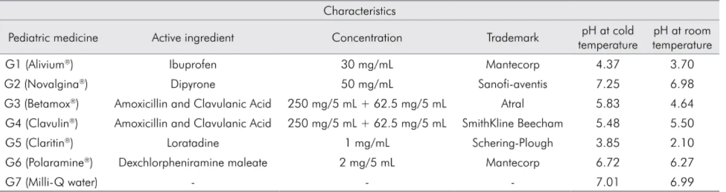
Table 1. Parameters of the pediatric medicines and control.
Characteristics
Pediatric medicine Active ingredient Concentration Trademark pH at cold
temperature
pH at room temperature
G1 (Alivium®) Ibuprofen 30 mg/mL Mantecorp 4.37 3.70
G2 (Novalgina®) Dipyrone 50 mg/mL Sanofi-aventis 7.25 6.98
G3 (Betamox®) Amoxicillin and Clavulanic Acid 250 mg/5 mL + 62.5 mg/5 mL Atral 5.83 4.64
G4 (Clavulin®) Amoxicillin and Clavulanic Acid 250 mg/5 mL + 62.5 mg/5 mL SmithKline Beecham 5.48 5.50
G5 (Claritin®) Loratadine 1 mg/mL Schering-Plough 3.85 2.10
G6 (Polaramine®) Dexchlorpheniramine maleate 2 mg/5 mL Mantecorp 6.72 6.27
transferred into artiicial saliva. All pediatric medicines were stored at 7°C, but removed 5 min before the treatment period. After block immersion, they were returned to the refrigerator until new treatment.
Two artiicial saliva solutions were prepared to simulate the oral environment and the pH changes that occur during the day, according to Queiroz et al.,12
with some modiications. The irst solution consisted of 0.05 mol/L acetate buffer, 1.28 mmol/L Ca, 0.74 mmol/L P, and 0.03 µg F/mL with pH 5.0 for 2 h (50 mL per block); and the second one contained 0.1 mol/L Tris buffer, 1.5 mmol/L Ca, 0.9 mmol/L P, 150 mmol/L KCl, and 0.05 µg F/mL with pH 7.0 for 22 h (25 mL per block).
The experimental protocol consisted of three periods of immersion in pediatric medicines and four periods in artiicial saliva per day: 1st period – 5 min in pediatric
medicines; 2nd period – 6 h in artiicial saliva (pH 7.0);
3rd period – 2 h in artiicial saliva (pH 5.0); 4th period – 5
min in pediatric medicines; 5th period – 8 h in artiicial
saliva (pH 7.0); 6th period – 5 min in pediatric medicines;
and 7th period – 8 h in artiicial saliva (pH 7.0).
The experiment was carried out at 37ºC. On the 4th day, the artiicial saliva solutions were replaced
with fresh ones in order to avoid oversaturation.
3D non-contact profilometry
The surface topography of the specimens was analyzed by a 3D proilometer (Nanovea PS50 Optical, NANOVEA Inc., Irvine, USA). The measurements of capture were performed with a chromatic confocal sensor with a white light axial source at a scan velocity of 2 mm/s and with a refractive index of 10,000.
3D non-contact proilometry was used to determine the primary outcome: tooth structure loss, i.e., the gap between the experimental and control areas (Gap) in each group; and the secondary outcome: surface roughness - linear surface roughness (Ra) and volumetric surface roughness (Sa). All comparisons between the exposed and unexposed areas of enamel were performed after the removal of the acid-resistant nail varnish.
The Gap was calculated from the step height difference between the unexposed and exposed enamel surfaces in each block; three linear measurements were made involving the unexposed and exposed areas. All measurements were done in triplicate, and the
mean values were used to represent the inal result of the surface proile.
To determine Ra, three linear measurements (one vertical, one horizontal and one transversal) were performed in each area (experimental window or sound window) of the enamel specimen. The average of these three linear measurements was used to determine Ra1 (surface linear roughness in the sound window) and Ra2 (surface linear roughness in the experimental window), and the Ra value for all groups was calculated as Ra = Ra1 – Ra2.
Also, three scan areas (200 µm × 200 µm) were obtained for each block in the sound and experimental window. The average of these three areas was used to determine Sa1 (surface roughness in the sound window) and Sa2 (surface roughness in the experimental window); and the Sa value for all groups was calculated as Sa = Sa1 – Sa2.
Scanning Electron Microscopy (SEM) and Energy Dispersive Spectrometry (EDS)
Three enamel blocks from each group were randomly selected and prepared for EDS and SEM under a scanning electron microscope (6460LV, JEOL, Tokyo, Japan). The blocks were mounted onto stubs with double-faced carbon tape and analyzed by EDS. EDS was performed to assess the mineral content of the enamel, identifying the chemical elements on its surface before and after the experimental protocol. Therefore, it was possible to compare the chemical elements found in treated (exposed) and untreated
(unexposed) areas.This analysis was performed with
a Kontron automatic image analyzer system, and the total area of the block was evaluated. The results were represented by the mean of the measured values.
Statistical analysis
The normal distribution of the data was checked for all tested variables, using the Shapiro Wilk test. A Student’s paired t-test was used to compare Ra1 and Ra2 and Sa1 and Sa2. Differences in Ra and Sa among all treatment groups were tested with the Kruskal-Wallis test, followed by the Mann-Whitney test. One-way analysis of variance (one-way ANOVA), followed by a post-hoc test (Tukey’s test), was used for Gap analysis. The SPSS software version 22.0 (IBM, Armonk, USA) was used for the statistical analysis. The signiicance level was set at 5%.
Results
Table 1 shows the pH values of pediatric liquid medicines at room and cold temperatures. The pH values of pediatric medicines at room temperature were lower than at cold temperature. G1 and G5 presented the lowest pH values.
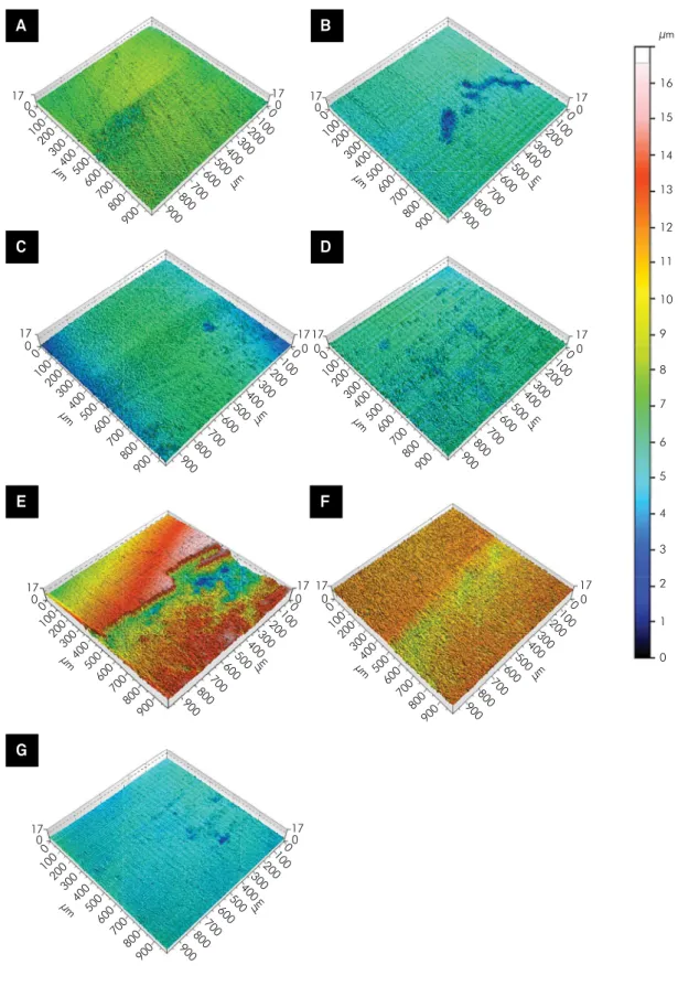
The results of 3D non-contact proilometry are summarized in Table 2 with mean Ra, Sa, and Gap for all groups. All pediatric medicines produced a signiicant alteration in surface roughness (Ra and Sa) values after 7 days (p < 0.05). These alterations were evidenced when the images of the sound and experimental areas were compared (Figure 1).
The group treated with Claritin® presented the
worst Ra and Sa values, being statistically different from the negative control (p < 0.05). Clavulin®,
Betamox®, and Alivium® also showed statistical
differences in Ra value when compared with the negative control (p < 0.05) (Table 2). Claritin® and
Clavulin® showed the worst alteration in surface
roughness (Sa value) when compared with the negative control (p < 0.05) (Table 2).
When differences in Gap values were evaluated
between the groups, only Claritin® presented a
statistically signiicant tooth structure loss when compared with the negative control (p < 0.05), but there was a similar trend among Claritin®, Clavulin®,
Polaramine®, and Alivium® since there was no
statistical difference between them (p > 0.05). (Table 2). Figure 1 shows only a change in surface roughness in some groups (Novalgina®, Polaramine® and Milli-Q
water), while in others, such as Claritin®, the loss of
tooth structure was evident in the exposed area. The same can be observed in the images obtained by SEM (Figure 2). The images of the negative control showed few signs of alteration; on the other hand, Claritin®
produced the worst alterations in enamel topography, causing loss of enamel structure (e.g., erosion).
The images of G1, G3, G4 and G5 showed that the enamel surfaces appeared to be more porous than the images of groups G2, G6 and G7.
The chemical analysis, made with EDS, revealed the presence of carbon, oxygen, sodium, phosphorus, calcium, chlorine, and aluminum. Exposed and unexposed areas retained high rates of carbon, oxygen, phosphorus, and calcium. In addition, it was possible to observe that calcium and phosphorus concentrations increased in exposed areas in all groups except for the
group treated with Polaramine®. The other elements
had almost the same rates when compared to the unexposed and exposed areas (Table 3).
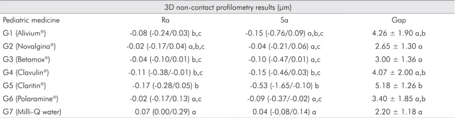
Table 2. Median (minimum/maximum value - µm) of surface roughness (Ra and Sa) and Media (µm) of Gap ± standard deviation between the unexposed and exposed enamel surface of enamel specimen groups.
3D non-contact profilometry results (µm)
Pediatric medicine Ra Sa Gap
G1 (Alivium®) -0.08 (-0.24/0.03) b,c -0.15 (-0.76/0.09) a,b,c 4.26 ± 1.90 a,b
G2 (Novalgina®) -0.02 (-0.17/0.04) a,b,c -0.04 (-0.21/0.06) a,c 2.65 ± 1.30 a
G3 (Betamox®) -0.04 (-0.10/0.01) b,c -0.10 (-0.47/0.01) a,c 3.00 ± 1.36 a
G4 (Clavulin®) -0.11 (-0.38/-0.01) b,c -0.15 (-0.46/0.03) b,c 4.07 ± 2.00 a,b
G5 (Claritin®) -0.17 (-0.28/0.05) b -0.53 (-1.65/-0.10) b 5.18 ± 1.26 b
G6 (Polaramine®) -0.02 (-0.17/0.13) a,c -0.09 (-0.37/-0.02) a,c 3.40 ± 1.85 a,b
G7 (Milli-Q water) 0.07 (0.00/0.29) a 0.04 (-0.08/0.14) a 2.20 ± 1.18 a
Figure 1. 3D profilometry scheme representation of enamel surfaces after treatment and pH cycling. left – sound window (unexposed area) and right – experimental window (exposed area). (A) G1: Alivium®, (B) G2: Novalgina®, (C) G3: Betamox®, (D) G4: Clavulin®, (E) G5: Claritin®, (F) G6: Polaramine®, and (G) G7: Milli-Q water (negative control).
17 17 17
Discussion
It is widely known that acidic medicines have certain characteristics, such as low pH, with the potential to produce alterations in the topography of enamel surface.3,4,8,13 Many of these medicines, such as antibiotics
and antihistamines, are usually used to treat children for long periods.9 So, acidic medicines likely promote
surface degradation of the tooth and increase dental roughness. Other pediatric medicines (e.g., analgesics) are largely used for children, and even though they are used
for short periods, it is also important to evaluate them once they could potentially cause surface alterations. Then, it is important to evaluate the effects of all these medicines chronically or usually used by children, on the topography of dental enamel surface.
For this reason, our selection of medicines was based on previous results8,9,10,11,14 as well as on prescriptions for children.
Our study is the irst in the searched literature that evaluated changes in surface enamel topography Table 3. Mean (%) of EDS analysis of surface of enamel blocks, chemical elements are shown according to the treatments and area (unexposed or exposed).
Groups Evaluated area C O Na Mg Al P Cl Ca
G1 Alivium® Unexposed 76.96 9.65 0.15 - 0.94 4.50 0.14 7.67
Exposed 62.62 15.77 0.18 - 0.48 7.21 0.16 13.59
G2 Novalgina® Unexposed 55.83 23.95 0.22 - 0.58 7.24 0.21 11.97
Exposed 50.51 26.34 0.39 - 0.28 8.54 0.21 13.73
G3 Betamox® Unexposed 45.78 23.67 0.45 - 0.67 7.45 0.15 11.98
Exposed 47.19 28.69 0.51 - 0.38 8.76 0.18 14.30
G4 Clavulin® Unexposed 65.63 18.23 - 0.27 0.46 5.74 0.16 9.51
Exposed 53.37 24.62 - 0.37 0.22 7.94 0.19 13.30
G5 Claritin® Unexposed 45.89 30.12 0.50 0.21 0.27 8.82 0.15 14.05
Exposed 44.53 29.46 - - 0.28 9.63 0.26 15.84
G6 Polaramine Unexposed 37.94 32.29 - 0.44 2.22 10.25 0.26 16.59
Exposed 56.93 22.13 - - 0.19 7.61 0.12 13.02
G7 Milli-Q water Unexposed 60.45 21.64 0.25 - 0.40 6.54 0.13 10.59
Exposed 47.05 28.76 0.43 - 0.34 8.81 0.13 14.61
-: Chemical element not found.
Figure 2. SEM images of enamel surfaces after treatment and pH cycling at 500X. Photomicrographs of the interface: left – sound window (unexposed area) and right – experimental window (exposed area). (A) G1: Alivium®, (B) G2: Novalgina®, (C) G3: Betamox®, (D) G4: Clavulin®, (E) G5: Claritin®, (F) G6: Polaramine®, and (G) G7: Milli-Q water (negative control).
×500 20 kV 50 µm
×500 20 kV 50 µm
×500
20 kV 50 µm 20 kV ×50050 µm
×5,000 20 kV 5 µm
×500 20 kV 50 µm
×500 20 kV 50 µm A
E
B
F
C
G
with regard to linear (Ra) and volumetric roughness (Sa) and between unexposed and exposed enamel surfaces (Gap). The primary outcome of the present study was the Gap formed between unexposed
and exposed enamel surface areas.This parameter
was very important to measure the magnitude of tooth structure loss in a direct comparison between unexposed and exposed areas. However, the Ra and Sa parameters were of great importance to determine the topographic alterations in each area. On the other hand, these parameters (Ra and Sa) cannot be used to compare the Gap.
We observed that the enamel blocks treated with
Claritin® presented the worst Ra and Sa values,
demonstrating a greater increase in roughness.
Only Claritin® presented significant Gap values.
Similar results were observed in previous studies that evaluated the effects of Claritin® on dental enamel.
Valinoti et al.10 evaluated the effect of three acidic medicines (Klaricid®, Claritin®, and Dimetapp®) and
showed Dimetapp® presented high changes in linear
roughness under normal pH-cycling conditions, but Claritin® had the worst linear roughness under erosive
pH-cycling conditions. By analyzing SEM images, the authors observed that the specimens exposed
to Dimetapp® presented the most severely eroded
areas, followed by Claritin®. Babu et al.11 observed
an irregular pattern in SEM, such as the erosion area caused by Claritin®. Costa et al.9 demonstrated that
the group treated with Claritin D® had signiicantly
lower enamel hardness.
The use of a 3D non-contact proilometer was very advantageous because it does not produce grooves on the surface of the samples and is more sensitive and speciic than a roughness tester.15,16,17 Non-contact
surface proilometry allowed for quantiication of tooth depth and its measurements can be compared to those of transverse microradiography (gold standard) for the quantiication of enamel changes in vitro.18 No
previous investigations had evaluated the action of pediatric medicines on roughness and tooth structure loss using 3D non-contact proilometry.
Salivary buffering and changes in oral pH are
complex,19,20,21 and many concomitant factors can
influence the potential of substances to promote changes in tooth topography, such as endogenous
pH lower than 5.5, low and salivary buffering, and higher titratable acidity.3,6,8,11,19,21 Nevertheless, the
evaluation of these factors alone cannot determine
whether a drug does have such potential.6,20
Previous studies1,4,8,11,14,22 evaluated various types of
medications, taking into consideration their chemical components, pH, or titratable acidity. Some authors
observed that Claritin® had low endogenous pH,
between 2.1 and 2.8,8,22 and that Polaramine® presented
the highest pH value (6.0).1 Tests using models that
may mimic oral conditions are required to evaluate the real action of drugs on tooth structure.23 In the
literature, there are few studies that test the action of pediatric medicines on teeth under conditions that mimic the oral cavity.9,10,19
In our study, we observed that the pH values of pediatric medicines at room temperature were
lower than at cold temperature, and that Claritin®
and Alivium® presented the lowest pH values. We
found a difference between pH values at room and cold temperatures. Since antibiotics could only be used at cold temperatures, the same conditions were utilized for all products. All the pediatric medicines were stored at 7°C between treatments and were only removed 5 min before block immersion.
The EDS revealed high rates of carbon, oxygen, phosphorus, and calcium elements in both (exposed and unexposed) areas. However, calcium and phosphorus levels were higher in unexposed than in exposed
areas only for the blocks treated with Polaramine®.
The drug composition itself can interfere with these results; however, this was not evaluated in the present study. The authors suggest that other studies be conducted in order to investigate the composition of these products for a better understanding of the interaction between the ion concentration of the medicines and tooth structure.
1. Neves BG, Farah A, Lucas E, de Sousa VP, Maia LC. Are paediatric medicines risk factors for dental caries and dental
erosion? Community Dent Health. 2010 Mar;27(1):46-51.
2. Moazzez R, Bartlett D. Intrinsic causes of erosion. Monogr
Oral Sci. 2014 Jun;25:180-96.
3. Hellwig E, Lussi A. Oral hygiene products, medications and drugs - hidden aetiological factors for dental erosion. Monogr
Oral Sci. 2014 Jun;25:155-62.
4. Maguire A, Baqir W, Nunn JH. Are sugars-free medicines
more erosive than sugars-containing medicines? An in vitro study of paediatric medicines with prolonged oral clearance
used regularly and long-term by children. Int J Paediatr Dent. 2007 Jul;17(4):231-8.
5. Linnett V, Seow WK. Dental erosion in children: a literature
review. Pediatr Dent. 2001 Jan-Feb;23(1):37-43.
6. Serra MC, Messias DCF, Turssi CP. Control of erosive tooth wear:
possibilities and rationale. Braz Oral Res. 2009;23 Suppl 1:49-55.
7. Bartlett DW. The role of erosion in tooth wear:
aetiology, prevention and management. Int Dent J. 2005 Aug;55 Suppl 4:277-84.
8. Arora R, Mukherjee U, Arora V. Erosive potential of sugar free and sugar containing pediatric medicines given regularly and
long term to children. Indian J Pediatr. 2012 Jun;79(6):759-63.
9. Costa CC, Almeida IC, Costa Filho LC. Erosive effect of an
antihistamine-containing syrup on primary enamel and
its reduction by fluoride dentifrice. Int J Paediatr Dent. 2006 May;16(3):174-80.
10. Valinoti AC, Pierro VS, Silva EM, Maia LC. In vitro alterations in dental enamel exposed to acidic medicines. Int J Paediatr
Dent2011 Mar;21(2):141-50.
11. Babu KL, Rai K, Hedge AM. Pediatric liquid medicaments--do
they erode the teeth surface? An in vitro study: part I. J Clin Pediatr Dent. 2008 Spring;32(3):189-94.
12. Queiroz CS, Hara AT, Paes Leme AF, Cury JA. pH-cycling
models to evaluate the effect of low fluoride dentifrice on
enamel de- and remineralization. Braz. Dent J. 2008;19(1):21-7.
13. Lodi CS, Sassaki KT, Fraiz FC, Delbem ACB, Martinhon CCR. Evaluation of some properties of fermented milk beverages that affect the demineralization of dental enamel. Braz Oral
Res. 2010 Jan-Mar;24(1):95-101.
14. Pierro VS, Abdelnur JP, Maia LC, Trugo LC. Free sugar
concentration and pH of paediatric medicines in Brazil. J Clin Pediatr Dent. 2005 Sep;22(3):180-3.
15. Attin T, Wegehaupt FJ. Methods for assessment of dental
erosion. Monogr Oral Sci. 2014 Jun;25:123-42.
16. Attin T, Becker K, Roos M, Attin R, Paque F. Impact of storage
conditions on profilometry of eroded dental hard tissue. Clin
Oral Investig. 2009 Dec;13(4):473-8.
17. Joniot SB, Gregoire GL, Auther AM, Roques YM.
Three-dimensional optical profilometry analysis of surface states obtained after finishing sequences for three composite
resins. Oper Dent. 2000 Jul-Aug;25(4):311-5.
18. Elton V, Cooper L, Higham SM, Pender N. Validation of
enamel erosion in vitro. J Dent. 2009 May;37(5):336-41.
19. Correa MC, Lerco MM, Cunha Mde L, Henry MA. Salivary parameters and teeth erosions in patients with gastroesophageal reflux disease. Arq Gastroenterol.
2012 Jul-Sep;49(3):214-8.
20. Hellwig E, Lussi A, Goetz F. Influence of Human Saliva
on the Development of Artificial Erosion. Caries Res.
2013 Jul;47(6):553-8.
21. Hall AF, Buchanan CA, Millett DT, Creanor SL, Strang R, Foye RH. The effect of saliva on enamel and dentine erosion.
J Dent. 1999 Jul;27(5):333-9.
22. Xavier AFC, Moura EFF, Azevedo WF, Vieira FF, Abreu
MHNG, Cavalcanti AL. Erosive and cariogenicity potential of pediatric drugs: study of physicochemical parameters.
BMC Oral Health. 2013 Dec;13:71.
23. Hara AT, Gonzalez-Cabezas C, Creeth J, Zero DT. The effect
of human saliva substitutes in an erosion-abrasion cycling
model. Eur J Oral Sci. 2008 Dec;116(6):552-6.
References
Notwithstanding, our indings are very important for future research, and these preliminary results can contribute to planning new in situ and in vivo studies in order to better understand the effect of the chemical and physical properties of pediatric medicines on enamel topography.
Conclusions
Most of the pediatric medicines analyzed in this study had a low pH, mainly at room temperature.
Claritin®, Clavulin®, Betamox®, and Alivium®
increased linear surface roughness, but only
Claritin® demonstrated the in vitro capacity to
create signiicant Gaps between unexposed and exposed enamel surfaces.
Acknowledgments
The authors acknowledge the inancial support from the Coordenação de Aperfeiçoamento de Pessoal de Nível Superior - CAPES, Fundação de Amparo à Pesquisa do Estado do Rio de Janeiro - FAPERJ, and Conselho Nacional