1School of Medicine, University of São Paulo (FMUSP) and LIM01/HCFMUSP, São Paulo, Brazil; 2D e p a rtment of Radiology of FMUSP; 3FMUSP and LIM01/HCFMUSP and London School of Hygiene and Tropical Medicine (Hon.Prof.) UK; 4A PAE-Jundiaí, Brazil. The pre
s-ent study was supported by a grant from FAPESP - Fundação de Amparo à Pesquisa do Estado de São Paulo. Received 22 June 2005, received in final form 23 November 2005. Accepted 28 November 2005.
Dr. Eduardo Massad - FMUSP - Av. Dr. Arnaldo 455 - 01246-903 São Paulo SP - Brasil. E-mail: [email protected]
MENTAL RETARDATION
A MRI study of 146 Brazilian children
Armando Freitas da Rocha
1,3, Cláudia da Costa Leite
2, Fábio Theoto Rocha
1,
Eduardo Massad
3, Giovanni Guido Cerri
2, Sueli Aparecida de Oliveira Angelotti
4,
Eloisa Helena Garcia Gomes
4, Carla Cristina M. Oliveira
4ABSTRACT - We re p o rt results of a magnetic ressonance imaging (MRI) study of 146 Brazilian childre n , whose intelligence quotient scored less than 70. 50% of MRI examinations did not exhibit any signal of structural lesion (N group), whereas a focal thinning at the junction of the body and splenium of the cor-pus callosum; ventricular asymmetry; periventricular leukomalacia; gliosis and arachnoid cysts were among the most frequent findings in the remaining of subjects (L group). Maternal stress and altered blood pre s-s u re were the mos-st frequent findings-s in the pre-natal his-story of both N and L children. Familial antecedents-s of mental deficiency were re p o rted in 30% of both groups, whereas familiar history of alcoholism was important in N group (60% in N versus 0% in L groups). Neuropsychomotor development was delayed in 80% of the children in both groups. Aggressiveness is the most frequent finding in the post-natal childre n history.
KEY WORDS: mental re t a rdation, magnetic resonance image, maternal stress, cerebral lesion, agre s s i v e n e s s .
Deficiência mental: um estudo de ressonância magnética em 146 crianças brasileiras
RESUMO - Estudamos, através de ressonância magnética (RM), 146 crianças com quociente de inteligência menor que 70. 50% das RM não exibiram nenhum sinal de lesão (grupo N), enquanto adelgaçamento focal da junção do corpo e esplênio do corpo caloso, assimetria ventricular, leucomalácia periventricular, gliose e cisto aracnóide foram os achados mais freqüentes no restante das crianças (grupo L). Estresse matern o e alteração da pressão arterial foram os achados mais freqüentes da história do pré-natal das crianças de ambos os grupos. Antecedentes familiares de deficiência mental apareceram em 30% de ambos os gru-pos. História de alcoolismo foi importante no grupo N. Atraso no desenvolvimento neuropsicomotor foi encontrado em 80% das crianças de ambos os grupos. Agressividade foi o achado mais freqüente na história pós-natal destas crianças.
PA L AV R A S - C H AVE: deficiência mental, ressonância magnética, estresse materno, lesão cerebral, agre s s i v i-dade.
Mental re t a rdation (MR) refers to organic brain dysfunction syndromes that are accompanied by sig-nificant cognitive limitation, reflected by an intelli-gence quotient (IQ) that is more than 2 standard devi-ations below the mean age on the test instru m e n t u s e d1. Others, however, call it an administrative
blan-ket term for a wide variety of diff e rent genetic, social and specific medical conditions sharing as common f e a t u re that affected individuals score below 70 on specific IQ tests2. DSM-IV definition of mental
defi-ciency re q u i res its onset to be established before age 18 years.
MR is frequently associated with other develop-mental disturbances, such as specific genetic syndro-mes, cerebral palsy, autism and communication dis-o rders. It may be assdis-ociated with psychiatric disdis-or- disor-ders or be the main manifestation of a brain malfunc-t i o n i n g1 - 2. Multiple etiological factors - including
g-nancy, labor and delivery has to be investigated for clues of both intrinsic and extrinsic influences on the fetus and infant development1. Intrauterine events
may account for more than half of the causes of men-tal re t a rdation, and they may be associated with ma-t e rnal malnuma-trima-tion, infecma-tion, alcoholism, healma-th pro-blems, etc1 - 3. Perinatal factors may include placental
i n s u ff i c i e n c y, complication of labor and delivery. Ho-w e v e r, perinatal events may be the consequence of p revious intrauterine fetal problems rather than the p r i m a ry cause of the brain malfunction resulting in mental retardation1,4.
When not associated to syndromes - e.g. Down and Cerebral Palsy - MR is diagnosed in children re-f e rred due to motor or language developmental de-lays, or school diff i c u l t i e s1 - 8. Is MR in these cases alw a y s
associated with macroscopic brain lesions detectable by such techniques? And why such lesions did not re-sult in any detectable signal or symptom? Or may it be the case that this type of MR results from cellular or synaptic abnormal development, undetectable by these techniques, without promoting any other vis-ible neurological deficit?
Animal experiments have demonstrated that pre-natal maternal stress affects pregnancy outcome and results in early programming of brain functions with p e rmanent changes in neural-endocrine re g u l a t i o n and behavior in off s p r i n g4. Prenatal stress is
associa-ted with a range of adverse outcomes in humans, with evidences supporting the link between stre s s and the adverse birth outcomes of low birth weight and pre - t e rm birt h9 , 1 0. There exists a trend for babies
of high cortisol level mothers to be delivered earlier than those from the low cortisol gro u p9. Incre a s e d
m a t e rnal stress during pregnancy seems to be one of the determinants of temperamental variation and delay of development of infants and may be a risk factor for developing psychopathologies later in life10,11.
The purpose of the present paper is to addre s s the above questions through a magnetic re s s o n a n c e imaging (MRI) study of 146 children that scored less than 70 either on Stanford-Binnet Scale or We s c h l e r Intelligence Scale, and whose main complaint were either motor and/or language development delays or school difficulties, and the analysis of their histo-ries of the pre g n a n c y, labor, delivery and postnatal life of the child.
METHOD
The present work studied 166 children aged from 7 to 19 years old, with IQ score less than 70 on the Stanford -Binnet Scale or Weschler Intelligence Scale for Children and
re f e rred to a special institution for education of MR peo-ple, due to delayed motor and/or language development or learning difficulties. The presence (either confirmed or suspected) of any genetic syndrome was an exclusion cri-teria. Learning difficulty was characterized by the incapac-ity of the child to follow the regular program of the ele-m e n t a ry school, as attested by the headele-master of the school where the child was enrolled.
The MRI studies were carried out in the Department of Radiology of the Clinics Hospital of the University of São Paulo in a 1.5 Tesla (Ge-Horizon) MR units. Children were not sedated for the MRI examination. Because of this 20 of these children were excluded because they did not coop-erated with the MR examination. The following pro t o c o l was used: a conventional spin echo sagittal T1-weighted image (TR=510-640, TE=12-15 msec), conventional spin echo axial T1-weighted image (TR=500-640, TE=12-15 msec), fast spin echo axial and coronal T2-weighted acquisitions (TR= 4400-4500, TE=100-120 msec, ET=8-16) and axial fluid atten-uated inversion re c o v e ry-FLAIR (TR=8000, TE=150, TI=2300 msec). The slice thickness was 5-6 mm on axial images and 3 mm on coronal plane, the interslice gap varied from 0.3 to 0.6 mm. The field of view varied from 18-24 cm, the ma-trix ranged from 179-256 X 224-256, and NEX=2. The images on the coronal plane were orientated perpendicular to the hippocampus. To evaluate corpus callosum atro p h y, the normal corpus callosum thickness was considered to be at the genu 1.1±0.2 cm, at the body 0.6±0.2 cm, and at the splenium 1.1±0.2 cm1 2. All MRI were classified by the same
radiologist (C. C. Leite).
Data from the histories of the pregnancy, labor, deliv-e ry, pos-natal lifdeliv-e and familial antdeliv-ecdeliv-eddeliv-ents of thdeliv-e childrdeliv-e n were obtained by interviewing the child relatives or care-takers. These interviews were carried out by the physician and psychologist of the institution, using a standard i z e d electronic protocol constructed by one (A. F. Rocha) of us. The presence or absence of a clearly MRI identified brain lesion was used to classify children in two groups, namely, L - children with a clear brain lesion and N - children with no visible MRI lesion. The frequency of all findings re l a t e d to the histories of the pre g n a n c y, labor, delivery, pos-natal life and familial antecedents of the children were calculat-ed for each group and comparcalculat-ed by means of Yates corre c-ted chi-squared. Since this is not intended to be a causal study we restricted the statistical analysis to univariate analysis, comparing both groups variable by variable with-out taking into account multiple comparisons for interac-tions between the variables. In a future work we intend to further the analysis of the present data set.
The study was approved by the ethical committees of the Hospital das Clínicas da FMUSP and APAE/Jundiaí.
RESULTS
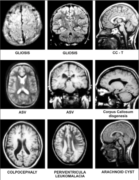
exam-inations and 75 other (51.4%) presented pathologic (L group) MRI (Fig 1).
The most frequent MRI finding was a focal thin-ning at the isthmus of the corpus callosum (CC-T in Figs 1 and 2 ), in 26 (35.2%) of L group children. It was the only finding in 15 of these children (57%); in two of them it was associated with leukomalacia; in four others with ventricular asymmetry, and one had gliosis.
The second most frequent MRI finding was ven-tricular asymmetry in the axial and coronal plane (Figs 1 and 2). It occurred in 22 (29.4%) of the L group chil-d ren: in 10 (45.4%) of the cases it was the only occur-rence, and in two cases it was associated with leuko-malacia.
The next most frequent MRI finding was leuko-malacia associated or not with other lesions in the Fig 1. MRI findings. N, normal examinations; P, MRI with pos
-itive findings; AR, arachnoid cist; ASV, ventricular asymmetry ; CCH, atrophy or disgenesis of the corpum callosum; CCT, thin -ning corpus callosum isthmus; CO, colpocephaly; GL, gliosis; LE, periventricular leukomalacia; OT, other fidings.
periventricular region (Figs 1 and 2). These lesions p resented hyperintensity on T2-weighted and FLAIR acquisitions. 17 children (22.6%) of the L group pre-sented this finding, and in three cases it was associ-ated with colpocephaly.
Another frequent finding identified in 12 (16%) of the L children was gliosis, defined as the pre s e n c e of a round or oval white matter lesions that pre s e n t-ed isointensity on T1-weightt-ed images and hyperin-tensity on T2-weighted and FLAIR images.
Arachnoid cysts were found in 6 (9% of the L g ro u p ) c h i l d ren. In three of the cases it was the only finding; in one case it was associated with corpus callosum dis-genesis, in another case with ventricular asymmetry and in the remaining case with unspecific white mat-ter lesion. The global atrophy or hypogenesis of the corpus callosum occurred in 4 children (5%): in thre e cases it was associated with periventricular leucoka-malacia and in one child with arachnoid cyst.
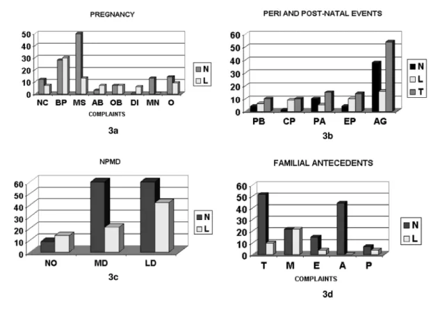
C h i l d ren of the N and L groups have diff e rent de-velopmental histories as illustrated by Figure 3 and the statistical analysis is summarized in Table 1.
70% of mothers of N children and 17% of those f rom the L group (p=0.00043, χ2=12.43) re p o rted e m
o-tional stress during pregnancy (N in Fig 3), related to domestic violence, economic distress, undesired pre g-n a g-n c y, ag-nd other familial problems. Spag-nkig-ng by hus-bands did not result in any reported relevant physi-cal trauma to the mother or the fetus. High or low blood pre s s u re (BP in Fig 3) was another common fin-ding in both groups affecting around 40% of the mo-thers of both groups. Malnutrition (MN in Fig 3) aff e c-ted 18% of mothers of the N group (p=0.0003, χ2=
12.90), and according to them it was associated to the emotional stress. In contrast, no mother of the L g roup re p o rted any nutrition problem. Abort i o n attempt (AB in Fig 3) attained a frequency of 4% in the N group and 9% in the L group (p=0.024, χ2=
claims (O in Fig 3) were associated with 14% and 19% of the L and N pregnancies (p=0.29 χ2=1.11). Only
one mother of each group re p o rted alcohol abuse during pregnancy.
Peri-natal anoxia (PA in Fig 3) was re p o rted by 14% and 7% of the mothers from groups N and L (p=0.229, χ2=1.45). Premature birth (PB) occurred in
6% and 8% of births in groups N and L (p =0.29, χ2=
1.11). Cerebral palsy (CP) was diagnosed in 1.4% and 12% of the children from groups N and L (p=0.013, Fisher exact test). Epilepsy (EP) was a manifestation in 6% of the children of the N group and in 13% of those in the L group (p=0.19, χ2=1.68). 53% of the
c h i l d ren on the N group and 21% of those from the g roup L (p=0.0001157, χ2=14.9) were re f e rred to the
psychiatrist because of aggressive behavior (AG) at the school or home.
Motor development delay (MD in Fig 3) aff e c t e d 85% of the N children and only 28% of those in L (p<
0.00000001, χ2=35). Language (LD) development
delay, however, affected 84% of the N children and 56% of those in L (p<0.0000001, χ2=29). 20% of the
L and 12% of the N children did not experience any motor or language development delay (p=0.14, χ2=
2.14).
The frequencies of the findings concerning famil-ial antecedents were diff e rent in N and L (p< 0.00000001, χ2=43). 75% of the children in the N
group and only 25% in the L group are reported to have at least one relative with a history of alcoholism (A), epilepsy (E), mental retardation and psychiatric diseases (P). Alcoholism attained a frequency aro u n d 62% in N and was absent in the L group (p< 0.00000001, χ2=56). Father alcoholism was a fre q u e n t
finding. History of mental re t a rdation in the family was present in 30% of each group. Epilepsy aff e c t-ed 22% of the relatives in the N group and 6% of those in L group (p=0.008, χ2=7). Psychiatric
ante-cedents exhibited a frequency of 9% in the case of the N group and 6% in the case of L group (p=0.28,
χ2=3.24).
DISCUSSION
H e re we observed that MR is associated in 51.4% of the children (L group) with structural lesions iden-tified by MRI such as, focal thinning of the corpus callosum at the isthmus level; asymmetry of the lat-eral ventricles; periventricular leukomalacia; atro p h y or dysgenesis of the corpus callosum; congenital mal-f o rmations; and round hyperintense lesions in the white matter identified on T2-weighted and FLAIR images.
Thompson et al.1 3demonstrated an import a n t
g rowth at the callosal isthmus between ages 6 and 15 years, suggesting that cort i c o - c o rtical networks s u p p o rting rapid associative relay and language func-tions may myelinate more extensively and over longer periods than rostral fiber system. Castro-Caldas and R e i s1 4showed a reduction of the size of the are a s
near the callosal isthmus in illiterate people when compared to literate ones. McLeod at al.12
hypothe-sized that the attenuations in the corpus callosum thickness may reflect areas where fusion was dimin-ished, and there were less fibers crossing to the oth-er coth-erebral hemisphoth-ere at that level. They found focal attenuations in the body of the corpus callosum in 35% of their patients, and considered this finding to represent a normal variant. Von Plessen et al.16
des-cribed a clear shape diff e rence in the posterior mid-body of the corpus callosum between dyslexic and c o n t rol subjects. Also, Krägeloh-Mann et al.1 7f o u n d
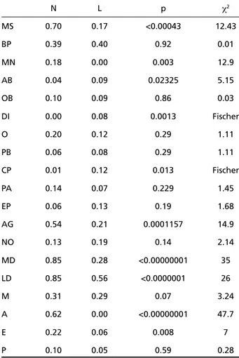
Table 1. Summary of the results.
N L p χ2
MS 0.70 0.17 <0.00043 12.43
BP 0.39 0.40 0.92 0.01
MN 0.18 0.00 0.003 12.9
AB 0.04 0.09 0.02325 5.15
OB 0.10 0.09 0.86 0.03
DI 0.00 0.08 0.0013 Fischer
O 0.20 0.12 0.29 1.11
PB 0.06 0.08 0.29 1.11
CP 0.01 0.12 0.013 Fischer
PA 0.14 0.07 0.229 1.45
EP 0.06 0.13 0.19 1.68
AG 0.54 0.21 0.0001157 14.9
NO 0.13 0.19 0.14 2.14
MD 0.85 0.28 <0.00000001 35
LD 0.85 0.56 <0.0000001 26
M 0.31 0.29 0.07 3.24
A 0.62 0.00 <0.00000001 47.7
E 0.22 0.06 0.008 7
P 0.10 0.05 0.59 0.28
a thinning of the corpus callosum in all children with mental re t a rdation in their study. A focal thinning at the isthmus of the corpus callosum was found in 15 (10.3% of all MRIs) of our children, a fact that s e e m s to confirm the pathological status of this anomaly of the corpus callosum. Also, global atrophy or hypo-genesis of the corpus callosum occurred in 4 childre n and associated to leukomalacia in three of them. These four children experienced a very poor cogni-tive development.
Asymmetry of the lateral ventricles was the only MRI finding in 10 of these children (6.8% of all MRIs). In addition, motor development delay was associat-ed with ASV in 60% of the cases and language devel-opment delay appeared in 70% of the ASV childre n . It seems there f o re that asymmetry of the lateral ven-tricles is clearly associated with mental re t a rd a t i o n in the present population. ASV has been associated with schizophre n i a1 8, but it has been claimed to be
a normal occurrence in the fetus and neonates1 9.
Gil-m o re et al.2 0re p o rted a mild enlargement of the
lat-eral ventricles on prenatal ultrasound, which persist-ed in childhood and was associatpersist-ed with attention deficit hyperactivity disorder (ADHD), autism and learning disorders.
The periventricular leukomalacia refers to the le-sions in the white matter dorsal and lateral to the e x t e rnal angle of the lateral ventricle, which are con-s i d e red to be vacon-scular bordercon-s zonecon-s at the pre m a-t u re infana-t2 1characterized by focal necrosis, part i
c-ularly at the level of the periventricular tissue at the level of the optic radiation adjacent to the trigone of the lateral ventricles and the frontal white mat-ter adjacent to the Monro foramen2 1. Periventricular
leukomalacia is manifested as hyperintense lesions on T2-weighted images, located in the peritrigonal regions with or without enlargement of the lateral ventricles. In some cases, the periventricular white matter in the frontal area is also aff e c t e d1 7.
Krägeloh-Mann et al.1 7associated periventricular brain
dam-age with mental re t a rdation. Periventricular leuko-malacia was identified in 17 of our 146 patients (11.7% of all MRIs).
Foci of hyperintense signal on T2-weighted and FLAIR images consist of small round or oval lesions located at the periventricular or subcortical white matter. In adults these lesions are common inciden-tal findings on MRI images of the brain of contro l subjects or patients with a variety of diseases2 2. These
lesions are believed to re p resent chronic low degre e of vascular insufficiency with sub-clinical
manifesta-t i o n2 3 , 2 4. This finding was identified in 12 of our
pa-tients (8.2% of the total population).
M a t e rnal stress during the pregnancy was the most f requent finding re p o rted by 63 mothers (43%) in the present study. There f o re, increased maternal s t re s s during pregnancy seems to be an important risk fac-tor for MR development in postnatal life. This is in accordance with the literature showing that mater-nal stress during pregnancy seems to be one of the determinants of delay developments of infants and may be a risk factor for development psychopathol-ogy later in life4 , 1 1 , 1 2 , 2 5. Maternal stress was a
com-plaint re p o rted by 70% of mothers from the N gro u p and by 17% of those from the L group (p<0.00043). T h e re f o re, it is a factor strongly associated with non-m a c roscopic brain insult, not detected by the MRI u s e d h e re. Both animal and human studies have convincin-gly demonstrated that prenatal maternal stress aff e c t s p regnancy outcome and results in early pro g r a m-ming of brain functions with permanent changes in neural-endocrine regulation and behavior in off-spring4,10-12,25.
The effect of maternal stress may be understood if it is accepted that the stress hormones from the mother induce an overproduction of fetal cort i s o l4.
This augmented fetal cortisol in turn may damage the brain and/or increase the damaging effects of other factors such as deficient blood and nutrient su-pplies, or it may act as a cofactor increasing the pa-thological effects of factors involved with epilepsy and other mental or psychiatric malfunctions re p o rt-ed in the family2 6 - 2 9. The maternal stress effects are
s t rongly influenced by hormones, and their outcomes a re gender dependent2 6. In agreement with this, it
must be remarked that 88 (60.2%) of tour childre n were male and 58 (39.8%) were female.
In the N group, maternal stress seemed to have magnified the effects of alcoholism, epilepsy, men-tal and psychiatric disturbs. It must be remarked that claims about these familial problems were low in the L group, and more than one type of claim were re-p o rted for more than one of the relatives in the N group. In particular, there is a high incidence (25%) of N alcoholic fathers and domestic violence was a major cause of maternal stress in this group. Indeed, Reichenheim, Moraes, and Hasselmann2 9re p o rt e d
pro-bably resulted in abnormal neurogenesis, explaining mental re t a rdation and promoting motor and lan-guage developmental delays in 80% of the N chil-d ren. Aggressiveness is also high among the chilchil-dre n of the N group. Indeed, many authors1 0 , 1 1have stre s
s-ed that increass-ed maternal stress during pregnancy seems to be one of the determinants of temperamen-tal variation and delay of development of infants and may be a risk factor for developing psychopathol-ogy later in life. It is interesting to remark here that only three children of the N group were re p o rted to be hyperactive.
M a t e rnal stress in the L group was less import a n t than in the case of the mothers of the N group. Abor-tion attempt and diabetes were also important occur-rences in the L group. Perhaps maternal stress associa-ted with blood pre s s u re problems, abortion attempt, obstetric problems and diabetes could be facilitated the occurrence of the macroscopic lesions detected in the L group. High levels of stress hormones could have magnified fetal suffering due to reduced oxy-gen and nutrients supplies associated with matern a l and placental hemodynamic disturbances. Such a com-posite effect might result into either gross lesions such as leukomalacia and ventricular asymmetry or more d i s c rete and diffuse neural losses as in the cases of the gliosis and thinning of the corpus callosum at the isthmus level. Such lesions were associated to a motor development delay in 28% of the L children and lan-guage development delay in 56% of these subjects. This higher incidence of language compared to motor p roblems may be understood by the high fre q u e n c y (17.8% of all MRIs) of corpus callosum isthmus anom-a l y. It must be re m e m b e red thanom-at Canom-astro-Canom-aldanom-as anom-and R e i s1 4showed a reduction of the size of the areas near
the callosal isthmus in illiterate people when com-p a red to literate ones. It is interesting to remark that perinatal anoxia was not a common event in the L g roup. Cerebral palsy occurred in 9 children of the L g roup and was predominant in this group in compar-ison with the N group. It seems that peri-natal anox-ia was not the main determinant of both the lesions and cerebral palsy in the L group. Also, the pre s e n t results did not point to a correlation between cere-bral palsy and motor development delay.
REFERENCES
1. A c c a rdo PJ, Capute AJ Mental re t a rdation. In Capute AJ, A c c a rdo PJ (eds) Developmental disabilities in infancy and childhood. Baltimore : Paul H. Brookes, Publishing Co, 1996.
2. A i c a rdi J. Mental re t a rdation in diseases of the nervous system in child-hood. Cambridge University Press, 1998.
3. Sundheim STPV, Ryan RM, Voeller KKS. Mental re t a rdation. In Coff e y CE, Brumback RA (eds.) Textbook of pediatric neuro p s y c h i a t r y. Wa-shington DC: American Psychiatric Press, Inc., 1998:649-690.
4. Mulder EJH, Robles de Medina PG, Hizink AC, Van den Bergh BRH, Buitelaar JK, Visser GHA. Prenatal maternal stress: effects on pre g n a n-cy and the (unborn)child. Eraly Human Dev 2002;70:3-14.
5. Harel S, Greenstein U, Kramer U, et al. Clinical characteristics of chil-d ren re f e r rechil-d to a chilchil-d chil-development center for evaluation of speech and communication disorders. Pediatric Neurol 1996;15:305-311. 6. Cans C, Wilhelm L, Baile MF, du Mazaubrun C, Grandjean H,
Rumeau-Rouquette C. Aetiological findings and associated factors in childre n with severe mental re t a rdation. Dev Med Child Neurol 1999;41: 233-239.
7. Denis D, Chateil J-F, Brun M, Brissau O, Lacomb D, Fontan D, Flurin V, Pedespan J-M. Schizencephaly: clinical and imaging features of 30 infantile cases. Brain Dev 2000,22:475-483.
8. S u resh P, Sebastian S. Developmental Gerstmann’s syndrome: a dis-tinct clinical entity of learning disabilities. Pediatric Neurol 2000;22: 267-278.
9. Glynn LM, Schetter CD, Wadhwa PD, Sandman CA. Pregnancy aff e c t s appraisal of negative life events. J Psychosom Res 2004;5:47-52. 10. Weerth C, Hees Y, Buitelaar JK. Prenatal maternal cortisol levels and
infant behavior during the first 5 months. Early Hum Dev 2003;74: 139-151.
11. Buitelaar JK, Huizink AC, Mulder EDUJ, Pascalle G, Medina R, Vi s s e r GHA. Prenatal stress and cognitive development and temperament in infants. Neurobiol Aging 2003;24: S53-S60.
12. McLeod NA, Williams P, Machen B, Lum GB. Normal and abnormal morphology of the corpus callosum. Neurology 1987;37:1240-1242. 13. Tompson PM, Diedd JN, Woods RP, MacDonald D, Evan AC, Toga AW.
G rowth patterns in the developing brain detected by using continnum mechanical tensor maps. Nature 2000;404:190-193.
14. C a s t ro-Caldas A, Reis A. Neurobiological substrates of illiteracy. Neuroscientist 2000;6:475-482.
15. Preis S, Steinmetz H, Knorr U, Jäncke L. Corpus callosum size in chil-dren with developmental language disorder. Cogn Brain Res 2000,10: 37-44.
16. Von Plessen K, Lundervold A, Duta N, et al. Less developed corpus callosum in dyslexic subjects: a structural MRI study. Neuro p s y c h o l o g i a 2002;40:1035-1044.
17. Krägeloh-Mann I, Toft P, Lunding J, A n d resen J, Pryds O, Lou HC. Brain lesions in preterms: origin, consequences, and compensation. Acta Paediatr 1999;88:897-908.
18. Chance SA, Esiri MM, Crow TJ. Ventricular enlargement in schizophre-nia: a primary change in the temporal lobe? Schizophrenia Res 2003;50: 123-131.
19. Ichihashi K, Iino M, Eguchi Y, Uchida A, Honma Y, Momoi M. Diff e-rence between left and right lateral ventricular sizes in neonates. Early Hum Dev 2002;68:55-64.
20. G i l m o re JH, van Tol JJ, Streicher HL, et al. Outcome in children with fetal mild ventriculogegaly: a case series. Schizophrenia Res 2001;48: 219-226.
21. Volpe JJ. Brain injury in the pre m a t u re infant: current concepts of patho-genesis and prevention. Biol Neonate 1992;62:231-242.
22. Yetkin FZ, Haughton VM, Fischer ME, et al. High-signal foci on MR images of the brain: observer variability in their quantification. A J R 1992;159:185-188.
23. Bryan RN, Wells SW, Miller TJ, et al. Infarctlike lesions in the brain: p revalence and anatomic characteristics at MR imaging of the elderly-data from the cardiovascular health study. Radiology 1997;202:47-54. 24. Yue NC, Arnold AM, Longstreth WT, et al. Sulcal, ventricular, and white matter changes at MR imaging in the aging brain: data from the car-diovascular health study. Radiology 1997;202:33-39.
25. Avishai-Eliner S, Brunson KL, Sandman CA, Baran TZ. Stre s s e d - o u t , or in (utero)? Trends in Neurosci 2002;25:518-524.
26. De Bellis MD, Keshavan MS, Shifflett S, et al. Brain stru c t u res in pedi-atric maltre a t m e n t - related posttraumatic stress disorder: a sociodemo-graphically matched study. Biol Psychiatry 2002;52:1066-1078. 27. Kaufer D, Friedman A, Soreq H. The vicious circle of st4ress and
anti-cholinesterase responses. Neuroscientist 1999;5:173-183.
28. Rubia K. Overmeyer S, Taylor E, et al. Functional frontalisation with age: mapping neurodevelopmental trajectories with fMRI. Neuro s c i Biobehav Review 2002;24:13-19.