Image quality in partially erased
DenOptix
storage phosphor plates
Abstract: This study aimed at investigating the effect of the partial eras-ing of DenOptix system storage phosphor plates on the image quality of digital radiographs. Standardized digital radiographs were acquired of a phantom mandible, using size 2 intraoral DenOptix storage phosphor plates (n =10). Subsequently, the active areas of the plates were placed in a viewing box with a constant light intensity of 1,700 lux for 130 sec-onds to achieve complete erasing (control plate), as well as for 0, 5, 10, 15, 20, 25, 34, 66, and 98 seconds, to compose the experimental group of partially erased plates. The same exposure settings were repeated us-ing the control and experimental plates, which were scanned at a resolu-tion of 300 dpi. Five radiologists independently examined the pairs of digital radiographs obtained with the control and partially erased plates, in random order, and indicated the best image for oral diagnosis. Co-chran-Mantel-Haenszel’s chi-square test, at a signiicance level of 5%, was used to compare the percentages of superior quality images in each combination of control and partially erased plates, subjectively assessed. No signiicant differences were found between radiographic images ac-quired with control and partially erased plates, except for the combina-tion of 0 second (30%) versus 130 seconds (70%), p = 0.0047. It can be concluded that, under adequate light intensity conditions, erasing intra-oral DenOptix storage phosphor plates may require time intervals of as little as 5 seconds.
Descriptors: Diagnosis; Radiography, dental, digital; Quality control.
Sérgio Lúcio Pereira de Castro Lopes(a)
Adriana Dibo da Cruz(a)
Rívea Inês Ferreira(b)
Frab Norberto Bóscolo(c)
Solange Maria de Almeida(d)
(a)PhD Students; (c)Professor, Chairman; (d)Associate Professor – Department of Oral Diagnosis, Piracicaba Dental School, State University of Campinas (UNICAMP).
(b)Associate Professor, Department of Pediatric Dentistry and Orthodontics, São Paulo City University (UNICID).
Corresponding author:
Solange Maria de Almeida
Faculdade de Odontologia de Piracicaba (FOP – UNICAMP)
Área de Radiologia Av. Limeira, 901, Areião Piracicaba - SP - Brazil CEP: 13414-903
E-mail: [email protected]
Introduction
At present, there are a number of digital radiog-raphy systems commercially available for dental use. In this context, studies comparing the performance of storage phosphor systems to that of conventional ilm and charge-coupled device (CCD) systems re-ported similar or better image quality with the for-mer.1-5 Furthermore, the storage phosphor systems
have a wider dynamic range and better low-contrast detectability in relation to the CCD systems.1,5,6
However, despite their relevant contributions in the technological advancement of the ield of oral and maxillofacial diagnosis, it should be emphasized that digital systems have their inherent practical limitations.4,7
There are storage phosphor plates (SPPs) hav-ing, approximately, the same size and lexibility as that of conventional ilm. This kind of sensor con-sists of a polyester base coated with a crystalline halide composed of europium-activated barium luorohalide compounds. When an image plate is irradiated, the absorbed X-ray energy is temporar-ily stored within the phosphor crystals. To read the stored information, a thin collimated helium-neon laser beam scans the plate surface and the energy is thereby released as luorescent blue light, which is detected by a photomultiplier and converted to electrical signals. An amount of the stored energy remains in the image plate even after scanning, but it is eliminated when the plate is exposed to strong light.8,9
With regard to DenOptix, the SPPs might be erased just before use. To completely erase the plates, it is recommended that they be exposed to direct and intense light.10,11 In the user manual and
installation guide of the DenOptix system, the manufacturer gives some instructions on how to erase the plates under special conditions of light in-tensity.10 Nevertheless, sometimes these
recommen-dations cannot be correctly followed, because of operational restrictions, such as the impossibility of measuring light intensity from a viewing box or in-candescent lighting. Thus, the purpose of this study was to investigate the effect of incomplete erasing of the DenOptix SPPs on the image quality of digital radiographs.
Material and Methods
Experimental design
In this experimental model, a phantom mandible was used to produce the radiographic images, in agreement with the current ethical principles (Reso-lution 196/96 of the National Health Committee/ Health Department, Brazil). The phantom presented suficient anatomical and pathological characteris-tics to simulate oral tissue images obtained in clini-cal settings (enamel, dentine, pulp cavity, periodon-tal ligament space, lamina dura, trabecular pattern, caries-like lesions, and radiolucencies in the periapi-cal region).
Standardized radiographs of different regions of the phantom were taken with DenOptix sys-tem (Denstply International/Gendex Dental X-ray Division, Des Plaines, IL, USA) size 2 intraoral SPPs completely erased (n =10), using a GE 1000 X-ray unit (General Electric Co., Milwaukee, WI, USA). The X-ray unit operated at 60 kVp, 10 mA, 2.5 mm total aluminum iltration, and a 40 cm fo-cus–receptor distance. The exposure time was set at 0.3 seconds, and the dose deined was 840PGy.
An acrylic device was manufactured to hold the phantom, X-ray beam indicator device and image plate in a reproducible relationship. After the ra-diographic exposures, the SPPs were not scanned, but remained sealed in their protective light-tight polymer envelopes until the erasing, which was the subsequent step.
The same exposure settings were repeated us-ing the control and experimental plates. Next, the SPPs were scanned at a standard resolution of 300 dpi and the digital radiographs were stored in compact disc-recordable media as 8-bit TIFF (tagged image ile format) images. The DenOptix SPP size 2 has an active area of 41 x 31 mm2, and at the resolution selected for scanning, pixel size is estimated at 85 x 85Pm2, resulting in a matrix
of 485 x 367 pixels with 8-bit quantifying gray levels, determining a spatial resolution of around 6 lp/mm.10
No time intervals were systematically allowed to elapse throughout the experiment with the DenOp-tix SPPs (irst exposure – erasing; erasing – second exposure; second exposure – scanning) to avoid any possible effects of time delay or storage conditions on image quality.
Image assessment
A panel of ive experienced oral and maxillofacial radiologists independently examined the resultant ten pairs of digital radiographs (obtained with the control and experimental plates) imported into Vix-Win (Gendex Division, Des Plaines, IL, USA), the software that manages capture, display, treatment, analysis, and archiving of images acquired with the DenOptix system.10 Digital radiographs were pre-sented in random order on an SVGA 17-inch moni-tor screen. Observers were blind to the experimental procedures used; therefore, the iles were named A
or B plus an identiication number unrelated to the erasing times, and the identiication numbers were not consecutive.
Only one pair of radiographs was displayed at a time and all viewing was performed under uni-form subdued lighting in a quiet, secluded room. The analog brightness and contrast controls on the monitor were kept constant during the assessments. Following a calibration session, the observers were instructed to use the software brightness and con-trast commands to manipulate image characteristics intuitively and better extract the diagnostic signals by reference to the abovementioned anatomical and pathological features. The observers were asked to compare the two radiographs (A and B) and
indi-cate the image that provided superior quality for oral diagnosis.
Data analysis
Two-dimensional cross-tabulation was per-formed on the radiographic interpretation data, us-ing the SAS 8.02 package (SAS Institute Inc., Cary, NC, USA). Differences between the percentages of superior quality images in each combination of con-trol and partially erased plates, subjectively assessed by the ive observers, were analyzed using the Co-chran-Mantel-Haenszel’s chi-square test. The level of signiicance was set at p =0.05.
Results
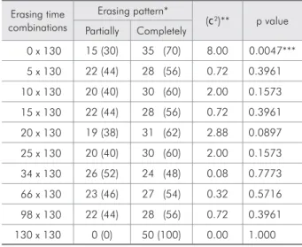
Table 1 shows the results of pair-wise compari-sons between images acquired with control and par-tially erased SPPs. Chi-square statistics demonstrat-ed a signiicant difference only between completely and not erased plates, i.e. between SPPs that were positioned in the viewing box for 130 seconds and 0 second, respectively (p =0.0047). There were subtle contrast differences between images acquired with the control and experimental plates. However, in most cases, the decreased image contrast in partially
Table 1 - Comparison of the diagnostic quality, expressed as percentages of selected digital radiographs by five ob-servers, between images acquired with completely and par-tially erased DenOptix SPPs.
Erasing time combinations
Erasing pattern*
(F2)** p value
Partially Completely
0 x 130 15 (30) 35 (70) 8.00 0.0047***
5 x 130 22 (44) 28 (56) 0.72 0.3961
10 x 130 20 (40) 30 (60) 2.00 0.1573
15 x 130 22 (44) 28 (56) 0.72 0.3961
20 x 130 19 (38) 31 (62) 2.88 0.0897
25 x 130 20 (40) 30 (60) 2.00 0.1573
34 x 130 26 (52) 24 (48) 0.08 0.7773
66 x 130 23 (46) 27 (54) 0.32 0.5716
98 x 130 22 (44) 28 (56) 0.72 0.3961
130 x 130 0 (0) 50 (100) 0.00 1.000
*Data are presented as n (% of total sample, considering five observ-ers). **(F2) indicates chi-square values. ***Highly significant difference,
erased SPPs did not impair radiographic diagnosis (Figure 1).
Discussion
Storage phosphor systems may be considered the most suitable commercially available substitutes for conventional radiography, since they provide all the image processing facilities of digital radiographs as well as high diagnostic accuracy1-5 and wide
dynam-ic range3,6,11,12 at the expense of low radiation doses.
However, there are some operational drawbacks that must be overcome in order to establish their use in clinical practice, despite the relatively higher cost in comparison with conventional radiography.
After exposure, it is recommended that the SPPs be scanned within a short period of time to prevent information loss that may occur due to degradation from surrounding light and freeing of some of the trapped electrons produced by the absorbed X-ray photons.2 Hildebolt et al.13 (2000) reported that
25-50% of the latent image stored in SPPs is lost within the irst hour after exposure, even though the rest of the radiographic information can persist for many days.
One of the main causes of the latent image fad-ing in SPPs is the time interval between exposure of the plate and its scanning. Because it may not al-ways be possible to perform the scanning procedure in clinical settings, previous studies were carried out
to assess the effects of different storage and scan de-lay conditions on image quality.1,2,9,14 According to
Akdeniz et al.1 (2005) and Martins et al.14 (2006),
it is reasonable to assume that after exposure, SPPs should be stored in a light-tight environment, un-der ambient or low humidity (60% and 26%, re-spectively) conditions at approximately 25qC, and
scanned no longer than 3 hours later.
Although the two well-known worldwide mar-keted storage phosphor systems, DIGORA and DenOptix, apply the same basic technology for capture and digitization of the radiographic informa-tion, they differ in the scanning procedure. Once the DIGORA SPPs are scanned, they are looded with light to erase any remaining image and to prepare them for the next exposure, whereas the DenOp-tix SPPs must be erased just before they are used. Scanning the DenOptix SPPs does not erase all the radiographic information.10 The need for an outside
erasing procedure may be considered an impractical time-consuming task.13 In addition, the lack of
ad-equately controlled light intensity and exposure time parameters during the erasing procedure may com-promise image quality, which in turn, might lead to under-diagnosing in a clinical situation.
Based on the manufacturer’s instructions, view-ing boxes typically give off between 1,000 and 5,000 lux. With the use of a viewing box that gives off 1,000 lux of luorescent light, the DenOptix
Figure 1 - Radiographic images acquired with the DenOptix£ storage phosphor plates. 0, not erased. 5, erasing time set at five seconds. 130, erasing time set at 130 seconds.
5
SPPs will be erased in one minute. At 2,000 lux or more, 30 seconds are suficient.10 Conversely,
Me-nig11 (1999) suggested that using a viewing box, a
two-minute time interval was necessary for com-pletely erasing the DenOptix SPPs. In fact, there is controversy about the extent to which reduced erasing times will affect the diagnostic information obtained with DenOptix radiographs. When as-sessing subjective image quality evaluated in digital radiographs acquired with partially erased DenOp-tix SPPs, it has been observed that exposure times as low as ive seconds may be suficient to yield an acceptable diagnostic signal, under controlled view-ing box luminosity of 1,700 lux. On the other hand, it should be emphasized that the DenOptix SPPs must deinitely be erased, even if only for a few sec-onds, in order to avoid signiicant information loss (Table 1).
Considering the time spent on scanning the Den-Optix SPPs, a reduction in erasing time would op-timize the radiographic settings. For example, scan-ning the top row of the DenOptix carousel takes less than one and a half minute at 300 dpi.10
Accord-ing to the indAccord-ings of this study, irrespective of the time spent on radiographic exposure of the patient, if an erasing time interval of ive seconds is achieved, the total operational setting may be accomplished in about two minutes. The proposed time interval is shorter than those previously suggested.10,11
Another issue that should be taken into account is the radiation dose.7 The higher the selected
radia-tion dose, the more stored X-ray energy has to be
eliminated by exposing the SPPs to light. In the pres-ent study, exposure time was intpres-entionally higher than that recommended by the manufacturer when anatomical regions of the mandible are supposed to be imaged, with the purpose of ensuring that the DenOptix SPPs were irradiated with a considerable X-ray dose.
This experimental investigation was conducted under controlled radiation exposure parameters, as well as lighting conditions for handling and erasing the DenOptix SPPs, and thus provided a real sci-entiic contribution to the amount of time that may be spent during the erasing procedure. Neverthe-less, subjective assessment of image quality prob-ably fails to detect small signs of image degradation due to an incomplete DenOptix SPPs erasing pro-cedure, as the human eye is unable to discern more than 32 gray levels.14 Although subjectively assessed
image quality may be an important aspect for clini-cal comparisons, it does not correspond most appro-priately to diagnostic accuracy.15 Therefore, future
studies should address the objective characteristics of DenOptix radiographs acquired with partially erased plates.
Conclusion
This study demonstrated that under adequate and controlled viewing box luminosity, the DenOp-tix SPPs can be erased using time intervals of as lit-tle as ive seconds, since incomplete erasing did not cause signiicant loss of diagnostic image quality in simulated clinical settings.
References
1. Akdeniz BG, Gröndahl HG, Kose T. Effect of delayed scanning of storage phosphor plates. Oral Surg Oral Med Oral Pathol Oral Radiol Endod. 2005;99(5):603-7.
2. Akdeniz BG, Gröndahl HG. Degradation of storage phos-phor images due to scanning delay. Dentomaxillofac Radiol. 2006;35(2):74-7.
3. Almeida SM, Oliveira AEF, Ferreira RI, Bóscolo FN. Im-age quality in digital radiographic systems. Braz Dent J. 2003;14(2):136-41.
4. Borg E, Attaelmanan A, Gröndahl HG. Subjective image quality of solid-state and photostimulable phosphor systems
for digital intra-oral radiography. Dentomaxillofac Radiol. 2000;29(2):70-5.
5. Ferreira RI, Haiter-Neto F, Tabchoury CPM, Paiva GAN, Bóscolo FN. Assessment of enamel demineralization using conventional, digital, and digitized radiography. Braz Oral Res. 2006;20(2):114-9.
6. Wenzel A, Gröndahl HG. Direct digital radiography in the dental office. Int Dent J. 1995;45(1):27-34.
8. Borg E, Attaelmanan A, Gröndahl HG. Image plate systems differ in physical performance. Oral Surg Oral Med Oral Pathol Oral Radiol Endod. 2000;89(1):118-24.
9. Martins MGBQ, Haiter Neto F, Whaites EJ. Analysis of digi-tal images acquired using different phosphor storage plates (PSPs) subjected to varying reading times and storage condi-tions. Dentomaxillofac Radiol. 2003;32(3):186-90.
10. Gendex Dental X-Ray Division. DenOptix digital imaging system – user manual and installation guide. Des Plaines: The Division; 1998.
11. Menig JJ. The DenOptix digital radiographic system. J Clin Orthod. 1999;33(7):407-10.
12. Berkhout WE, Beuger DA, Sanderink GC, van der Stelt PF. The dynamic range of digital radiographic systems: dose
re-duction or risk of overexposure? Dentomaxillofac Radiol. 2004;33(1):1-5.
13. Hildebolt CF, Couture RA, Whiting BR. Dental photo-stimulable phosphor radiography. Dent Clin North Am. 2000;44(2):273-97.
14. Martins MGBQ, Whaites EJ, Ambrosano GMB, Haiter Neto F. What happens if you delay scanning Digora phosphor stor-age plates (PSPs) for up to 4 hours? Dentomaxillofac Radiol. 2006;35(3):143-6.