Braz Dent J 16(3) 2005
Adenomatoid odontogenic tumor 251
Adenomatoid Odontogenic Tumor
-A Rare Cause of Jaw Swelling
Sonu NIGAM Sanjeev Kumar GUPTA K. Uma CHATURVEDI
Department of Pathology, Maulana Azad Medical College, New Delhi, India
Adenomatoid odontogenic tumor (AOT) is an uncommon tumor of odontogenic origin, characterized histologically by the formation of ductlike structures with amyloid-like deposits. Histogenesis of AOT is still uncertain and it is often considered as a hamartomatous lesion rather than a true neoplasm. AOT has a benign behavior and conservative surgical enucleation or curettage is sufficient. We report a case of AOT in a 15-year-old female who presented with left-sided jaw swelling with tooth resorption. Histopathology revealed intraosseus follicular variant of AOT. A brief review of literature is also discussed.
Key Words: adenomatoid odontogenic tumor, jaw swelling.
Correspondence: Dr. Sonu Nigam, C-367, Saraswati Vihar, Pitampura, 110034 Delhi, Índia. e-mail: [email protected]
ISSN 0103-6440
INTRODUCTION
Adenomatoid Odontogenic Tumor (AOT) is an uncommon tumor of odontogenic origin, constituting only 3% of all odontogenic tumors (1,2). It was first described by Stafne in 1948 (2). AOT is characterized histologically by the formation of ductlike structures with amyloid-like deposits. It is a very uncommon cause of jaw swelling. We present a case of intraosseus follicular AOT causing jaw swelling in a young female.
CASE REPORT
A 15-year-old female presented with painless slowly increasing swelling in left upper jaw for nine months. There was no history of trauma, pain, discharge or any other swelling in the body. Facial symmetry was maintained. On examination, a single 4 x 4 cm swelling was seen on labial aspect of left maxillary alveolus in relation to teeth 21 to 25. The nodular swelling had ill-defined margins with normal overlying mucosa. On palpation, the swelling was bony hard and non-tender. Fine needle aspiration yielded only blood. X-ray showed circumscribed radiolucent area in relation to teeth 22 to 24. The left upper canine was impacted and the lateral incisor and first premolar were resorbed. The
possibilities of dentigerous cyst, apical cyst and ameloblastoma were considered clinically.
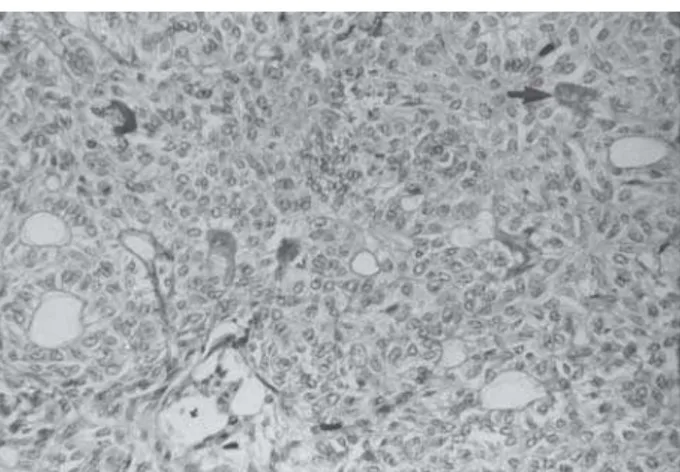
Peroperatively, a single fluid-filled sac was seen in left maxillary alveolus. Gross examination of the specimen showed a single grey-brown soft tissue measuring 2.5 x 2 cm. Cut section revealed a cyst with wall thickness of 0.1-0.5 cm with an impacted canine tooth. Microscopy revealed a cystic space lined by focal solid areas of the tumor showing polyhedral to spindle cells forming nests, cords and ducts (Fig. 1).
Figure 1. Section from solid area of tumor with cells arranged in nests and glands. Amyloid-like material (arrow) is also seen. Hematoxylin-eosin staining (original magnification X250).
Braz Dent J 16(3) 2005
252 S. Nigam et al.
In some areas, the ducts were filled with eosinophilic (amyloid-like) material (Fig. 1).The final diagnosis of intraosseus follicular variant of AOT was obtained.The patient is healthy and has not shown any signs of recurrence two years after surgery.
DISCUSSION
Adenomatoid odontogenic tumor is a rare tumor that comprises only 0.1% of tumors and cysts of the jaw and 3% of all odontogenic tumors (1). It is an uncommon cause of jaw swelling. Common non-neoplastic causes of jaw swelling in this age group are apical cyst, dentigerous cyst, calcifying epithelial odontogenic cyst, odontogenic keratocyst, periapical granuloma and central giant cell granuloma.
There are three clinicopathologic variants of AOT, namely intraosseus follicular, intraosseus extrafollicular and peripheral, all with identical histology. The follicular type is a central intraosseus lesion associated with an impacted tooth, while extrafollicular intraosseus AOT has no relation with an unerupted tooth. In spite of this, it is often located between, above or superimposed upon the roots of adjacent erupted teeth. The peripheral variant appears as a gingival fibroma or epulis attached to the labial gingival (3).
The follicular and extrafollicular variants account for 96% of all AOT cases (of which 71% are follicular). Follicular and extrafollicular variants together are more commonly found in the maxilla than in the mandible (2.1:1 ratio). More than two thirds are diagnosed in the second decade, mosty in the 13-19 year age group. The female:male ratio is 1.9:1 (3). Even higher ratios are found in Asian populations, the highest incidence being observed in Sri Lanka (3.2:1) (4) and Japan (3:1) (5). The tumor is usually associated with unerupted teeth, frequently canines or lateral incisors. Irregular root resorption is seldom reported (1,6). The patient we describe in this report also presented resorption of the upper left lateral incisor and first premolar, together with impaction of the canine. Radiologically, it should be differentiated from dentigerous cyst, which most frequently occurs as a pericoronal radiolucency in the jaws. Dentigerous cyst encloses only the coronal portion of the impacted tooth, whereas AOT shows radiolucency usually surrounding both the coronal and radicular aspects of the involved tooth (2)
Common neoplastic causes, such as
ameloblastoma, calcifying epithelial odontogenic tumor (CEOT), ameloblastic fibroma and ameloblastic fibro-odontoma are easily differentiated on histology. CEOT shows larger and more numerous calcifying spherules within eosinophilic cytoplasm of large cells along with smaller cells with hyperchromatic nuclei. Amyloid-like eosinophilic material is also present. Ameloblastoma has characteristic lining and arrangement with stellate reticulum besides usual location in mandible and posterior maxilla in contrast to AOT that is located in anterior maxilla (7). Areas of CEOT-like tissues have been described in classic AOT.
Immunohistochemical and ultrastructural findings have shown that the eosinophilic deposits (amyloid-like material) most probably represent some form of enamel matrix (8). The histogenesis of AOT is still uncertain, although recent findings strongly indicate that AOT is derived from a complex system of dental laminae or its remnants (8). It is often considered as a hamartomatous lesion rather than a true neoplasm (8). All variants of AOT are well encapsulated and show an identical benign behavior. Conservative surgical enucleation or curettage is the treatment of choice with only rare recurrence (3). The patient we described in this case report is healthy without recurrence and is under follow-up after local excision.
We conclude that AOT should also be considered in the differential diagnosis of radiolucent jaw swellings, although its incidence is low.
RESUMO
O tumor odontogênico adenomatóide (TOA) é um tumor i n c o m u m d e o r i g e m o d o n t o g ê n i c a , c a r a c t e r i z a d o histologicamente pela formação de estruturas tubulares com depósitos do tipo amilóide. A histogênese do TOA ainda é indeterminada e este tumor é frequentemente considerado mais como uma lesão hamartomatosa do que propriamente um neoplasma. O TOA tem comportamento benigno, sendo suficiente a enucleação cirúrgica conservadora ou curetagem. Neste artigo, é descrito um caso de TOA em paciente de 15 anos do sexo feminino, que exibia um edema no lado esquerdo da mandíbula com reabsorção dental. A histopatologia revelou uma variante folicular intra-óssea do tumor odontogênico adenomatóide. Uma breve revisão da literatura é também apresentada.
ACKNOWLEDGEMENTS
Braz Dent J 16(3) 2005
Adenomatoid odontogenic tumor 253
REFERENCES
1 . Dayi E, Gurbuz G, Bilge OM, Ciftcioglu MA. Adenomatoid odontogenic tumour (adenoameloblastoma). Case report and review of the literature. Aust Dent J 1997;42:315-318. 2 . Lee JK, Lee KB, Hwang BN. Adenomatoid odontogenic tumor:
A case report. J Oral Maxillofac Surg 2000;58:1161-1164. 3 . Philipsen HP, Reichart PA. Adenomatoid odontogenic
tumour: facts and figures. Oral Oncol 1998;35:125-131. 4 . Mendis BRRN, MacDonald DG. Adenomatoid odontogenic
tumour: A survey of 21 cases from Sri Lanka. Int J Oral Maxillofac Surg 1990;19:141-143.
5 . Toida M, Hyodo I, Okuda T, Tatematsu N. Adenomatoid odontogenic tumor: report of two cases and survey of 126 cases in Japan. J Oral Maxillofac Surg 1990;48:404-408. 6 . Nomura M, Tanimoto K, Takata T, Shimosato T. Mandibular
adenomatoid odontogenic tumor with unusual clinicopathologic features. J Oral Maxillofac Surg 1992;50:282-285.
7 . Rosai J. Mandible and Maxilla. In: Ackerman’s Surgical Pathology. Rosai J (Editor). 8th ed. St. Louis: Mosby; 1996. p. 257-288.
8 . Philipsen HP, Samman N, Ormiston IW, Wu PC, Reichart PA. Variants of the adenomatoid odontogenic tumor with a note on tumor origin. J Oral Pathol Med 1992;21:348-352.