Case Report
Co-occurrence of Calcifying Odontogenic Cyst, Aggressive Central
Giant Cell Granuloma and Central Odontogenic Fibroma: Report of a
Very Rare Entity and Its Surgical Management
Touraj Vaezi
1, Jahanshah Salehinejad
2, Mansoreh Darijani
3, Hamed
Ebrahimnejad
4, Javad Darijani
5, Masoud Nazeri
6, Mahtab Vakili
71
Fellowship of Traumatology, Oral and Maxillofacial Surgery Department, Tehran University of
Medical Sciences, Tehran, Iran
2
Professor, Dental Research Center, Department of Oral Pathology, Faculty of Dentistry, Mashhad
University of Medical Sciences, Mashhad, Iran
3
Postgraduate Student of oral and maxillofacial Radiology , School of Dentistry, Mashhad
University of Medical Sciences and Health Services, Mashhad, Iran.
4
Oral and Maxiollofacial Radiologist
5
Undergraduate Student of medicine, Kerman University of Medical Sciences, Kerman, Iran.
6
Resident of Oral Medicine, Chronic Headache and Orofacial Pain Clinic and Oral Medicine
Department, School of Dentistry, Kerman University of Medical Sciences, Kerman, Iran.
7
Oral and Maxillofacial pathologist, Kermanshah University of Medical Science, Kermanshah,
Iran.
Received 10 January 2016 and Accepted 12 April 2016
Abstract
Calcifying odontogenic cyst (COC), Central odontogenic fibroma (COF) and aggressive central giant cell granuloma (CGCG) are rare pathologic diseases affecting the jaws. While the Co-existence of two of them is reported in the literature, existence of all three conditions in one patient is an extremely rare entity. In the present report, initial biopsy revealed fibrosarcoma, therefore mandibular resection was performed for the subject. Sectional Histopathologic evaluation revealed the co-existence of three conditions through histopathologic evaluation. This report emphasizes the importance of precise microscopical evaluation of jaw lesions and thorough sectional examination of the lesions to reach the precise diagnosis. Treatment modalities and follow-up radiographs are also provided to help clinicians manage these entities.
Keywords: Calcifying odontogenic cyst, Central giant cell granuloma, Central odontogenic fibroma, combined lesion, surgical management
--- Vaezi T, Salehinejad J, Darijani M, Ebrahimnejad H, Darijani J, Nazeri M, Vakili M. Co-occurrence of Calcifying odontogenic cyst, aggressive central giant cell granuloma and central odontogenic fibroma: Report of a very rare entity and its surgical management. . J Dent Mater Tech 2016; 5(3): 153-7.
Introduction
Calcifying odontogenic cyst (COC) was introduced to the literature in 1962 by Gorlin et al. (1). The lesion does not demonstrate any specific sex or location predilection and mostly occurs in the canine-incisor region. About 79-85% of the cases are intraosseous (central), while 15-21% are extraosseous (peripheral) (2). Though it is considered as a benign condition, multiple variants including cystic, neoplastic (solid) and malignant (aggressive) exist with distinct histopathological and clinical features that complicate the process of diagnosis and treatment (3).
Conclusion regarding sex and location distribution. It is characterized histologically by the presence of variable amounts of inactive looking odontogenic epithelium scattered through a collagenous fibrous connective tissue (4). It is described as an asymptomatic expansion of the lingual or buccal cortical plate of the mandible and maxilla that is accidentally discovered in radiographic examinations (5).
Central giant cell granuloma (CGCG) is a relatively common reactive tumor of the jaws and rarely demonstrates an aggressive behavior. Differentiation between aggressive and non-aggressive subtypes could be made both clinically and radiographically. It is characterized histologically by osteoclast-like giant cells scattered in a cellular fibrovascular stroma (6). CGCG is associated with several other lesions, including aneurysmal bone cyst (ABC), odontogenic
fibroma, cherubism and Paget’s disease of the bone (7).
A very few number of case series have demonstrated the association of CGCG with Central ossifying fibroma(COF) and declared that the clinical progress of the combined lesion is dependent upon the behavior of CGCG (8).
While there are no reports on association of CGCG with COF and COC, here we present a 10- year old child whom was presented with a single lesion of the mandible that was diagnosed as fibrosarcoma in initial biopsy. Mandibular resection was performed and
sectional precise evaluation of the lesion eventually led to the diagnosis of COC, COF and aggressive CGCG. This very rare incident emphasizes the need for a more thorough histological examination of specimen diagnosed with CGCG, COF or COC.
Case report
The patient was a 10 year-old boy referring to oromaxillofacial surgery department of Mashhad dental school, Mashhad, Iran. Past medical and familial history was clear. The patient had no systemic disease and did not use any drugs.
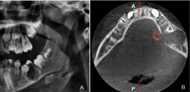
Chief complaint of the patient was pain and swelling in the left side of the face which started from about two weeks prior to admission. Extra and intra oral examination revealed a nodular swelling with 2 * 3 cm dimension in the buccal vestibule of the mandibular left first molar tooth. Swelling could be palpated extraorally and had a bony hard consistency. Radiographic imaging (Panoramic and CBCT) was ordered for the patient. In preoperative panoramic image, a well-demarcated lesion was observed in the tooth region which had led to tooth resorption and adjacent tooth movement. CBCT revealed a radiolucent lesion with well-defined borders which had led to the adjacent tooth resorption and buccal and lingual expansion. Lingual plate perforation was also observed in CBCT (Fig. 1.A and B).
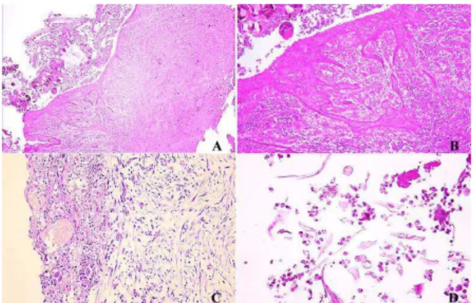
Figure 2. A-D) Initial biopsy revealed neoplastic proliferation of fibroblasts, several mitosis, a vast number of blood vessels and foreign body giant cells.
The diagnosis based on these observations was Fibrosarcoma.
The most relevant differential diagnosis based on the clinical and radiographic characteristics included: COC, COF and aggressive CGCG. Therefore, the affected tooth was extracted and sent to the pathologist for further evaluation. Histopathologic studies revealed a neoplastic proliferation of fibroblasts (Herring bone formation), several mitosis (atypical mitosis and pleomorphism was observed as well), a vast number of blood vessels and foreign body giant cells were also present in the pathological sample. The diagnosis based on the biopsy was fibrosarcoma (Fig. 2.A- 2.D)
The patient was referred to oral surgeon for the surgical removal of the lesion. Partial resection of the mandible was performed under general anesthesia and samples were sent for pathological evaluation.
In the pathological sample, three distinct entities were observed which altered the final diagnosis of lesion. Sectional analysis of the sample revealed the following pathologies: The first entity observed in histological evaluation was COC. The lumen was obstructed by pus and inflammatory cells and instead capsule was against
Cell lesion (Fig 3.A). Intraluminal calcification, pus and inflammatory cells and hyperplastic arched shaped epithelium was also observed in the section (Fig.3.B) and ghost cells were observed in the lesion (Fig.3.C) Spindle-shaped fibroblast that were present with hyperchromic and multinuclear giant cells (Fig 4.A, B). Furthermore, CGCG was observed adjacent to the striated muscle bundles (Fig. 4.C, D) to the mandibular nerve trunk (Fig.4.E) that revealed aggressive nature of this lesion.
Figure. 3: A) Calcifying odontogenic cyst. The lumen of the cyst was obstructed by pus and inflammatory
cells. B) Intraluminal calcification, pus and inflammatory cells and hyperplastic arched shape epithelium was observed in a higher magnitude. C-D)
Ghost cells were observed in thelining and lumen.
Figure 4. A and B) Spindle-shaped fibroblast present with hyperchromic and multinuclear giant cells are observed in the lesion. (C and D) CGCG adjacent to the striate muscle bundles and mandibular nerve trunk
was also observed in this section.
The other entity observed was central odontogenic fibroma (COF). This lesion was adjacent to oral mucosa and odontogenic epithelial islands were observed in the lesion (Fig.5.A and B).
Figure 5. A and B) Central odontogenic fibroma (COF) adjacent to the oral mucosa and odontogenic
Following partial mandibular resection, reconstructive surgery was performed to rehabilitate function and aesthetics. Mandibular plates were placed and screwed to the remaining structure (Fig. 6).
Figure 6. Post-operative radiograph demonstrating the bony reconstruction.
Post-operative radiographs were obtained and the patient was followed-up for three years. No recurrence was observed in this observation period.
Discussion
Most cases of COC are asymptomatic and diagnosed accidentally in radiographic examinations. Radiographically, the lesion is presented as a unilocular or multilocular radiolucency with well-demarcated borders, with calcifications of variable density present in one third to half of COC cases. Clinical presentation of COC is usually accompanied by an asymptomatic swelling in both intraosseous and extraoseous lesions with expansion of the buccal and/or lingual cortical plates (9). Root resorption and divergence and association with an impacted teeth is a common finding in COC cases (10) .
Histopathologic features of COC are variable, but the main finding is a fibrous capsule with four to six layers of odontogenic epithelium. Cyst lining consists of an outer layer composed of columnar basaloidodontogenic epithelium and an inner layer resembling stellate reticulum of the enamel organ. The characteristic feature of COC is the presence of ghost cells and calcification occurring within the fibrous capsule and the cyst lining (10).
Another lesion manifested in the patient was COF, which is an extremely rare condition. It is a benign tumor of the mesodermal origin and due to its paucity of cases; its characteristics are not well described in the literature. This tumor occurs mostly in the mandible and there is no age or sex predilection. This lesion is also asymptomatic and grows very slowly; therefore, it is discovered incidentally in radiographs taken for other reasons. Association of this tumor with other
diagnosed histopathologically through observation of a dense fibrous connective tissue stroma with mature spindle-shaped fibroblasts. Odontogenic epithelium is also observed with one to two layers of cells thickness. Treatment of choice for the COF includes enucleation and curettage since recurrence and tendency to undergo malignant transformation is very rare in this entity(12). Therefore, isolated cases of COF do not need an aggressive surgical procedure. But, it is important to mention that cases of combined COF with another lesions need special considerations and treatment planning is based on the prognosis of other lesions presented with COF (5).
Aggressive CGCG is a rare condition affecting jaws, displacing teeth and usually leading to occlusal disturbances as a consequence. Radiological findings are diverse from a small unilocular lesion to a large multilocular lesion displacing teeth and cortical plate perforation(13). Histopathologic features of CGCG are pathognomonic and are characterized by a highly cellular fibroblastic stroma with spindle-shaped cells with high mitotic rate and a high vascular density(14). Multinucleated giant cells are noticeable throughout the fibroblastic stroma. Giant cells are distributed irregularly and are mostly found in hemorrhage areas (14). There are some reports regarding the association of CGCG with other lesions, including central ossifying fibroma. As Kaplan et al. (2007) have demonstrated in their case series, the clinical behavior of this type of lesions is determined by the more aggressive component, e.g. CGCG in the current case (8). Treatment of choice for aggressive CGCG is surgical curettage and resection in aggressive cases. Other treatment options include injection of corticosteroids into the lesions or subcutaneous administration of calcitonin or interferon alpha, though there is no randomized clinical trial demonstrating their efficacy (15,16).
Referencess
1. Neuman AN, Montague L, Cohen D, Islam N,
Bhattacharyya I. Report of Two Cases of
Combined Odontogenic Tumors: Ameloblastoma
with Odontogenic Keratocyst and Ameloblastic
Fibroma with Calcifying Odontogenic Cyst. Head
and neck pathology. 2015;9(3):417-420.
2. Saghafi S, Zare-Mahmoodabadi R, Salehinejad J,
Kadeh H, Afzal-Aghaee M. Immunohistochemical
analysis of p53 and PCNA expression in calcifying
odontogenic cyst. Journal of oral science.
2010;52(4):609-613.
3. Habibi A, Saghravanian N, Salehinejad J,
Jafarzadeh H. Thirty years clinicopathological
study of 60 calcifying cystic odontogenic tumors
in Iranian population. The journal of contemporary
dental practice. 2011;12(3):171-173.
4. Salehinejad J, Ghazi N, Heravi F, Ghazi E.
Concurrent central odontogenic fibroma (WHO
type) and odontoma: Report of a rare and unusual
entity. Journal of Oral and Maxillofacial Surgery,
Medicine, and Pathology. 2015;27(6):888-892.
5. Santoro A, Pannone G, Ramaglia L, Bufo P,
Muzio LL, Saviano R. Central odontogenic
fibroma of the mandible: A case report with
diagnostic considerations. Annals of Medicine and
Surgery. 2016;5:14-18.
6. Jadu F, Pharoah M, Lee L, Baker G, Allidina A.
Central giant cell granuloma of the mandibular
condyle: a case report and review of the literature.
Dentomaxillofacial Radiology. 2011 ; 40 (1):
60-64.
7. Crusoé-Rebello I, Torres M, Burgos V, Oliveira C,
dos Santos J, Azevedo R, et al. Hybrid lesion:
central giant cell granuloma and benign
fibro-osseous lesion. Dentomaxillofacial Radiology.
2009 ; 38(1): 421-425.
8. Kaplan I, Manor I, Yahalom R, Hirshberg A.
Central giant cell granuloma associated with
central ossifying fibroma of the jaws: a
clinicopathologic study. Oral Surgery, Oral
Medicine, Oral Pathology, Oral Radiology, and
Endodontology. 2007;103(4):35-41.
9. Lee J, Song Y-G, Moon S-Y, Choi B, Kim BC,
Yoon J-H. Calcifying cystic odontogenic tumor
associated with ameloblastic fibro-odontoma of
the anterior mandible. Journal of Craniofacial
Surgery. 2014;25(3):259-260.
10. Phulambrikar T, Kant SV, Kode M, Magar S.
Cone Beam Computed Tomography Findings in
Calcifying Cystic Odontogenic Tumor Associated
with Odontome: A Case Report. Journal of
Dentistry. 2015;16(4):374-379.
11. Khajeh Ahmadi S, Rahpeyma A. Central
Odontogenic Fibroma. Iranian Journal of
Pathology. 2013;8(2):131-134.
12. Araki M, Nishimura S, Matsumoto N, Ohnishi M,
Ohki H, Komiyama K. Central odontogenic
fibroma with osteoid formation showing atypical
radiographic appearance. Dentomaxillofacial
Radiology. 2009; 38 (6): 426-430.
13. Tarsitano A, Del Corso G, Pizzigallo A, Marchetti
C. Aggressive Central Giant Cell Granuloma of
the Mandible Treated With Conservative Surgical
Enucleation and Interferon–α-2a: Complete
Remission With Long-Term Follow-Up. Journal of
Oral and Maxillofacial Surgery.
2015;73(11):2149-2154.
14. Kaur S, Ahluwalia SS, Singh P. Case report of
aggressive Central Giant Cell Granuloma-A
diagnostician's approach. International Journal of
Medical Research & Health Sciences.
2014;3(3):743-747.
15. Yüzbasioglu E, Alkan A, Özer M, Bayram M.
Multidisciplinary approach for the rehabilitation of
central giant cell granuloma: A clinical report.
Nigerian journal of clinical practice.
2014;17(4):528-533.
16. Neville BW, Damm DD, Allen CM, Chi AC.
"Bone Pathology". In: Oral and Maxillofacial
Pathology. BW Neville et al. 4th ed. Missouri:
Elsevier, 2016. 585-586.
Corresponding Author: Mansoreh Darijani,
Postgraduate Student, School of Dentistry,
Mashhad University of Medical Sciences and Health Services, Mashhad, Iran.