214 Radiol Bras. 2013 Jul/Ago;46(4):214–220
Screening of breast lesions: a comparative study between
mammography, B-mode ultrasonography, sonoelastography
and histological results
*
Rastreio de lesões mamárias: estudo comparativo entre a mamografia, ultrassonografia modo-B, elastografia e resultado histológico
Raquel Constantino Pardal1, António Fernando Lagem Abrantes2, Luís Pedro Vieira Ribeiro3, Rui Pedro Pereira Almeida4, Kevin Barros Azevedo5, Teresa Leonor Figueiredo6, Sónia Isabel Rodrigues7
Objective: To compare the capacity of mammography, sonoelastography, B-mode ultrasonography and histological analysis to differentiate benign from malignant breast lesions. Materials and Methods: A total of 12 histopathologically confirmed breast lesions were documented. The lesions were assessed by means of mammography, B-mode ultrasonography and sonoelastography, and histopathological analysis was utilized as a gold standard. Sensitivity and specificity were calculated. A receiver operating characteristic (ROC) curve was constructed to evaluate the diagnostic performance of the mentioned techniques. Results: Sensitivity and specificity in the differentiation between benign and malignant lesions were respectively 100% and 50% for mammography, 100% and 71% for B-mode ultrasonography, and 67% and 83% for sonoelastography. The area under the ROC curve was calculated for the three imaging modalities and corresponded to 0.792 for mammography, 0.847 for B-mode ultrasonography, and 0.806 for sonoelastography. Conclusion: Sonoelastography demonstrated higher specificity and lower sensitivity as compared with mammography and B-mode ultrasonography. On the other hand, B-mode ultrasonography had the largest area under the ROC curve. Sonoelastography has demonstrated to be a promising technique to detect and evaluate breast lesions, and could potentially reduce the number of unnecessary biopsies.
Keywords: Breast B-mode ultrasonography; Breast lesions; Mammography; Sensitivity; Specificity; Sonoelastography.
Objetivo: Comparar a capacidade de diferenciação de lesões benignas versus malignas por parte da mamografia, ul-trassonografia modo-B e elastografia. Materiais e Métodos: Um total de 12 lesões mamárias confirmadas histologi-camente foi documentado. A avaliação das lesões foi realizada por meio da mamografia, ultrassonografia modo-B e elastografia. Os resultados histopatológicos foram utilizados como técnica padrão ouro. As sensibilidades e as especifi-cidades foram calculadas. A curva receiver operating characteristic (ROC) foi realizada para avaliar o desempenho diag-nóstico das técnicas utilizadas. Resultados: A sensibilidade e a especificidade na diferenciação entre lesões mamárias benignas e malignas foram 100% e 50%, respectivamente, para a mamografia, e 100% e 71% para a ultrassonografia modo-B. A elastografia obteve sensibilidade de 67% e especificidade de 83%. A área abaixo da curva ROC foi calculada para as três técnicas imaginológicas, sendo 0,792 para a mamografia, 0,847 para a ultrassonografia modo-B e 0,806 para a elastografia. Conclusão: A elastografia mostrou ter maior especificidade e menor sensibilidade comparativa-mente à mamografia e à ultrassonografia modo-B. A ultrassonografia modo-B foi a técnica que demonstrou maior área abaixo da curva ROC. A elastografia mostra-se uma técnica promissora na detecção de doença mamária e, potencial-mente, poderá reduzir biópsias desnecessárias.
Unitermos: Elastografia; Especificidade; Lesões mamárias; Mamografia; Sensibilidade; US modo-B mamária.
Abstract
Resumo
* Study developed at Escola Superior de Saúde da Univer-sidade do Algarve (ESSUAlg), Algarve, Portugal.
1. Licenciate, Collaborator at Department of Radiology, Es-cola Superior de Saúde da Universidade do Algarve (ESSUAlg), Algarve, Portugal.
2. PhD, Member of Centro de Investigação Cesnova – Uni-versidade Nova de Lisboa, Professor and Member of Centro de Estudos em Saúde (CES) – Escola Superior de Saúde da Univer-sidade do Algarve (ESSUAlg), Algarve, Portugal.
3. PhD, Member of Centro de Investigação do Desporto e da Actividade Física (CIDAF) – Universidade de Coimbra, Profes-sor and Member of Centro de Estudos em Saúde (CES) –
Es-Pardal RC, Abrantes AFL, Ribeiro LPV, Almeida RPP, Azevedo KB, Figueiredo TL, Rodrigues SI. Screening of breast lesions: a comparative study between mammography, B-mode ultrasonography, sonoelastography and histological results. Radiol Bras. 2013 Jul/Ago;46(4):214– 220.
cola Superior de Saúde da Universidade do Algarve (ESSUAlg), Algarve, Portugal.
4. Postgraduate degree, Member of Centro de Estudos em Saúde (CES), Professor, Department of Radiology, Escola Supe-rior de Saúde da Universidade do Algarve (ESSUAlg), Algarve, Por-tugal.
5. Licenciate, Member of Centro de Estudos em Saúde (CES), Professor, Department of Radiology, Escola Superior de Saúde da Universidade do Algarve (ESSUAlg), Algarve, Portugal.
6. Master, MD, Radiologist, Professor of the Integrated Master Degree Programme of Medical and Biomedical Sciences, Universidade do Algarve (DCBM-UAlg), Algarve, Portugal.
7. Master, Radiology Technician at Hospital de Faro, Profes-sor, Department of Radiology, Member of Centro de Estudos em Saúde (CES) – Escola Superior de Saúde da Universidade do Algarve (ESSUAlg), Algarve, Portugal.
Mailing Address: Raquel Pardal. Departamento de Radiolo-gia da Escola Superior de Saúde da Universidade do Algarve. Avenida Doutor Adelino da Palma Carlos, 8000-510. Faro, Por-tugal. E-mail: [email protected].
INTRODUCTION
Breast cancer is the most common neo-plasm in the female population, compris-ing about 16% of all tumors affectcompris-ing women. Its incidence is quite variable worldwide. In North America, its incidence has been calculated to be 99.4 cases per 100,000 women. In regions such as East-ern Europe, South America, the SouthEast-ern African region and Western Asia, the re-ported incidence is moderate, however, it is increasing(1). Such a fact is followed by a decrease in mortality, which is intimately related to early detection of breast diseases and to the increasing availability of appro-priate treatment(2).
In Portugal, the recorded incidence is approximately 4,000 new cases/year(3).
Mammography is a diagnostic tech-nique aimed at producing detailed images from the internal structures of the breast so as to allow for early diagnosis of breast diseases. The mammographic study com-prises the acquisition of two basic views, namely, craniocaudal and mediolateral ob-lique views. Supplementary views may be acquired whenever the presence of a sus-pected lesion is detected(4).
In the diagnosis of breast lesions, B-mode ultrasonography (US) has demon-strated to be quite relevant as a complement with other diagnostic modalities. Whenever possible, the sonographic study should al-ways be preceded by a complete mammo-graphic study(5). It is a screening method for younger women with dense breasts at mam-mography (BI-RADS 3 and 4)(6). The ac-quisition technique varies among patients, indications, location and type of lesion. Sonographic findings should be docu-mented in two orthogonal planes (longitu-dinal and cross-sectional) to allow the vi-sualization of all their characteristics.
Currently, sonoelastography has shown to be a promising technique in the follow-up of suspicious breast lesions in elderly patients, and in the intervals of the mam-mographic follow-up, thus allowing for guidance in diagnosis and prognosis. At younger ages, such a technique allows the addition of information to the diagnosis of solid lesions, avoiding unnecessary biop-sies. It is complementary with B-mode US in the diagnosis of breast diseases. Its main
advantages include absence of ionizing radiations, providing more specific data on the possible lesion, besides not requiring a significant increase in time for the investi-gation of suspected lesions(7). Such a method allows for quantifying the tissues elasticity degree by means of pressure ex-erted on them(6,8). The lesions are quantified according to a color scale. A transducer that superimposes the color data on the B-mode US images is utilized(9). The color scale ranges from red to blue, and is associated with the benignity/malignancy degree, with the absence of deformation being charac-terized by the blue color associated to the malignancy degree(6,10).
Breast biopsy is defined as an invasive procedure aimed at confirming the breast lesion detection. Such a procedure relies on the collection of breast tissue specimens which are then histologically analyzed. There are several types of biopsies, which are utilized according to lesion type, size and depth in relation to the skin surface. The post-procedural follow-up depends upon the type of histological finding(11).
Thus, the present study is aimed at de-termining the capability of mammography, B-mode US and sonoelastography to differ-entiate benign from malignant breast le-sions relying on histological results as the gold-standard technique.
The present study reveals itself as an opportune investigation, considering that breast sonoelastography is a relatively re-cent technique whose diagnostic accuracy is scarcely described in the literature. In case the reliability of the technique in dif-ferentiating benign from malignant lesions is proven, the number of unnecessary breast biopsies currently performed could be re-duced.
MATERIALS AND METHODS
The present study population comprised a group of female patients with unilateral or bilateral breast disease who were submit-ted to imaging studies at the Radiology Services of Hospital do Faro, E.P.E. and at H.P.P. – Hospital Santa Maria de Faro. The sample comprised 12 breast lesions docu-mented by means of mammography, B-mode US and sonoelastography images. The histological results from the biopsies
performed on the studied lesions were con-sidered as the gold-standard, so as to in-crease the specificity of the results. The lack of any of the previously mentioned diag-nostic imaging study was defined as an exclusion criterion for the present study.
The mean age of the patients was 54.8 ± 10.4 years, ranging from 43 to 73 years. Tables were prepared to simplify the data collection. Such tables include the essential characteristics of the lesions which allowed their classification as benign or malignant with basis on the four diagnostic modali-ties. By means of observation and analysis of the images from the three imaging meth-ods, as well as the correct filling of the above mentioned tables, it was possible to establish the degree of probability of benig-nity/malignancy of the studied lesions.
The collected data were exclusively and solely utilized in the present study, and the patients’ identity, anonymity and confiden-tiality were safeguarded.
Mammography
All the patients were submitted to mam-mography. The images were acquired in GE Medical System Senographe DMR® and
GM Medical System Apollon®
mammog-raphy apparatuses, with craniocaudal and oblique mediolateral acquisitions, which allowed for the demonstration of the outer and inner quadrants, the upper and lower quadrants, and the inframammary angle, respectively.
The mammographic images were ana-lyzed according to the Breast Imaging Re-porting and Data System (BI-RADS®) from
the American College of Radiology. By means of such a system, it is possible to standardize the terms utilized in mammog-raphy reports, making it clear, concise and easily understandable(8).
The classification of mammographic findings comprises seven categories di-rectly related to the approach recommenda-tions, as follows(8):
• Category 1 – Refers to images negative for malignancy, with no evidence of sig-nificant focal radiographic alterations. • Category 2 – Utilized in situations
where the mammographic findings are characteristically benign.
• Category 3 – Mammographic findings with high probability of benignity, with positive predictive value (PPV) ≥ 98%. • Category 4 – Lesions presenting prob-ability of malignancy, but with no typi-cal characteristic of carcinoma. • Subcategory 4a – Lesions with
interme-diate malignancy suspicion, with indi-cation for biopsy for histological corre-lation.
• Subcategory 4b – Amorphous calcifica-tions, nodules with partially circum-scribed and partially indistinct contours. • Subcategory 4c – Lesions under moder-ate suspicion of malignancy, with ex-pectation of histological result positive for malignancy.
• Category 5 – Lesions whose malignancy probability is high (PPV > 95%). • Category 6 – Lesions whose malignancy
has already been previously histopatho-logically confirmed and that have not been submitted to any definitive treat-ment yet.
B-mode US and sonoelastography
The B-mode US and sonoelastography images were obtained by GE Medical Sys-tem Logic 9® and Siemens Acuson Antares®
US apparatuses, with the utilization of a high-frequency linear transducer. The fre-quencies ranged between 7.5 and 14 MHz, depending upon the depth of the lesion and breast thickness. The lesions under study were documented in two orthogonal planes (longitudinal and cross-sectional).
Once the elastography mode is selected, the scale of colors is superimposed over the B-mode image(9). Both images are simul-taneously presented on the equipment screen in real time, with the B-mode being presented on the left side and the sonoelas-togram being presented at the right side of the screen.
As regards sonoelastography, measure-ments were performed before and after uniform tissue compression, so as to evalu-ate the multi-directional deformation of the suspicious breast tissue.
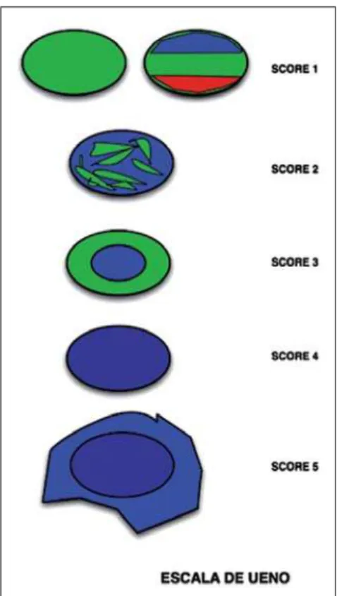
The evaluation of the B-mode US im-ages, similarly to mammography, is per-formed according to the BI-RADS classi-fication. On the other hand, sonoelasto-graphic images, namely the elastic tissue properties, are quantitatively analyzed ac-cording to the Ueno elasticity five-score system (Figure 1)(8,12–15), as follows:
• Level 1 – Uniformly elastic lesion, green colored. A variation of this type is the diagnostic image of a cyst (1*), where three layers are presented (blue, green and red).
• Level 2 – Fundamentally elastic lesion, with some zones of elasticity absence, characterized by a green and blue mo-saic pattern.
• Level 3 – Peripheral elasticity and ab-sence of elasticity in the central region of the lesion, with green color at the pe-ripheral zone and blue color within the lesion.
• Level 4 – Absence of elasticity in the en-tire lesion, which is visualized enen-tirely in the blue color.
• Level 5 – Absence of elasticity not only in the entire lesion but also in surround-ing tissues, with a blue region more extensive than the lesion itself being visualized.
The levels attributed at sonoelasto-graphy may be compared with the BI-RADS classification. Levels 1 and 2 at sonoelastography correspond to BI-RADS category 2. The remaining levels at sono-elastography present a one-to-one corre-spondence with the BI-RADS classifica-tion(9). Thus, level 1 at sonoelastography represents findings negative for malig-nancy, level 2, benign findings, level 3, probably benign findings, level 4 demon-strates findings suspicious for malignancy, and level 5 represents findings that are highly suspicious for malignancy(16). There-fore, one can affirm that the elasticity level of lesion in intimately correlated with the BI-RADS classification, since low elastic-ity levels correspond to high BI-RADS categories. The contrary is also true(17).
Histology
The histological results were available for all patients. Percutaneous US-guided core biopsies with automated biopsy device were performed in nine patients (75%) and
stereotactic biopsies were performed in three patients (25%). The specimens were collected and histologically analyzed in a laboratory.
Statistical analysis
The obtained results were analyzed with the softwares Microsoft Excel 2010® and
Statistical Package for the Social Sciences (SPSS) V 20.0®.
The Microsoft Excel 2010® software
was utilized for the calculation of sensitivi-ties, specificisensitivi-ties, PPVs and negative pre-dictive values (NPVs) for the three imag-ing methods.
difference of the mean values, considering an alpha of 0.05.
RESULTS
Twelve breast lesions were identified in female patients by means of mammogra-phy, b-mode US and sonoelastography (Figures 2 and 3). The histological result was obtained for all patients, with identi-fication of six benign and six malignant lesions measuring, on average, 15.1 ± 10.6 mm and 13.8 ± 3.8 mm, respectively.
In order to quantify the lesions detected by the imaging methods under study, the frequencies were calculated according to their classification. Mammography identi-fied three lesions classiidenti-fied as BI-RADS 3;
five lesions as BI-RADS 4b; and four le-sions as BI-RADS 4c, with a mean value of 5.8 ± 1.2. B-mode US detected four le-sions classified as BI-RADS 3; one lesion classified as BI-RADS 4a; two as BI-RADS 4b; and 5 as BI-RADS 4c, with a mean val-ues of 5.7 ± 1.4. Sonoelastography on its turn, detected 7 lesions quantified as level 2; one lesion as level 3; and four lesions as level 4. Thus the mean value was 2.8 ± 1. With the objective of identifying the number of benign and malignant lesions correctly diagnosed by the imaging meth-ods, with basis on the histological results, the frequencies were calculated. Mammog-raphy correctly identified nine of the 12 lesions, three of them classified as BI-RADS 3, three as BI-BI-RADS 4b, and three
as BI-RADS 4c. B-Mode US correctly identified seven of the 12 lesions (four clas-sified as BI-RADS 3, one as BI-RADS 4b, and one classified as BI-RADS 4c). Finally, sonoelastography correctly identified 10 of the 12 lesions, five of them level 2, one level 3, and four lesions level 4.
Thus, in order to better understand the diagnostic accuracy of the imaging meth-ods under study, their respective sensitivi-ties, specificisensitivi-ties, PPVs and NPVs were calculated (Table 1).
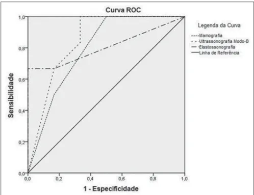
In order to quantify the performance of the imaging methods under study, a ROC curve was built, representing the sensitiv-ity values on the ordinate axis and the val-ues of 1-specificity on the abscissa axis (Figure 4).
According to Figure 4, the area under the mammography ROC curve was calcu-lated as being 0.792. B-mode US presented an area of 0.847 under the ROC curve, while sonoelastography presented an area of 0.806 under the ROC curve.
Also, the mean values and respective standard deviations of the Ueno classifica-tions for both benign and malignant lesions were calculated for sonoelastography. Mean values of 2.17 ± 0.408 for benign lesions, and 3.33 ± 1.033 for malignant lesions were observed. In order to deter-mine if the mean value of the Ueno classi-fication for malignant lesions is signifi-cantly higher than the mean Ueno classifi-cation for benign lesions, the t-test was applied for the difference between mean
values, and a p < 0.05 value was obtained, thus a higher and statistically significant mean value was found for the Ueno classi-fication of malignant lesions.
DISCUSSION
Considering that an incorrect diagnosis of breast diseases is many times directly related to a failure in perception of the le-sion by the investigator, it becomes essen-tial to evaluate the diagnostic capability of the imaging methods for detecting true-positive results (sensitivity) and true-nega-tive results (specificity).
With the progress in medical technolo-gies, it is already possible to utilize sono-elastography in routine diagnoses. Such
method presents some advantages, namely, the data obtained is immediately evaluated and superimposed over the B-mode US images, and it does not require more time than conventional B-mode US.
Previous studies demonstrated low sen-sitivity and high specificity of sonoelasto-graphy as compared with B-mode US(9, 14,18). As regards mammography, data on the
calculation of sensitivity and specificity in comparison with other imaging methods are still scarce in the literature.
Based on the above mentioned data and according to the present results, one ob-serves that sonoelastography presents sen-sitivity and specificity to differentiate breast lesions, and also that the three im-aging methods under study do not present the same sensitivity and specificity for the same lesions.
The present study is in agreement with the results reported by Thomas et al.(9), since B-mode US obtained higher sensitiv-ity (100% in the present study and 94% as reported by Thomas et al.(9)) and higher
Figure 3. Female 56-year-old patient with a recent history of mastitis in the right upper outer quadrant. Mammography with craniocaudal (a) and oblique mediolateral (b) views revealing an irregular hyperdense region with partially obscured contours, classified as BI-RADS 4b. Sonographic image (c) revealed an ovoid hypodense area with partially obscured margins, causing a acoustic shadow cone, classified as BI-RADS 3. Sonoelastography (d) demonstrated a fundamentally elastic lesion, with some elasticity absence zones, classified as level 2 according the Ueno score system. Histopathological analysis demonstrated the presence of fibroadipose tissue.
Table 1 Sensitivity, specificity, PPV and NPV for the imaging methods under study.
Mammography B-mode US Sonoelastography
Sensitivity
100% 100% 67%
Specificity
50% 71% 83%
PPV
67% 71% 80%
NPV
NPV (100% in the present study and 95% reported by Thomas et al.(9)), while sono-elastography showed lower specificity (83% in the present study and 87% reported by Thomas et al.(9)). As regards PPV, the study developed by Thomas et al.(9) is in disagreement with the present study, since according to those authors, mammography obtained the highest value (89%), while in the present study sonoelastography pre-sented the highest value (80%). The study developed by Lee et al.(14) is in agreement with the present study, since B-mode US obtained the highest sensitivity value (95.8%) and sonoelastography obtained the highest specificity value (45.7%), and PPV, 23.7%. Only the NPV is different from the present study results, with a higher value being obtained by sonoelastography (97.6%), as compared with B-mode US result of 100% obtained in the present study(14). The study developed by Mansour et al.(16) is completely in disagreement with the present study, since the results obtained by those authors are contradictory, namely the higher specificity (86.2%) and higher PPV (81.4%) were obtained with B-mode US and the higher sensitivity (92.3%) and higher NPV (93.4%) were obtained with sonoelastography.
Thus, it is possible to conclude that the association of the various techniques al-lows for the improvement of the accuracy in breast lesions diagnosis, since all the imaging methods contribute to the diagno-sis. Such association also allows for the reduction of the number of biopsies cur-rently performed.
The mean values of the Ueno classifi-cations for benign and malignant lesions at sonoelastography were calculated, and the
t-test was utilized to calculate the difference between mean values. The results from present study are in agreement with those reported by Lee et al.(14), demonstrating lower mean values (1.72 ± 0.78 for benign lesions, and 3.02 ± 1.33 for malignant le-sions as compared with the mean values of 2.17 ± 0.408 for benign lesions and 3.33 ± 1.033 for malignant lesions in the present study). After the t-test, a p value lower than alpha (p < 0. 001 as compared with the p < 0.05 obtained in the present study), also confirming a statistically significant differ-ence between the mean values for malig-nant and benign lesions.
The ROC curve represents a powerful tool for quantifying the performance of imaging methods. One verified that B-mode US was the method with the largest
area under the ROC curve (0.847), fol-lowed by sonoelastography (0.806) and mammography (0.792). According to the classification attributed to the areas under the ROC curve, mammography presented a reasonable performance, while both B-mode US and sonoelastography perfor-mances were classified as good. It is pos-sible to observe that the values obtained with B-mode US and sonoelastography are quite similar, allowing the assertion that based on such analysis, both methods present a similar diagnostic performance. The results from the present study are dif-ferent from those reported by Lee et al.(14), with the area under the ROC curve for B-mode US (0.616) lower than that from sonoelastography (0.784), with a weak and reasonable performance, respectively for the two methods. The results reported by Schaefer et al.(12) are also different from the results from the present study, with an area under the ROC curve for B-mode US (0.820) lower than that for sonoelastography (0.884), with both methods presenting a good performance.
CONCLUSION
The present study allows for the conclu-sion that sonoelastography presents a good diagnostic sensitivity and high diagnostic specificity in the differentiation between benign and malignant lesions, thus allow-ing for a reduction in the current number of breast biopsies. Although the imaging methods described in the present study do not present the same sensitivity and speci-ficity for the same lesions, their combina-tion may clearly improve the accuracy of the diagnosis of breast lesions.
The main limitation of the present study was the reduced sample size, conditioned by the reduced number of breast biopsies performed over the data collection period. As a recommendation for future studies, the authors suggest that prospective stud-ies are undertaken about the theme in ques-tion with a larger sample and in different clinical centers, in order to determine whether a quantitative analysis of the im-ages may be useful to overcome some shortcomings of the method. The authors also wish to propose that breast MRI be included in the studies, so as the compari-Figure 4. ROC curves representing the sensitivity and specificity values for mammography, B-mode US
son can cover the whole range of imaging methods.
REFERENCES
1. World Health Organization. Breast cancer: pre-vention and control. World Health Organization. [Online] 2012. [cited 2012 Sept 5]. Available from: h t t p : / / w w w. w h o . i n t / c a n c e r / d e t e c t i o n / breastcancer/en/index1.html.
2. Oliveira FGTF, Fonseca LMB, Koch HA. Respon-sabilidade civil do radiologista no diagnóstico do câncer de mama através do exame de mamogra-fia. Radiol Bras. 2011;44:183–7.
3. Ministério da Saúde. Guia de apoio à mulher com cancro da mama. Direcção Geral de Saúde. [On-line] 2011. [cited 2012 Sept 5]. Available from: http://www.dgs.pt/.
4. Kopans DB. Imagem da mama. 2ª ed. Rio de Ja-neiro: Medsi; 2000.
5. Dronkers DJ, Hendriks JHCL, Holland R, et al. The practice of mammography. New York: Thieme; 2002.
6. Fleury EFC, Rinaldi JF, Piato S, et al. Apresenta-ção das lesões mamárias císticas à ultra-sonogra-fia utilizando a elastograultra-sonogra-fia. Radiol Bras. 2008; 41:167–72.
7. Stavros AT, Rapp CL, Parker SH. Breast ultra-sound. Philadelphia: Lippincott Williams & Wil-kins; 2004.
8. Camps J, Sentis C. Elastosonografia mamaria. Rev Chil Radiol. 2008;14:122–7.
9. Thomas A, Kümmel S, Fritzsche F, et al. Real-time sonoelastography performed in addition to B-mode ultrasound and mammography: im-proved differentiation of breast lesions? Acad Radiol. 2006;13:1496–504.
10. Kemp C, Baracat FF, Rostagno R. Lesões não palpáveis da mama: diagnóstico e tratamento. Rio de Janeiro: Revinter; 2003.
11. Aguillar V, Bauab S, Maranhão N. Mama – diag-nóstico por imagem. Rio de Janeiro: Revinter; 2009.
12. Schaefer FK, Heer I, Schaefer PJ, et al. Breast ultrasound elastography – results of 193 breast
lesions in a prospective study with histopatho-logic correlation. Eur J Radiol. 2011;77: 450–6. 13. Tan S, Teh HS, Mancer JF, et al. Improving B mode ultrasound evaluation of breast lesions with real-time ultrasound elastography – a clinical approach. Breast. 2008;17:252–7.
14. Lee JH, Kim SH, Kang BJ, et al. Role and clini-cal usefulness of elastography in small breast masses. Acad Radiol. 2011;18:74–80. 15. Itoh A, Ueno E, Tohno E, et al. Breast disease:
clinical application of US elastography for diag-nosis. Radiology. 2006;239:341–50.
16. Mansour SM, Omar OS. Elastography ultrasound and questionable breast lesions: does it count? Eur J Radiol. 2012;81:3234–44.
17. Ueno E, Iboraki P. Clinical application of US elastography in the diagnosis of breast disease. European Congress of Radiology. 5–9 March 2004; Vienna, Austria.