Letters to the Editor
Radiol Bras. 2016 Jan/Fev;49(1):56–64
61
Dear Editor,
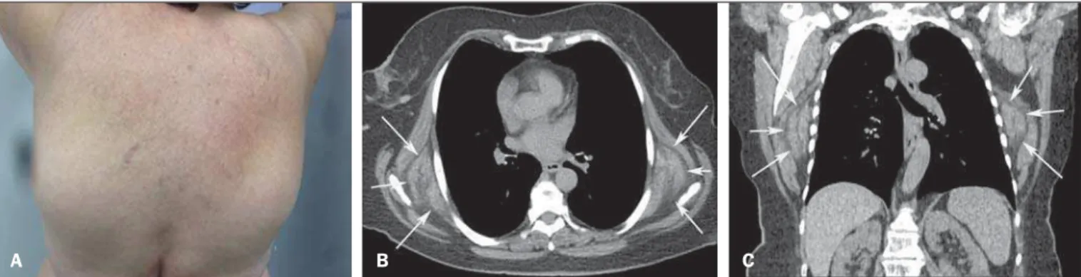
A 76-year-old female patient with a previous history of surgi-cally resected rectal cancer for seven years was admitted for diag-nostic investigation of bilateral and symmetrical dorsal masses. She reported chronic pain in the thoracic spine. At clinical exami-nation, the masses were solid, mobile, located subcutaneously and inferiorly to the scapulae (Figure 1A). Computed tomography (CT) showed the presence of bilateral soft tissue masses in the infrascapular region (Figures 1B and 1C). On the basis of the clinical findings and the images, the diagnosis of elastofibroma dorsi (ED) was established.
ED is a slow growing soft tissue pseudotumor incidentally di-agnosed during routine imaging studies, that may also cause chronic scapular pain(1). It is a benign fibroelastic tumor
inferi-orly located in the infrascapular region, between the scapula and the thoracic wall deeply to the serratus and latissimus dorsi muscles, possibly inserting into the periosteum of the posterior ribs. Coin-cidentally such type of tumor has been detected at CT in up to 2% of elderly patients(1,2). It is most frequently found in elderly women
(female to male ratio 5:1) in the age range between 65 and 70 years at the moment of the diagnosis(3).
Unilateral masses are slightly more prevalent at the right side, but up to 60% of EDs are bilateral(3). Other reported sites include
deltoid muscle, axillae, ischial tuberosity, olecranon, hands and feet, among others(4). It is also characterized by symptoms
ab-sence at early phases. With the disease progression, there is an increase in the mass volume, possibly limiting the upper limb motion, principally in the upward movements of the arm which require sliding of the scapula in relation to the thoracic wall. Such a movement may cause pain(5). Macroscopically, ED is
charac-terized by an ill defined mass with fibrous tissue and internal adi-pose tissue. Histopathological analysis demonstrates non-encap-sulated hypocellular mass composed of benign fibroblasts, eosi-nophilic collagen bundles and apparently fragmented elastic fi-bers, with groups of interposed mature adipocytes(1,3,6).
Although in most cases of thoracic investigation magnetic resonance imaging (MRI) is indicated to evaluate extrapulmonary lesions, and CT remains reserved for investigation of parenchy-mal diseases(7–11), CT may be diagnostic in cases where the
le-sion presents as an infrascapular or subscapular ill defined, non-encapsulated soft parts mass, isoattenuating to the muscles (fi-brous tissue), interspersed with fat attenuation strips or lines. Homogeneity may be observed in cases of smaller masses(1,3).
Bilateral elastofibroma dorsi
Elastofibroma dorsi bilateral
Juliana Pessoa1, Aline Amaral Dal Sasso1, Miriam Menna Barreto1, Gláucia Maria Ribeiro Zanetti1, Edson Marchiori1
1. Universidade Federal do Rio de Janeiro (UFRJ), Rio de Janeiro, RJ, Brazil. Mailing Address: Dr. Edson Marchiori. Rua Thomaz Cameron, 438, Valparaíso. Petrópolis, RJ, Brazil, 25685-120. E-mail: edmarchiori@gmail. com.
http://dx.doi.org/10.1590/0100-3984.2015.0137
Figure 1.A: Photo of the patient’s dorsal region showing the appearance of infrascapular tumors. B,C: Computed tomography, axial (A) and coronal (B) sections showing bilateral, symmetrical masses in the infrascapular region (arrows).
A B C
MRI is the method of choice for the diagnosis and demonstrates an expansile, solid, ill defined, non encapsulated and heteroge-neous mass, with predominance of isosignal in relation to the muscles (fibrous tissue) and, typically, intermingled with hypersig-nal lines on T1- and T2-weighted sequences (fat tissue)(1).
Re-cently, reports about ED detection at positron emission tomogra-phy were published in the literature. Mild or moderate fluorodeoxy-glucose uptake was frequently observed and should not be inter-preted as a malignant finding(2). In cases of asymptomatic lesions,
there is no need for excision. Surgical resection in indicated in cases were pain and discomfort are present(12).
REFERENCES
1. Britto AVO, Rosenfeld A, Yanaguizawa M, et al. Imaging assessment of the scapular girdle elastofibromas. Bras J Rheumatol. 2009;49:321–7. 2. Hochhegger B, Marchiori E, Soares Souza L. MR diffusion in
elasto-fibroma dorsi. Arch Bronconeumol. 2011;47:535–6.
3. Ochsner JE, Sewall SA, Brooks GN, et al. Best cases from the AFIP: Elastofibroma dorsi. Radiographics. 2006;26:1873–6.
4. Naylor MF, Nascimento AG, Sherrick AD, et al. Elastofibroma dorsi: radiologic findings in 12 patients. AJR Am J Roentgenol. 1996;167: 683–7.
5. Carrera EF, Matsumoto MH, Netto NA, et al. Elastofibroma dorsi: relato de casos e revisão da literatura. Rev Bras Ortop. 2004;39:468–75. 6. Chandrasekar CR, Grimer RJ, Carter SR, et al. Elastofibroma dorsi: an
uncommon benign pseudotumour. Sarcoma. 2008;2008:756565. 7. Amorim VB, Rodrigues RS, Barreto MM, et al. Computed tomography
findings in patients with H1N1 influenza A infection. Radiol Bras. 2013;46:299–306.
8. Francisco FAF, Rodrigues RS, Barreto MM, et al. Can chest high-reso-lution computed tomography findings diagnose pulmonary alveolar mi-crolithiasis? Radiol Bras. 2015;48:205–10.
9. Batista MN, Barreto MM, Cavaguti RF, et al. Pulmonary artery sar-coma mimicking chronic pulmonary thromboembolism. Radiol Bras. 2015;48:333–4.
10. Franco RM, Guimaraes MD, Moreira BL, et al. Enhancing survival with early surgical resection of endobronchial metastasis in a follow-up of ovarian carcinoma. Radiol Bras. 2015;48:130.
11. Nishiyama KH, Falcão EAA, Kay FU, et al. Acute tracheobronchitis caused by Aspergillus: case report and imaging findings. Radiol Bras. 2014;47:317–9.