HEALING OF ROOT PERFORATIONS TREATED WITH
MINERAL TRIOXIDE AGGREGATE (MTA) AND
PORTLAND CEMENT
REPARO DE PERFURAÇÕES RADICULARES TRATADAS COM AGREGADO TRIÓXIDO
MINERAL (MTA) E CIMENTO PORTLAND
Norberto JUÁREZ BROON1, Clovis Monteiro BRAMANTE2, Gerson Francisco de ASSIS3, Eduardo Antunes BORTOLUZZI4, Norberti BERNARDINELI2, Ivaldo Gomes de MORAES5, Roberto Brandão GARCIA6
1- Master of Science in Dentistry, Area of Endodontics, Bauru Dental School, University of São Paulo, Brazil. 2- PhD, Professor of the Discipline of Endodontics, Bauru Dental School, University of São Paulo, Brazil. 3- PhD, Associate Professor of the Discipline of Histology, Bauru Dental School, University of São Paulo, Brazil. 4- DDS, Master of Science in Dentistry, Area of Endodontics, Bauru Dental School, University of São Paulo, Brazil. 5- PhD, Associate Professor of the Discipline of Endodontics, Bauru Dental School, University of São Paulo, Brazil. 6- PhD, Assistant Professor of the Discipline of Endodontics, Bauru Dental School, University of São Paulo, Brazil.
Corresponding address: C.D.M. Norberto Juárez Broon - Joaquín Musel Acereto, Edificio 8, Departamento 401. Colonia Héroes de la Revolución, Naucalpan, Estado de México, México - CP 53840 - Tel: +52 (55) 55898719, e-mail: [email protected] - [email protected]
Received: January 3, 2005 - Modification: May 23, 2005 - Accepted: August 25, 2006
F
A
ABSTRACT
RESUMO
ourteen root perforations were performed for microscopic evaluation of the repair of interradicular tissue in dogs’ teeth. These perforations were accomplished at low-speed with a STP 58 bur at the cervical third of the mesial root toward the furcation under irrigation with saline solution, followed by immediate sealing with ProRoot MTA, MTA-Angelus and white Portland cement. The dogs were killed after 90 days, revealing good results. The Kruskal-Wallis test did not demonstrate any statistically significant difference. It was concluded that the three materials showed good sealing in mineralized tissue, with complete closure, and they were free of inflammation in most teeth.
Uniterms: Mineral Trioxide Aggregate; Root perforations; White Portland cement.
valiou-se o reparo de perfurações em dentes de cães, tratadas com ProRoot MTA, MTA Angelus e cimento Portland branco. As perfurações foram feitas na região de furca de premolares, superiores e inferiores, com broca STP 58 sob refrigeração com soro fisiólogico. Os animais foram mortos após 90 dias e os dentes foram preparados para análise microscópica pela coloração da hematoxilina e eosina. Os três materiais propiciaram o selamento da perfuração com tecido mineralizado e o teste de Kruskal-Wallis demostrou não haver diferença estatística entre eles.
Unitermos: Agregado de Trióxido Mineral, Perfuração dental; Cimento Portland.
INTRODUCTION
Perforations may occur during endodontic treatment and bring about difficulties for its completion. The material employed for sealing is one of the important factors for prognosis that directly interfere with the repair of these defects4. Several materials have been proposed for sealing
of perforations. However the divergent outcomes have demonstrated that so far no ideal sealing material has been
achieved, i.e. a material that may provide optimal sealing, easy manipulation, biocompatibility and ability of induction of osteogenesis and cementogenesis3,9,26. Mineral Trioxide
and bismuth oxide, which provides it with radiopacity17.
However, Wucherpfenning and Green31 emphasized that
MTA and the Portland cement that are available for civil construction are similar as to their chemical composition and biocompatibility. Estrela, et al.7 observed that the
difference between those materials is the presence of bismuth oxide in MTA, which is used to provide radiopacity. The MTA-Angelus (Angelus – Soluções em Odontologia, Londrina-PR, Brazil) has been marketed in Brazil since 200116,
competing with ProRoot MTA3. Considering that MTA is
similar to the Portland cement, this aim of this study was to evaluate the response of interradicular periodontal tissues of dogs’ teeth exposed to root perforations immediately sealed with ProRoot MTA, MTA-Angelus and white Portland cement (WPC).
MATERIAL AND METHODS
Fiveteen teeth of four young adult dogs aged 18-24 months, weighing 10 to 20 Kg were used and submitted to general anesthesia with tiletamine-zolazepam for the procedures. Rubber dam was placed and the pulp was removed, root canal instrumentation was carried out and obturation, for the experiment ProRoot MTA, MTA-Angelus white WPC were used and distributed in 3 experimental groups (Table 1). After cleaning the pulp chamber, perforation was performed at low-speed under irrigation with saline solution using a STP 58 bur (2.15 X 0.585mm) at the cervical third of the mesial root of each tooth the hemorrhage was control with irrigation of saline solution and sterilized cotton balls. The perforations were immediately sealed with
ProRoot MTA, MTA-Angelus and WPC in relation 1:1 of powder/liquid, employing a micro-port-amalgam and the condensation was accomplished with the endodontic plugger. With the spatula of Hollemback 3S (SS White), the excess was eliminated and with the Dycal instrument (SS White) the material was polished, finally, with cotton balls sterilized the material excess was removed that by chance had stayed in the chamber pulp. The coronal openings were sealed with glass ionomer Vitromolar DFL and were obtained the x-rays of the teeth involved in the research. The distribution was according to anatomical characteristics of the roots of the teeth. In the Table 2 it is the distribution of the perforations in agreement with the materials in test. After 90 days, the animals were killed by perfusion with 10% buffered formalin as described by Bramante, et al.4 and
specimens remained in fixation for one week in 10% buffered formalin; followed by demineralization in EDTA solution (4.3% and pH-7.2) without temperature control and shaking. The demineralization, verified by the radiographic exam of each piece, it happened in approximately 3 to 9 months, depending on the size of each piece. After, the specimens were embedded in paraffin the blocks were serially sectioned from mesiodistal direction with 5ìm of thickness and stained with hematoxylin and eosin (HE) and Masson trichrome. The microscopic events investigated in the interradicular periodontal tissues adjacent to the perforations were inflammatory infiltrate and sealing with mineralized tissue. Based on these data, scores were assigned for evaluate trh inflammation and mineralized sealing according with the following criteria Statistical analysis was performed by the non-parametric Kruskal-Wallis test, due to the small sample with differences in the variables. Inflammation: 0 – absence
Groups Material Manufacturers Composition
Group 1 ProRoot MTA Dentsply & Tulsa Dental, Tulsa Portland cement – 75% –Oklahoma, USA. Bismuth oxide – 20%
Calcium sulfate – 5% Group 2 MTA-Angelus Angelus Company, Soluções em Portland cement – 80%
Odontología, Londrina – PR, Brazil. Bismuth oxide – 20% Group 3 White Portland cement Irajazinho – Votorantim, of Votoram, Portland cement – 100%
Cimentos Company – São Paulo, Brazil.
TABLE 1- Distribution of experimental material, manufacturers and composition
Material Dog 1 Dog 2 Dog 3 Dog 4 Total of perforations
Group 1 - ProRoot MTA 1 1 1 2 5 Group 2 - MTA-Angelus 1 2 1 1 5 Group 3 - WPC 1 1 1 1 4 Total of teeth in each dog 3 4 3 4 14
of inflammatory cells or in a non-signifying number localized in the perforation; 1 – mild numbers of inflammatory cells localized in the perforation and the adjacent area; 2 – moderate number of inflammatory cells localized in the perforation and the half of the adjacent area; 3 – intense number of inflammatory cells localized in the perforation and beyond the adjacent area with formation of abscess. Mineralized sealing: 0 – absence mineralized tissue (biologic sealant) in the perforation; 1 – mild incomplete formation of mineralized tissue (biologic sealant) in a 1/3 in the perforation; 2 – moderate, incomplete formation of mineralized tissue (biologic sealant) in 2/3 of the perforation; 3 – intense, complete formation of mineralized tissue in the perforation (biologic sealant).
RESULTS
After the study period (90 days), all teeth demonstrated complete or incomplete mineralized sealing, besides moderate to mild inflammation in the teeth with intense overflow of sealing material.
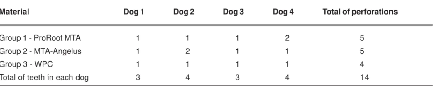
Group 1 - ProRoot MTA, there were 3 teeth without inflammatory infiltrate and 2 with inflammation; 3 teeth demonstrated complete mineralized sealing and 2 incomplete mineralized sealing. Figure 1 exhibits one tooth with complete mineralized sealing and without inflammation, vessels, organization and insertion of periodontal ligament fiber in the mineralized tissue noe-formated, scant of cementoblast-like placing mineralized matrix and normal conditions of the osseous tissue next to the perforation
Group 2 - MTA-Angelus, there were 4 teeth presented inflammation, being moderate to mild and 1 without inflammation; 2 teeth revealed complete mineralized sealing and 2 incomplete mineralized sealing. Figure 2 shhows one of the teeth with mineralized sealing without inflammation, organization of more than 2/3 of the periodontal ligament fibers and insertion in the mineralized barrier new-formed vessels and normal condition of osseous tissue next to the perforation.
Group 3 – WPC, there were 2 teeth showed inflammation, being moderate to mild and 2 without inflammation and the mineralized sealing was complete in 2 and incomplete in 2 teeth analyzed. In this group presented intense extrusion of material, because the operator did not have an appropriate control with the sealing. Figure 3 shows one tooth with mineralized sealing and moderate inflammation restricted to the area adjacent to the defect, increase of vascularization, intense activity of cementoblast-like cells depositing of matrix in the defect, scant of organization of periodontal ligament fibers..
Although the sealant perforation by WPC and MTA-Angelus showed more than number of teeth with inflammation in comparation with the closed con ProRoot MTA, The non-parametric Kruskal-Wallis test did not demonstrate any statistically significant difference (p>0.05) between the materials employed (Tables 3 and 4).
DISCUSSION
The ProRoot MTA is 75% of Portland cement, bismuth oxide (20%) as radiopacificator and calcium sulfate (5%) for improve the management. The MTA-Angelus is constituied by 80% of Portland cement and bismuth oxide (20%), both are white and grey. The color grey of the clincker of Portland cement is due to iron and manganese, for this reason when decrease the concentration of clincker’s iron there are
FIGURE1- ProRoot MTA, revealing the mineralized sealing
at the perforation (P), with no inflammation and with organization of the periodontal ligament. Hematoxylin and eosin – Olympus 10X
FIGURE 2- MTA-Angelus, demonstrating new formation of
production of clear color, in addition, during the fabrication of white cement is using clay, and carbonate stone without iron as main material. In the studies of Holland, et al.13 in
2002 was evaluated the reaction of the connective tissue in rats in the dentin tubes filled with white and grey MTA, obtaining similar results. Nevertheless, Chakmakchi, et al.5
compared the capacity of the sealant grey and white MTA with the Portland cement in the furca perforation of extracted human teeth, showing differences only between the white MTA and Portland cement, but, no differences between the groups white and grey MTA. Diamanti, et al.6 analized the
chemical composition, pH and the characteristics of the surface of grey MTA with white MTA (ProRoot MTA). They demostrated that both are the same, only different in its
chemical composition, such as iron oxide (Fe2O3) which was absent in the white MTA and the calcium sulfate (CaSO4) which was absent in the grey MTA. The Portland cement was used in this study, because is the base of MTA7,20,34.
The WPC is classifying in two sub-groups, structural and non-structural, Non-structural is rich in carbonic materials, (ground stones), including mainly calcium carbonate, which is use to give back the concrete, and easy mix, in addition the WPC is use in the paste of the ceramic title, to make hydraulic bricks, it is mean non-structural applications. According with Bernabé and Holland3 there are more types
of cements, Nonetheless, which have association with our interest in investigation.
Several materials have been investigated to find the ideal material for sealing of perforations. However none of them has met all requirements to be considered ideal3,9,27. Pitt Ford,
et al.21 investigated perforations in dogs’ teeth immediately
sealed or contaminated with MTA or amalgam and observed 6 cases without inflammation and 1 case with moderate inflammation after 4 months in the specimens sealed immediately. Holland, et al.10 observed root perforations in
dogs’ teeth immediately sealed with MTA and Sealapex, it was demonstrated that after 30 days, 4 specimens with no inflammation and 3 teeth with scarce inflammatory cells. At 180 days, 10 specimens presented no inflammation and 2 teeth demonstrated overflow of material and moderate chronic inflammatory reaction with presence of giant cells. In the present study, with ProRoot MTA group, there were 3 teeth without inflammatory infiltrate and 2 with inflammation. MTA-Angelus group, there were 4 teeth presented inflammation, being moderate to mild and 1 without inflammation. WPC group, all teeth presented moderate to mild inflammation (4 teeth). Although the static study Kruskal-Wallis used in our small simples with variability of results, showed that among of the material did not exist singificative differences in the number, even there was moderated to mild inflammation in the four specimens sealant with WPC and MTA-Angelus in comparation with ProRoot MTA that had 3 teeth with inflammation, the clinical application should be more in those 4 specimens with partial or completed reparation. Some cases try to repair the peridontal tissue. We believe that the inflammatory infiltrate observed in this study was associated to dispersion of sealing material may be for lack of control about its chemical component of WPC, situation that no occurred with the
Group Median Sum of scores Mean score Values
Angelus 1.000 37.500 7.500 5 WPC 2.000 43.500 10.875 4 ProRoot MTA 0.000 24.000 4.800 5
TABLE 3- Non-parametric Kruskal-Wallis test demonstrating analysis of the inflammatory infiltrate according to the three materials employed
Hc= 5.265000 Exact probability = 0.063270
Chi-square at 2 degrees of freedom Probability = 0.071898 Study groups without statistical difference (p>0.05)
FIGURE 3- WPC, with complete new formation of
ProRoot MTA and MTA-Angelus, which approved the quality norm, quality of control to be use as dental products. According with Holland, et al.10 mention that the over-filling
is responsible for the chronic inflammation. The present study revealed that, even though there was overflow in some cases, the perforations were sealed and there was new formation of mineralized tissue around the over-flown material. The postoperative radiographs revealed presence of overfilling. However, after 90 days, the material was totally or partially resorbed in some cases. This resorption requires more time, since 18.8% of MTA is insoluble in water25 and
according to Bernabé and Holland3, the insoluble product
in MTA is the bismuth oxide, a chemical product with high molecular weight. The overflow presents a tendency toward chronic inflammation, as revealed by Holland, et al.10 For
that reason, Bernabé and Holland3, Bramante, et al.4; Silva
and Moraes26; Weldon, et al.33; Hardy, et al.8 recommend the
fabrication of a calcium hydroxide plug or a matrix to restrict the MTA only to perforation area. Arens and Torabinejad1;
Torabinejad and Chivian29 recommend that MTA should be
carefully applied under minimum pressure. Bernabé and Holland3 highlight that the material should not fill the
periodontal space. However, Balto2 observed that MTA did
not cause any citotoxic effect when applied on culture of the human periodontal ligament. All study groups, demonstrated the presence of repair in resorption areas in dentin and cementum, as well as mineralized sealing in the 14 teeth. Some teeth without complete sealing exhibited an attempt of sealing of the defect, initiated below the defect, with deposition over the existing cementum. These data agreed with Pitt Ford, et al.21, they conducted an
investigation with perforation of dogs’ teeth immediately sealed with MTA and observed deposition of cementum over the material in 5 teeth after 4 months. Teeth with contamination presented new formation in only 2 teeth, revealing that cases with incomplete new formation require more time for observation of complete sealing. In the present study showed cases with reparation associated with a severe chronic inflammation, it could be suggest the present of bacteria. Nevertheless, no special study was performed (Brown and Breen), for bacteria identification, because the cutting were in series and the blocks were run out during the process of repair. The process of reparation in the sealant teeth wit MTA which present inflammation, according with Pitt Ford, et al.22, Lemon and Torabinejad18, Holland, et al.15
y Thomson, et al.28 it is due to the capacity of MTA for
stimulate neo-formation of mineralized tissue according with Holland, et al.15 and Bernabé and Holland3, it is for mechanism
of action that is similar to calcium hydroxide. Holland, et al.10 observed that 9 out of 10 teeth with presences of new
cementum formation at 180 days, some being with tunnel-shaped irregular defects containing connective tissue, studies done by Holland, et al.11, Holland, et al.12, Holland,
et al.13, Holland, et al.14 demonstrated that the calcium
hydroxide, MTA and Portland cement, determined the formation of calcic granules and mineralized tissue, in the adjacent to the dentin tubes implanted in the sub-coetaneous tissue rats. According with those authors, the mechanism of action during the mix of MTA with water becomes in calcium hydroxide while it has contact with the tissue fluids, it is associated with ions of calcium and hydroxyl. The ions de calcium react with the carbonic gas in the carbonic of the tissue, creating calcita granules, which came from with accumulation of fibronectina, produced by fibroblastos, macrophages and endothelials cells3,32.
According with Seux, et al.24, the fibronectina is responsible
for migration and adhesion of periodontal cells, that sensitize and deposit collagen type I, forming organic extracellular matrix, inducing in the cell differentiation of the cementoblast, responsible for the deposition of the mineralized tissue in the areas of reabsortion3. Thomson, et
al.28 evaluated the capacity of differentiation of the
cementoblast in the surface, demonstrated that the material promoted the production of osteocalcina and stimulate the production of mineralized matrix, considering the MTA as a cementocondutor material . Moretton, et al.19 (2000), after
the implantation of MTA in the osseous tissue and sub-coetaneous conjunctive of the rats, is consider osteoindutor. Nonetheless, more than cementoconductor or osteoinductor, we believe, that the MTA create the ideal condition of fisical sealant, its mean, no soluble, even with the presence of blood30; its high pH and high realize of ions calcium31, stop
the growing and pass of bacteria toward to periodontal tissue to the local perforation because this mechanism of actions, high alkaline, fisical properties, chemical and biologic, the organism react, stimulate the process of reparation, as clinically evidence in the majority of the perforation sealant biologically in this study. Other aspect that was remarkable by some author as Saidon, et al.12 who
considered the Portland cement has the potential to be used
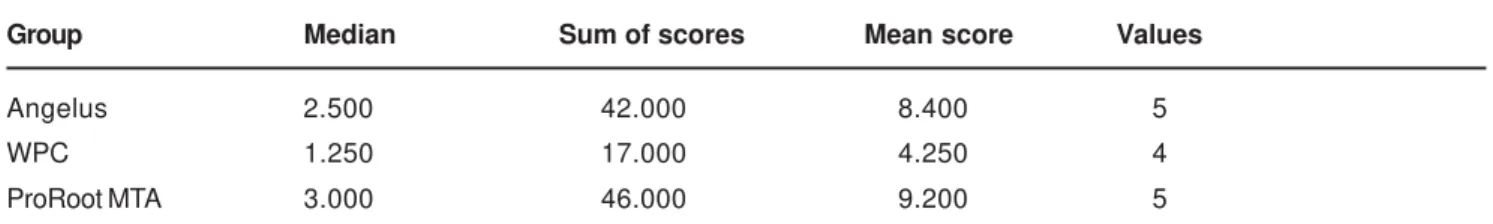
Group Median Sum of scores Mean score Values
Angelus 2.500 42.000 8.400 5 WPC 1.250 17.000 4.250 4 ProRoot MTA 3.000 46.000 9.200 5
TABLE 4- Non-parametric Kruskal-Wallis test demonstrating analysis of the mineralized sealing according to the three
materials employed
Hc= 3.664733 Exact probability = 0.162354
as a cheaper restorative material, however it should not be used in patients yet. Bernabé and Holland3, the utilization
of Portland cement still involve ethic and juridical principles and the authors do not agree with the use of the WPC in the patients because, it could cause some reaction in contact with the periodontal tissue, in the same way presented in the teeth of the dogs.
CONCLUSIONS
According to the methodology used in this study and considering the results, it was concluded that the three materials stimulated new cementum formation on root perforations in dogs’ teeth; when present inflammation was associated to overflow of sealing material to the periodontal tissues.
ACKNOWLEDGMENTS
To Drs. Ernesto Garcia Yañez and Alejandro Bates Souza, of Dentsply – Mexico, DF – Mexico.
To Drs. Roberto Queiroz Martins Alcantara and Lygia Madi Kranz, of Angelus – Soluções em Odontologia – Londrina, PR, Brazil.
REFERENCES
1- Arens DE, Torabinejad M. Repair of furcal perforations with mineral trioxide aggregate: two cases reports. Oral Surg Oral Med Oral Pathol Oral Radiol Endod. 1996;82(1):84-8.
2- Balto HA. Attachment and morphological behavior of human periodontal ligament fibroblasts to mineral trioxide aggregate: a scanning electron microscope study. J Endod. 2004;30(1):25-9.
3- Bernabé PFE, Holland R. Cirurgia paraendodôntica: como practicá-la com embasamento científico. In: Estrepracticá-la C. Ciência endodôntica. São Paulo: Artes Médicas; 2004. p. 657-797.
4- Bramante CM, Berbert A, Bernardineli N, Gomes de Moraes I, Garcia RB. Acidentes e complicações no tratamento endodôntico: soluções clínicas. 2a.ed. São Paulo: Ed. Santos; 2004.
5- Chakmakchi M, Karoni C, Meliou H, Kerezoudis NP. Sealing effectiveness of white ProRoot MTA in furcation perforations [abstract n. R75]. Int Endod J. 2003;36(12):945.
6 - Diamanti E, Kerezoudis NP, Gakis DB, Tsatsas V. Chemical composition and surface characteristics of grey and new white ProRoot MTA [abstract n. R81]. Int Endod J. 2003;36(12):946-7.
7 - Estrela C, Bammann LL, Estrela CRA, Silva RS, Pecora JD. Antimicrobial and chemical study of MTA, Portland cement, calcium hydroxide paste, sealapex and Dycal. Braz Dent J. 2000;11(1):3-9.
8 - Hardy I, Liewehr FR, Joyce AP, Agee K, Pashley DH. Sealing ability of one-up bond and MTA with and without a secondary seal as furcation perforation repair materials. J Endod. 2004;30(9):658-61.
9 - Hartwell GR, England MC. Healing of furcation perforation in primate teeth repair with decalcified freeze-dried bone. J Endod. 1993;19(7):357-61.
10- Holland R, Otoboni JA Filho, Souza V, Nery MJ, Bernabé PFE, Dezan E Jr. Mineral trioxide aggregate repair of lateral root perforations. J Endod. 2001;27(4):281-4.
11- Holland R, Souza V, Nery MJ, Otoboni Filho JA, Bernabé PF, Dezan E Jr. Reaction of rat connective tissue to implanted dentin tubes filled with mineral trioxide aggregate or calcium hydroxide. J Endod. 1999;25(3):161-6.
12- Holland R, Souza V, Nery MJ, Faraco IM Jr, Bernabé PF, Otoboni JA Filho, et al. Reaction of rat connective tissue to implanted dentin tube filled with mineral trioxide aggregate, portland cement or calcium hydroxide. Braz Dent J. 2001;12(1):3-8.
13- Holland R, Souza V, Nery MJ, Faraco IM Jr, Bernabé PF, Otoboni JA Filho, et al. Reaction of rat connective tissue to implanted dentin tubes filled with a white mineral trioxide aggregate. Braz Dent J. 2002;13(1):23-6.
14- Holland R, Souza V de, Nery MJ, Bernabé PFE, Otoboni JA Filho, Dezan E Jr, Murata SS. Calcium salts deposition in rat connective tissue after the implantation of calcium hydroxide-containing sealers. J Endod. 2002;28(3):173-6.
15- Holland R, Souza V; Mérida Delgado RJ; Murata SS. Agregado de trióxido mineral (MTA): Composição, mecanismo de ação, comportamento biológico e emprego clínico. Rev Ciênc Odontol. 2002;5(5):7-21.
16- Kranz ML. MTA–Angelus: relatório técnico. Londrina; 2004 (MT003).
17- Lee SJ, Monsef M, Torabinejad M. Sealing ability of a mineral trioxide aggregate for repair of lateral root perforations. J Endod. 1993;19(11):541-4.
18- Lemon RR, Torabinejad M. Procedural accidents. In: Torabinejad M, Walton RE. Principles and practice of endodontics. 3rd ed. Philadelphia: Saunders; 2002. cap. 18, p. 310-30.
19- Moretton TR, Brown CE Jr, Legan JJ, Kafrawy AH. Tissue reactions after subcutaneous and intraosseous implantation of mineral trioxide aggregate and ethoxybenzoic acid cement. J Biomed Mater Res. 2000;52(3):528-33.
20- MTA Angelus. Cimento reparador: indicações e técnica de uso. Londrina: Soluções em Odontologia; 2003.
21- Pitt Ford TR Torabinejad M, McKendry DJ, Hong C, Kariyawasam ST. Use of mineral trioxide aggregate for repair of furcal perforations. Oral Surg. Oral Med. Oral Pathol. 1995;79(6):756-62.
22- Pitt Ford TR. Surgical treatment of apical periodontitis. In: Ørstavik D, Pitt Ford TR. Essential endodontology: prevention and treatment of apical periodontitis. London: Blackwell Science; 1998. cap.12, p. 278-307.
23- Saidon J, He J, Zhu Q, Safavi K, Spångberg LSW, Conn F. Cell and tissue reactions to mineral trioxide aggregate and Portland cement. Oral Surg Oral Med Oral Pathol. 2003;95(4):483-9.
24- Seux D, Couble ML, Hartmann DJ, Gauthier JP, Magloire H. Odontoblast-like cytodifferentiation of human dental pulp cells in vitro in the presence of a calcium hydroxide containing cement. Arch Oral Biol. 1991;36(2):p.117-28.
26- Silva Neto UX. Capacidade seladora e adaptação marginal proporcionadas por alguns materiais quando utilizados em perfurações na região de furca de molares humanos. Bauru; 2002. [Dissertação (Mestrado) – Faculdade de Odontología de Bauru da USP].
27- Sluyk SR, Moon PC, Hartwell GR. Evaluation of setting properties and retention characteristics of mineral trioxide aggregate when used as a furcation perforation repair material. J Endod. 1998;24(11):768-71.
28- Thomson TS, Berry JE, Somerman MJ, Kirkwood KL. Cementoblasts maintain expression of osteocalcin in the presence of mineral trioxide aggregate. J Endod. 2003;29:407-12.
29- Torabinejad M, Chivian N. Clinical applications of mineral trioxide aggregate. J Endod 1999;25(3):197-205.
30- Torabinejad M, Higa RK, McKendry DJ, Pitt Ford TR. Dye leakage of four root end filling materials: effects of blood contamination. J Endod. 1994;20(4):159-63.
31- Torabinejad M, Hong CU, McDonald F, Pitt Ford TR. Physical and chemical properties of a new root-end filling material. J Endod. 1995;21(7):349-53.
32- Trowbridge H.O, Emiling R.C. Inflammation: a review of the process. 5th ed. Chicago: Quintessence Books; 1997.
33- Weldon JK, Pashley DH, Loushine RJ, Weller RN, Kimbrough WF. Sealing ability of mineral trioxide aggregate and Super-EBA when used as furcation repair materials: a longitudinal study. J Endod. 2002;28(6):467-70.