ABSTRACT
http://dx.doi.org/10.1590/1678-7757201302117
Minimal alterations on the enamel surface by
micro-abrasion:
in vitro
roughness and wear
assessments
Marcela Charantola RODRIGUES1, Rafael Francisco Lia MONDELLI2 3, Eduardo Batista
FRANCO4, Wagner BASEGGIO5, Linda WANG2
1- MSc, PhD student, Department of Operative Dentistry, Dental Materials and Endodontics, Bauru School of Dentistry, University of São Paulo, Bauru, SP, Brazil. 2- DDS, MSc, PhD, Associate Professor, Department of Operative Dentistry, Dental Materials and Endodontics, Bauru School of Dentistry, University of São Paulo, Bauru, SP, Brazil.
3- MSc, Department of Operative Dentistry, Dental Materials and Endodontics, Bauru School of Dentistry, University of São Paulo, Bauru, SP, Brazil. 4- DDS, MSc, PhD, Full Professor, Department of Operative Dentistry, Dental Materials and Endodontics, Bauru School of Dentistry, University of São Paulo, Bauru, SP, Brazil.
5- DDS, MSc, PhD, Department of Operative Dentistry, Dental Materials and Endodontics, Bauru School of Dentistry, University of São Paulo, Bauru, SP, Brazil.
Corresponding address: Prof. Dr. Rafael Francisco Lia Mondelli - Departmento de Dentistica, Endodontia e Materiais Odontológicos, Faculdade de Odontologia de Bauru, Universidade de São Paulo - Alameda Dr. Octávio Pinheiro Brisolla, 9-75 - Cx Postal 73 - 17012-901 - Bauru - SP - Brazil - Phone: 55 14 32358265 - Fax: 55 14 32261495 - e-mail: [email protected]
!"#$"%&& '$ %&&
O
bjective: To evaluate the in vitro changes on the enamel surface after a micro-abrasion treatment promoted by different products. Material and Methods: Fifty (50) fragments ! "# $%&'#*+,/ +1234637 448 '#+1%193; '#<= > #8 ?1@#1% 6A? responsible variables used to analyze these surfaces in four stages: baseline, 60 s and 120 s after the micro-abrasion and after polishing, using a Hommel Tester T1000 device. G ' ' GI=&G JA A= KGI=&G and Tukey tests were applied for individual comparisons between the products in eachJAA? "1 Pm) after
"#,A*LAN2+AL*AL,'#**A*AL*2+,A<<+A++' #+ANAP2+<AP+LAP*'#<AP*A*P2+NA<*AL#APNA+2++A<*ALLA At 60 seconds, all products tended to produce less surface roughness with a variable AG ' between the groups, except for G1. Independent of the product utilized, the enamel wear occurred after the micro-abrasion. Conclusions: In this in vitro study, enamel micro-abrasion presented itself as a conservative approach, regardless of the type of the paste compound utilized. These products promoted minor roughness alterations and minimal wear. The use of phosphoric acid and pumice stone showed similar results to commercial products for the micro-abrasion with regard to the surface roughness and wear.
Key words: Micro-abrasion. Enamel. Roughness. Wear.
INTRODUCTION
Many patients consider enamel staining unpleasant, leading them to seek treatment in
order to remove it2,5,9.
G
a successful approach, as different levels of compromised dental structures require distinct decisions to avoid sub or over-treatments. It is extremely relevant that these white spots are not related to caries activity, such as for patients who
have undergone orthodontic treatment8,10,19.
most common etiologic factors that cause color
alterations3,9. These characteristics correspond to
the clinical manifestation of a defective process during the enamel maturation and mineralization
' \ 3,9.
However, other clinical situations may also cause enamel staining, such as hypo-calcification (imperfect formation of enamel) with an irregular texture8,10,19.
For this purpose, slurries made of the mixture of different acid and abrasive systems were combined
in a technique called enamel micro-abrasion2,5.
$ ; 9]4 (1998)
description: superficial layers of enamel with by selective removal utilizing an association of an erosive agent (mainly hydrochloric or phosphoric acids) with an abrasive agent (pumice paste or silicone carbide). A sub layer is exposed with normal characteristics.
The effectiveness of a removal technique depends on the level of the compromised substrate. _ ' and is an easier and more conservative procedure, which results in a more appealing appearance. Also,
this selection seems to be acidic-type dependent1.
9
of superficial enamel removal9. Despite the
advantages and available resources for this procedure, there is still a lack of knowledge about the consequences of this approach.
The purpose of this study was to clarify the \ of acid (hydrochloric or phosphoric) with different abrasives (pumice or silicone carbide) on the enamel by means of roughness and wear assessments.
MATERIAL AND METHODS
This in vitro experimental design involved two
" stages of treatment (in four levels).
Figure 1 presents the main information about
the products used in this study, highlighting the acid and abrasive components.
Preparation of specimens was conducted
according to Mondelli12 (2009). Fifty (50) bovine
incisors were selected, excluding teeth with severe wear, fracture or other visible alterations. The roots were discarded and the crowns were cut with a diamond disc, using a low speed cutting machine (Isomet 1000/Buehler, Lake Bluff, IL, USA) to obtain blocks of 15 mm x 5 mm. To obtain parallel surfaces, one metallic matrix was used and the opposite dentin surfaces were cleaned, acid-etched for 15 s and restored with a dentin bonding system (Adper Single Bond 2, 3M ESPE, St. Paul, MN, USA) and the Filtek Z250 (3M ESPE, St. Paul, MN, USA) composite resin. Next, all the enamel surfaces were a mechanical polishing machine (APL 4, Arotec, Cotia, SP, Brazil). A water-cooled sequence of #320, #600, #800 and #1200 abrasive silicone carbide discs (Extec Corp., Enfeld, CT, USA) were used under a constant load of 172 g for 30 s each. G ' ' IL, USA) was applied with a felt disc and a 10 min-ultrasonic bath in deionized water was employed to remove all residues on the surface.
The roughness and wear were assessed using a basic Hommel Tester T100 (Hommelwerke GmbH ref. #240851, Schwenningen, Germany). The roughness assessments were standardized with parameters of $A '$NA ' '<AA*K' $ ' ' 'KA@ readings were taken for each surface.
When the wear was assessed, all readings were performed from the control side of the surface to the micro-abraded side. Thus, wear reading was performed from the reference area (control side- not challenged for none groups) to treated area. The difference determines the provoked wear. The $N ' $< ' ' P
Groups Comercial brand Erosive agent Abrasive agent
G1 Silicone Polisher (Optimize System – TDV, Pomerode, SC, Brazil)
aluminium oxide
G2 - phosphoric acid 37% pumice stone (SSWhite, Rio de Janeiro, RJ, Brazil) G3 Micropol (DMC Equipments LTDA, São Carlos,
SP, Brazil)
hydrochloridric acid 6.6%
silicon carbide
G4 Opalustre (Ultradent, South Jordan, UT, USA) hydrochloridric acid 6.6%
silicon carbide
G5 Whiteness RM (FGM Dental Products, Joinville, SC, Brazil)
hydrochloridric acid 12%
silicon carbide
AKA
The specimens were randomly assigned into 5 '@ A specimen surface was protected with adhesive tape (3M do Brasil Ltda., Sumaré, SP, Brazil), and acted as the reference control side. Only the other half was than treated with one of the techniques under evaluation. The recommendations of each manufacturer were followed. In Group 2, 37% phosphoric acid was mixed with the same volume of pumice, resulting in homogeneous slurry. During K' the same way to a metallic base to be abraded under constant pressure of 217 g. This procedure was performed using a low speed and a torpedo-shaped siliconee rubber cup for 30 s. Another 30 s-application was performed, for a total of 60 s.
Next, the slurry was washed out with an air-water spray for 30 s. A new series of roughness and wear assessments was performed. The same steps were repeated to obtain 120 s-registrations. In the end, the surfaces were polished with felt discs and polishing paste (Diamond Excel/ FGM 6 =' ' 49' ! 30 s at low speed.
After testing, the normal distribution of data was A ? GI=&G were used to compare each product in different stages (p<0.05). One-way ANOVA and Tukey tests
were applied for individual comparisons between the products in each stage (p<0.05).
RESULTS
Tables 1 and 2 summarize the means and standard deviations of roughness and wear, respectively.
Regarding the roughness, as presented in Table 1, G1 (control group), treated with siliconee polisher presented no differences among different treatment stages overtime (p>0.05). The overall results showed that for the other groups, the roughness tended to decrease over time after polishing. All to their initial situations and didn’t differ from each other, except for G1, which was comparatively rougher.
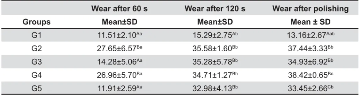
Table 2 shows that the micro-abrasion was able to provoke wear in all the groups, including G1, treated with silicone polisher only. After polishing, #< respective 120 s-assessment. When the groups were compared at each stage, G2 and G4 presented L A G * ' when compared to G1 (control), according to the particular comparison between the baseline and after polishing stages, considering neither the 60
Initial Roughness Roughness after 60 s Roughness after 120 s Roughness after polishing
Groups Mean±SD Mean±SD Mean±SD Mean±SD
G1 7.29±1.57Aa 7.16±1.26Aab 7.06±1.39Aa 7.26±1.81Aa
G2 6.69±1.60Aa 4.63±1.05Ab 3.60±1.54Bb 2.02±0.62Bc
G3 6.96±2.12Aa 8.57±3.74Ba 5.51±3.42ACa 1.81±0.91Bb
G4 6.63±2.61Aa 4.62±0.77Ab 3.32±0.57BCbc 1.92±0.29Bc
G5 6.61±1.83Aa 7.40±2.75Aab 2.18±0.47Bb 1.98±0.53Bb
Different lower case letters indicate differences between columns and different capital letters indicate differences between rows
Table 1- Mean and standard deviation (SD) of Ra (μm) of initial surface roughness and roughness after 60 seconds, 120 seconds, and polishing
Wear after 60 s Wear after 120 s Wear after polishing
Groups Mean±SD Mean±SD Mean ± SD
G1 11.51±2.10Aa 15.29±2.75Ab 13.16±2.67Aab
G2 27.65±6.57Ba 35.58±1.60Bb 37.44±3.33Bb
G3 14.28±5.06Aa 35.28±5.78Bb 34.93±6.92Bb
G4 26.96±5.70Ba 34.71±1.27Bb 38.42±0.65Bc
G5 11.91±2.59Aa 32.98±4.13Bb 33.45±2.66Cb
Different lower case letters indicate differences between columns and different capital letters indicate differences between rows
s nor 120 s time evaluation, for all groups, except for G1.
DISCUSSION
Investigations about the consequences to the enamel surface from different chemical-mechanical challenges have been extensively performed using bovine teeth since it can represent human type
tissue12,14. For many of the evaluated properties,
\
wear assessments, as previously reported14,15. Since
the enamel presents a hierarchical and regular
distribution7' \
\ A
Many factors are reported that can interfere with the enamel surface after micro-abrasion, such as manual or mechanical techniques, amount of application, interval between applications, mechanical speed, and pressure. More particularly, acid type and concentration, and type and granulation of the abrasive particles are also relevant to determine the effectiveness and
consequences to the micro-abraded enamel2,4,11,21.
In the present study, technical factors were standardized as the amount of applications, intervals, and pressure. The slurries, which varied according to the chemical and abrasive characteristics, are presented in Table 1.
It can be observed from Table 2 that the initial roughness for all specimens did not differ from each other, regardless of the treatment. It is important to highlight that all the data is reliable because all treatments began from a standardized condition. Also, it is particularly noted that the control group (G1), which had an enamel surface that was solely mechanically treated, presented the same roughness through all of the evaluated stages. This suggests that the chemical features were determinant.
Specimens treated with phosphoric acid (G2) or hydrochloric acid in different concentrations (G3, G4, and G5) produced different outcomes after the micro-abrasion. However, all produced smoother surfaces. After polishing, all treatments obtained smoother surfaces when compared to their initial assessment. The results recorded are in accordance with previous studies that reported a glass-like
surface, called the enamel glaze effect1,3,5.
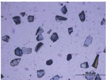
Figure 2- Micropol - irregular silicone carbide particles Figure 3- Opalustre - irregular silicone carbide particles
Figure 4- Whiteness RM - irregular silicone carbide particles
According to previous studies2,18,different
acids promote distinct demineralization patterns on enamel surfaces, which can, in part, explain the distinct reactions of the specimens treated with phosphoric or hydrochloric acids. In general, phosphoric acid promotes a less aggressive ' A = other hand, hydrochloric acid was not selective, dissolving the entire enamel surface after the micro-A@ ' \ materials also needs to be considered. Figures 2 to 5 illustrate the abrasive particles presented in the tested products. Except for pumice, all other particles were greater and with a similar magnitude. Another difference is that pumice was associated with phosphoric acid while the other abrasives were associated with hydrochloridric acid.
When the wear was observed, all products showed enamel loss, which was significantly greater after 120 s. After polishing, all products, #<' A
In the literature11, these amounts varied greatly,
being reported at 142.87 mm and 295.5 mm for the pumice + 37% phosphoric acid and pumice + 18% hydrochloric acid, respectively after 10 5 s-applications, totaling 50 s. This may explain the greater amount of wear when compared to the present study.
Previous studies highlighted a more aggressive
action of HCl-based products compared to H3PO4,
which was not observed in the present study. This may be attributed to the difference of the abrasive content. In previous studies, the pumice was
combined to HCl instead of silica carbide2,11.
Abrasives play a relevant role in the clinical performance of the slurry, allowing greater attrition
enamel layer, called “enamel glaze”1. It was
highlighted in the present study that the size and \ the enamel. Figures 2 to 4 illustrate, respectively, Micropol, Opalustre and Whiteness RM, which are based on irregular silicone carbide particles with similar size. All are greater when compared to the pumice dimensions (Figure 5).
The larger and irregular abrasive agents determined the worn surfaces. When we evaluated the proportion of wear to enamel thickness, we observed about 10% of enamel wear to all tested groups, which suggests a safe and conservative procedure. These results agree with previous studies that assessed the enamel wear using scanning electronic microscopy, which observed * <L ' performed between 1 and 10 5-second applications
with HCl and pumice16. Using the same slurry,
this assessment, under polarized microscopy, was evidenced by enamel wear ranges between 25 and
< +' 20.
? '
to decrease with multiple micro-abrasions11, as was
also noted in the present study. After the micro-abrasion, the enamel surfaces became smooth
and lusterous8. This is favorable, as it can reduce
bacteria colonization on the enamel surface, mainly
S. mutans17A ;
when enamel surface free energy is reduced, as well
as diminishing bacterial adhesion to the surface13.
The enamel characteristics change after the micro-abrasion, resulting in a different optical ' ! that refracts light in a different way, and is able to
mask the spot16. This may occur due to a gradual
formation of a compact, mineralized, and polished ' “enamel glaze”. This enamel glaze is about 15 μm thick, and is composed of a mixture of residues of abrasives and a smear layer that impregnates K K
enamel created during acid erosive action6.
Based on the results, all tested abrasive agents/ techniques showed the potential to determine a safe and conservative wear and the ability to modify the surface roughness, resulting in a smoother surface.
CONCLUSIONS
Within the limitations of this in vitro study,
enamel micro-abrasion seems to be a conservative approach, regardless of the type of the paste compound. The use of phosphoric acid and pumice stone showed similar results to commercial products for the micro-abrasion with regard to the surface roughness and wear.
ACKNOWLEDGMENTS
This investigation was supported in part by CNPq 19? *,AA*NNA*ALA
REFERENCES
1- Croll TP. Enamel microabrasion: the technique. Quintessence Int. 1989;20:395-400.
2- Croll TP. Enamel microabrasion: concept development. In: Croll TP, editor. Enamel microabrasion. Chicago: Quintessence Publishing; 1991. p. 37-41.
3- Croll TP. Enamel microabrasion: observations after 10 years. J Am Dent Assoc. 1997;128(Suppl):45S-50S.
<K9$6A3 \\K like enamel dysmineralization. J Esthet Dent. 1998;10:21-9. 5- Croll TP, Helpin ML. Enamel microabrasion: a new approach. J Esthet Dent. 2000;12:64-71.
7- Cui FZ, Ge J. New observations of the hierarchical structure of human enamel, from nanoscale to microscale. J Tissue Eng Regen Med. 2007;1:185-91.
8- Killian CM, Croll TP. Enamel microabrasion to improve enamel surface texture. J Esthet Dent. 1990;2:125-8.
9- Limeback H, Vieira AP, Lawrence H. Improving esthetically \ technique. Eur J Oral Sci. 2006;114(Suppl 1):123-6; discussion 7-9, 380.
10- Meireles SS, Andre DA, Leida FL, Bocangel JS, Demarco FF. Surface roughness and enamel loss with two microabrasion techniques. J Contemp Dent Pract. 2009;10:58-65.
11- Mendes RF, Mondelli J, Freitas CA. Avaliação da quantidade de desgaste do esmalte dentário submetido à microabrasão. Rev Facul Odont Bauru. 1999;7:6.
12- Mondelli RF, Azevedo JF, Francisconi PA, Ishikiriama SK, Mondelli J. Wear and surface roughness of bovine enamel submitted to bleaching. Eur J Esth Dent. 2009;4:396-403. 13- Quirynen M. The clinical meaning of the surface roughness and the surface free energy of intra-oral hard substrata on the microbiology of the supra- and subgingival plaque: results of in vitro and in vivo experiments. J Dent. 1994;22(Suppl 1):S13-6.
<K ? %' 1' 1 G9' 4 41' % G9' Machado MA, et al. Scanning electron microscopic study of the in situ effect of salivary stimulation on erosion and abrasion in human
and bovine enamel. Braz Oral Res. 2008;22:132-8.
15- Rios D, Santos FC, Honorio HM, Magalhães AC, Wang L, Machado MAAM, et al. An in situ/ex vivo comparison of the ability
of regular and light colas to induce enamel wear when erosion is combined with abrasion. Quintessence Int. 2011;42:e44-50. 16- Segura A. Acid-abrasive enamel reduction for tooth color correction. Am J Dent. 1991;4:103-4.
17- Segura A, Donly KJ, Wefel JS, Drake D. Effect of enamel microabrasion on bacterial colonization. Am J Dent. 1997;10:272-4.
18- Silverstone LM, Saxton CA, Dogon IL, Fejerskov O. Variation in the pattern of acid etching of human dental enamel examined by scanning electron microscopy. Caries Res. 1975;9:373-87. 19- Sundfeld RH, Croll TP, Briso AL, Alexandre RS, Sundfeld Neto D. Considerations about enamel microabrasion after 18 years. Am J Dent. 2007;20:67-72.
20- Sundfeld RH, Komatsu J, Russo M, Holland C Jr, Castro MAM, Quintella LPAS, et al. Removal of enamel stains: clinical and microscopic study. Rev Bras Odontol. 1990;47:6.
21- Waggoner WF, Johnston WM, Schumann S, Schikowski E. Microabrasion of human enamel in vitro using hydrochloric acid