203
Importance of gallium-67 scintigraphy in primary cutaneous B-cell lymphoma
Radiol Bras. 2010 Mai/Jun;43(3):203–206 Case Report • Relato de Caso
Importance of gallium-67 scintigraphy in primary
cutaneous B-cell lymphoma: report of two cases*
Importância da cintilografia com gálio-67 no linfoma cutâneo primário de células B: relato de dois casosCyomara Sanches Attab1, Sônia Marta Moriguchi2, Eduardo José Alencar Paton3, Mário Henrique Leite de Alencar4, Euclides Timóteo da Rocha5
The authors describe two cases of cutaneous B-cell lymphoma where correct staging, treatment and follow-up could be achieved through a combination of conventional imaging studies and gallium-67 scintigraphy.
Keywords: Lymphoma; Scintigraphy; Gallium.
Os autores relatam dois casos de linfoma cutâneo de células B, nos quais o correto estadiamento, tratamento e seguimento foram possíveis graças à combinação de exames convencionais e a cintilografia com gálio-67.
Unitermos: Linfoma; Cintilografia; Gálio. Abstract
Resumo
* Study developed at Hospital de Câncer de Barretos – Fun-dação Pio XII, Barretos, SP, Brazil.
1. MD, Nuclear Physician, Assistant at Centro Brasileiro de Medicina Nuclear – Cebramen, Goiânia, GO, Brazil.
2. PhD, Coordinator for the Course of Specialization in Nuclear Medicine, Hospital de Câncer de Barretos – Fundação Pio XII, Barretos, SP, Brazil.
3. MD, Hematologist, Coordinator for Division of Hemotherapy and Bone-Marrow Transplant, Hospital de Câncer de Barretos – Fundação Pio XII, Barretos, SP, Brazil.
4. MD, Oncologic Surgeon, Clínica de Prevenção em Câncer, Goiânia, GO, Brazil.
5. PhD, MD, Nuclear Physician, Head for Department of Nu-clear Medicine, Hospital de Câncer de Barretos – Fundação Pio XII, Barretos, SP, Brazil.
Mailing address: Dra. Cyomara Sanches Attab. Rua T 038, nº 1069, ap. 1101, Residencial Bella D’oro, Setor Bueno. Goiâ-nia, GO, Brazil, 74223-040. E-mail: [email protected] Received October 23, 2008. Accepted after revision January 30, 2009.
sive to home treatment. Later, the patient had observed the emergence of nodules in the inguinal region. No fever or weight loss was observed.
Clinical examination demonstrated the presence of an ulcerovegetating tumor with 15 cm in diameter on the medial aspect of the left thigh, besides bilateral painless in-guinal lumps, the ipsilateral one with 6 cm, and the other, at right, with 3 cm, with no sign of inflammation and mobile to palpa-tion (Figure 1). Tests and imaging studies were performed for diagnosis and staging, as follows: chest, abdomen and pelvis com-puted tomography, myelogram, protein electrophoresis blood test, open biopsy of the lesion and gallium-67 scintigraphy.
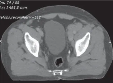
Anatomopathological and immunohis-tochemical results were compatible with diffuse large B-cell non-Hodgkin’s lym-phoma. Tomographic images demonstrated only a group of coalescent, ovoid nodules in the left internal iliac chain, the largest one measuring 2.8 × 2.5 cm, and a lobu-lar, homogeneous mass in the inguinal re-gions, the largest one at left (7.4 × 4.3 cm) (Figure 2). Gallium-67 citrate scintigraphy was performed, with images being ac-quired 48 hours after radiotracer injection at a dose of 296 MBq (8 mCi), and dem-onstrated an unusual radiotracer uptake, from the inguinal-pelvic to the medial as-pect of the left knee, besides multiple
hyper-Attab CS, Moriguchi SM, Paton EJA, Alencar MHL, Rocha ET. Importance of gallium-67 scintigraphy in primary cutaneous B-cell lymphoma: report of two cases. Radiol Bras. 2010;43(3):203–206.
raphy in cases of cutaneous B-cell lym-phoma has been poorly described in the literature(1), particularly after the adoption of 18F-fluorodeoxyglucose positron emis-sion tomography in the clinical practice of great European centers that demonstrated the high sensitivity and specificity of the method(2). Gallium-67 scintigraphy still remains as a relevant, complementary re-source in the pre- and post-treatment clini-cal management, allowing a functional and metabolic tissues characterization, as a complement to anatomical data. Addition-ally to the diagnosis and staging, informa-tion provided by gallium-67 scintigraphy is useful in the follow-up and evaluation of the therapeutic response of patients with lymphoma. An evaluation of anatomical characteristics alone is insufficient in the presence of a residual mass following che-motherapy, or small-sized lymph nodes in-filtration. Such evaluation can be completed with the utilization of gallium-67 scintig-raphy, considering that PET-FDG is not easily accessible yet in Brazilian centers.
CASES REPORT
Case 1
A male, white, 65-year-old patient re-ferred to the Division of Hematology, pre-senting a lesion on the left thigh with pro-gressive growth for one year and
unrespon-0100-3984 © Colégio Brasileiro de Radiologia e Diagnóstico por Imagem INTRODUCTION
Lymphomas may affect the skin either as a primary or secondary disease. Cutane-ous T-cell lymphoma is the most common type of lymphoproliferative disorder, with mycosis fungoides and Sézary syndrome being main representative examples of this disease. Cutaneous B-cell lymphoma, a rare nosological entity, is observed in less than 10% of cases o non-Hodgkin lympho-mas(1).
scintig-204
Attab CS et al.
Radiol Bras. 2010 Mai/Jun;43(3):203–206
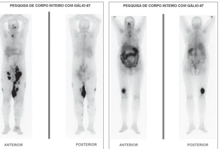
uptake areas in the left maxilla, manu-brium, left inguinal-pelvic region, right gluteus, at left in the intergluteal area, thighs, legs and left ankle (Figure 3).
A total coincidence was observed be-tween the lesions detected at physical ex-amination and the scintigraphic findings. Bone marrow biopsy as well as protein electrophoresis results were normal. The proposed chemotherapy scheme included cyclophosphamide, hydroxydoxorubicin (doxorubicin), vincristine (Oncovin) and prednisone (CHOP), the whole regimen comprising eight cycles. After chemo-therapy completion, a new staging was made by means of the same tests and im-aging studies previously performed. Noninvasive imaging methods demon-strated the presence of amorphous tissue with soft tissue density in the left inguinal region (4.5 × 1.5 cm) and at right (1.8 ×
1.2 cm), besides the absence of pelvic ad-enopathy. Follow-up gallium-67 scintigra-phy results were normal, demonstrating the disappearance of the lesions. Complete re-gression of the thigh lesion was observed, with substitution by cicatricial tissue con-firmed at gallium-67 scintigraphy.
Case 2
A female, white, 71-year-old patient was referred with a history of a left thigh lesion with progressive growth for three months. The patient denied fever and night sweats. Her previous pathological history did not include any surgery; the patient only
had hypertension and diabetes, with a fa-milial history negative for neoplasia.
At clinical examination, the patient pre-sented a good general condition, with body mass índex of 26 and performance status of 1. There was a hard patchy lesion mea-suring 10 × 9 cm, on the posterior aspect of her thigh, with infiltration into deep planes, and with no satellite adenopathy. Incisional skin biopsy result was consistent with cu-taneous B-cell lymphoma. Conventional imaging methods (chest and abdomen com-puted tomography) did not demonstrate the presence of disease in other sites.
Gallium-67 scintigraphy with high-en-ergy collimator, with images acquisition 48 hours after 67-gallium citrate injection at a dose of 296 MBq (8 mCi), demonstrated an abnormal radiotracer uptake on the dis-tal third of the right leg (Figure 4). Bone marrow biopsy and protein electrophoresis results were normal.
The patient received six cycles of CHOP chemotherapeutic regimen, additionally to locoregional radiotherapy (total dose = 3600 cGy (20 × 180 cGy)). The patient pro-gressed with improvement and complete lesion regression after the treatment. Post-therapy follow-up gallium-67 scintigraphy did not demonstrate any abnormality.
DISCUSSION
Cutaneous involvement by a lymphoma may be primary or secondary to a metastatic disease. Primary cutaneous lymphoma
cor-responds to the second most frequent group of extranodal involvement of non-Hodgkin lymphomas, preceded only by the gas-trointestinal tract (about 10%)(3). It is clas-sified according to the involved cell type, i.e., T-cell (CTCL) with 65% of cases, and B-cell (CBCL) with 20–25% of cases. Cutaneous T-cell lymphomas predomi-nantly affect men with ages ranging from 40 and 60 years. Both tumor types present a histological architecture similar to the systemic lymphoma, but with a completely different prognosis and behavior. Usually, such tumors originate from mature cutane-ous T-helper cells, with mycosis fungoides and Sézary syndrome being the most fre-quent ones in this group. In this case, the disease tends to spare the bone marrow, is hematogenically disseminated, and pre-sents a high rate of recurrence. On the other hand, cutaneous B-cell lymphomas origi-nate from centrofollicular cells and are characterized by their locoregional extent. These tumors are rarely found (8%) and, in truth, they represent a secondary cutaneous involvement by a systemic lymphoma in most of cases(3). The lesions are character-ized by nodules with a glossy surface, and rare desquamation or ulceration. The sur-rounding skin may be hyperemic, with small papular lesions or plaque infiltrates, with dimensions ranging from small sizes to 15 cm(1). Generally, these tumors present an indolent pattern, and may remain stable for months or years before presenting an abrupt development and growth.
205
Importance of gallium-67 scintigraphy in primary cutaneous B-cell lymphoma
Radiol Bras. 2010 Mai/Jun;43(3):203–206
Extracutaneous dissemination is not fre-quently observed, but is described in up 13% of cases, usually limited to the site of lymph node drainage(1). The correct deter-mination of the disease extent and classi-fication plays a relevant role in the defini-tion of cutaneous lymphomas management and prognosis; therefore, several diagnos-tic modalities should be utilized in the at-tempt to rule out extracutaneous lesions. According to the Ann Arbor system, the staging itself justifies the utilization of imaging methods, considering that this sys-tem is based on the analysis of the disease extent. Computed tomography, as com-pared with magnetic resonance imaging, allows an early and comprehensive staging, because of the rapid images acquisition(4). However, in spite of computed tomography being the method of choice in these cases, the presence of a post-chemotherapy re-sidual mass or small-sized lymph nodes infiltration constitute examples of
situa-tions where an evaluation of anatomical characteristics alone is insufficient, besides failing in the provision of accurate infor-mation about malignant cutaneous le-sions(5,6). The mechanism by which gal-lium-67 scintigraphy demonstrates the le-sion is based on the gallium ion transport into the cell through the plasmatic mem-brane, after gallium-transferrin complex binds to transferrin receptors on the tumor cell surface(7). Factors such as vasculariza-tion, vascular patency and increased ion-exchange due to increased membrane po-rosity, contribute for the final gallium up-take by the malignant cell. Gallium-67 scintigraphy demonstrated multiple uptake areas with a good correlation in all lesions of both patients, being useful in the diag-nosis, staging and therapy planning(6), con-sidering that, in association with conven-tional imaging methods, gallium-67 scin-tigraphy constitutes a relevant complemen-tary method for detecting occult lesions and
guiding changes in the management of patients with post-chemotherapy residual tumor activity. Therefore, the present cases demonstrate the relevant role of gallium-67 scintigraphy in the detection of the disease extent and in the evaluation of the patient’s response to therapy, by demonstrating, or not, the tumor activity(8). In the authors’ institution gallium-67 scintigraphy is also utilized in the follow-up of these patients for monitoring residual disease. Many of gallium-67 scintigraphy and computed to-mography limitations can be resolved with the utilization of PET-FDG that has dem-onstrated high accuracy, sensitivity and specificity in the detection of both nodal and extranodal involvement in patients with lymphoma. However, only a restricted number of studies with few patients and variable results have reported the utiliza-tion of PET-FDG in the diagnosis of cuta-neous lymphoma, as well as the difficult access of patients to the procedure. In sum-Figure 3. Gallium-67 scintigraphy. Early staging demonstrates gallium
up-take by lesions on the medial aspect of left thigh, extending from the in-guinal-pelvic region to the knee.
206
Attab CS et al.
Radiol Bras. 2010 Mai/Jun;43(3):203–206
mary, PET-FDG has demonstrated a poten-tial value in the early staging as well in the post-therapy restaging in these patients.
Although a consensus on this theme is still to be achieved by specialists, and the low number of cases reported is still a lim-iting factor, gallium-67 scintigraphy re-mains among the diagnostic methods in the protocol for both the initial staging and post-therapy restaging for cutaneous B-cell lymphomas, to differentiate between the presence of residual disease and possible sequelae, thus allowing an early
interven-tion in accordance with the therapy re-sponse.
REFERENCES
1. Assassa GS, Siegel ME, Chen DC, et al. Ga-67 uptake in cutaneous B-cell lymphoma. Clin Nucl Med. 1994;19:614–6.
2. Sapienza MT, Marone MMS, Chiattone CS. Con-tribuição da medicina nuclear para a avaliação dos linfomas. Rev Bras Hematol Hemoter. 2001; 23:79–92.
3. Kumar R, Xiu Y, Zhuang HM, et al. 18F-fluoro-deoxyglucose-positron emission tomography in evaluation of primary cutaneous lymphoma. Br J Dermatol. 2006;155:357–63.
4. Borba AMV, Monteiro AMV, Lima CMAO, et al.
Aspectos da tomografia computadorizada no lin-foma em pacientes abaixo de 20 anos de idade. Radiol Bras. 2007;40:87–92.
5. Delcambre C, Reman O, Henry-Amar M, et al. Clinical relevance of gallium-67 scintigraphy in lymphoma before and after therapy. Eur J Nucl Med. 2000;27:176–84.
6. Israel O, Front D, Lam M, et al. Gallium 67 im-aging in monitoring lymphoma response to treat-ment. Cancer. 1988;61:2439–43.
7. van Amsterdam JA, Kluin-Nelemans JC, van Eck-Smit BL, et al. Role of 67Ga scintigraphy in lo-calization of lymphoma. Ann Hematol. 1996;72: 202–7.