Edited by: Joao Santana Silva, University of São Paulo, Brazil
Reviewed by: Peter Kraiczy, Goethe University Frankfurt, Germany X Frank Yang, Indiana University School of Medicine, USA
*Correspondence: Ryan O. M. Rego [email protected]
Received:06 February 2017 Accepted:22 March 2017 Published:07 April 2017
Citation: de la Fuente J, Antunes S, Bonnet S, Cabezas-Cruz A, Domingos AG, Estrada-Peña A, Johnson N, Kocan KM, Mansfield KL, Nijhof AM, Papa A, Rudenko N, Villar M, Alberdi P, Torina A, Ayllón N, Vancova M, Golovchenko M, Grubhoffer L, Caracappa S, Fooks AR, Gortazar C and Rego ROM (2017) Tick-Pathogen Interactions and Vector Competence: Identification of Molecular Drivers for Tick-Borne Diseases. Front. Cell. Infect. Microbiol. 7:114. doi: 10.3389/fcimb.2017.00114
Tick-Pathogen Interactions and
Vector Competence: Identification of
Molecular Drivers for Tick-Borne
Diseases
José de la Fuente
1, 2, Sandra Antunes
3, Sarah Bonnet
4, Alejandro Cabezas-Cruz
4, 5, 6,
Ana G. Domingos
3, Agustín Estrada-Peña
7, Nicholas Johnson
8, 9, Katherine M. Kocan
2,
Karen L. Mansfield
8, 10, Ard M. Nijhof
11, Anna Papa
12, Nataliia Rudenko
5, Margarita Villar
1,
Pilar Alberdi
1, Alessandra Torina
13, Nieves Ayllón
1, Marie Vancova
5,
Maryna Golovchenko
5, Libor Grubhoffer
5, 6, Santo Caracappa
13, Anthony R. Fooks
8, 10,
Christian Gortazar
1and Ryan O. M. Rego
5, 6*
1SaBio. Instituto de Investigación en Recursos Cinegéticos CSIC-UCLM-JCCM, Ciudad Real, Spain,2Department of
Veterinary Pathobiology, Center for Veterinary Health Sciences, Oklahoma State University, Stillwater, OK, USA,3Global
Health and Tropical Medicine, Instituto de Higiene e Medicina Tropical, Universidade Nova de Lisboa, Lisboa, Portugal,
4UMR BIPAR INRA-ANSES-ENVA, Maisons-Alfort, France,5Biology Centre, Czech Academy of Sciences, Institute of
Parasitology, Ceske Budejovice, Czechia,6Faculty of Science, University of South Bohemia, ˇCeské Bud ˇejovice, Czechia, 7Facultad de Veterinaria, Universidad de Zaragoza, Zaragoza, Spain,8Animal and Plant Health Agency, Surrey, UK,9Faculty
of Health and Medicine, University of Surrey, Guildford, UK,10Institute of Infection and Global Health, University of Liverpool,
Liverpool, UK,11Institute for Parasitology and Tropical Veterinary Medicine, Freie Universität Berlin, Berlin, Germany, 12Department of Microbiology, Medical School, Aristotle University of Thessaloniki, Thessaloniki, Greece,13National Center of
Reference for Anaplasma, Babesia, Rickettsia and Theileria, Intituto Zooprofilattico Sperimentale della Sicilia, Sicily, Italy
Ticks and the pathogens they transmit constitute a growing burden for human and animal
health worldwide. Vector competence is a component of vectorial capacity and depends
on genetic determinants affecting the ability of a vector to transmit a pathogen. These
determinants affect traits such as tick-host-pathogen and susceptibility to pathogen
infection. Therefore, the elucidation of the mechanisms involved in tick-pathogen
interactions that affect vector competence is essential for the identification of molecular
drivers for tick-borne diseases. In this review, we provide a comprehensive overview of
tick-pathogen molecular interactions for bacteria, viruses, and protozoa affecting human
and animal health. Additionally, the impact of tick microbiome on these interactions was
considered. Results show that different pathogens evolved similar strategies such as
manipulation of the immune response to infect vectors and facilitate multiplication and
transmission. Furthermore, some of these strategies may be used by pathogens to
infect both tick and mammalian hosts. Identification of interactions that promote tick
survival, spread, and pathogen transmission provides the opportunity to disrupt these
interactions and lead to a reduction in tick burden and the prevalence of tick-borne
diseases. Targeting some of the similar mechanisms used by the pathogens for infection
and transmission by ticks may assist in development of preventative strategies against
multiple tick-borne diseases.
INTRODUCTION
Ectoparasites that derive nutrition through blood feeding
(haematophagy) are efficient vectors of disease. Ticks are
haematophagous ectoparasites of vertebrates. Approximately
10% of the 900 currently known tick species are of significant
medical or veterinary importance. Besides causing direct damage
associated with blood feeding and in some cases through the
excretion of toxins within their saliva, the main relevance
of ticks lies in the wide variety of pathogens they can
transmit, including bacteria, viruses, protozoa, and helminths
(
Jongejan and Uilenberg, 2004
). The continuous exploitation of
environmental resources and the increase in human outdoor
activities, which have allowed for the contact with tick vectors
normally present in the field, has promoted the emergence and
resurgence of tick-borne pathogens (
Jongejan and Uilenberg,
2004
).
As previously discussed (
Beerntsen et al., 2000
), the terms
“vectorial capacity” and “vector competence” are often used to
describe the ability of an arthropod to serve as a disease vector.
However, while vectorial capacity is influenced by behavioral
and environmental determinants affecting variables such as
vector density, longevity, and competence, vector competence
is a component of vectorial capacity that depends on genetic
factors affecting the ability of a vector to transmit a pathogen
(
Beerntsen et al., 2000
,
Box 1
). These genetic determinants affect
traits such as tick host preferences, duration of tick attachment,
tick-host-pathogen and microbiome-pathogen interactions, and
susceptibility to pathogen infection (
Ramamoorthi et al., 2005;
Hajdušek et al., 2013; Narasimhan et al., 2014; Nuttall, 2014;
Rynkiewicz et al., 2015; Vayssier-Taussat et al., 2015
). Therefore,
the elucidation of the mechanisms involved in tick-pathogen
BOX 1 | Important determinants influencing the acquisition, maintenance and transmission of pathogens by ticks.
Host range Ticks with a wide host range such asI. ricinus, are naturally exposed to a greater variety of pathogens compared to ticks with a narrow host range such asR. microplus(Estrada-Peña et al., 2015).
Number of hosts The potential transmission of pathogens could be limited when considering the host contact rate of 1- and 2- host ticks vs. 3-host ticks. This effect may however be partially annulled by the phenomenon of transovarial passage, when pathogens are passaged from the female to her eggs and offspring, which can subsequently infect new hosts. Argasid ticks of which the nymphs and adults take several blood meals, have a high host contact rate and could theoretically acquire or transmit pathogens from and to multiple hosts.
Midgut infection and escape barrier
The pathogen needs to pass through the midgut to reach the salivary glands and be transmitted with tick saliva, and for migration of some pathogens to the ovaries to allow transovarial pathogen passage. Mechanisms to pass the midgut infection barrier may depend on the presence and structure of specific surface receptors, such as TROSPA, to which OspA fromB. burgdorferiadheres, allowing the spirochete to colonize the midgut (Pal et al., 2004).
Innate immune response
Pathogens need to overcome tick defense mechanisms, such as the phagocytosis of microbes by hemocytes, antimicrobial peptides and RNA interference, in order to be transmitted with tick saliva (Hajdušek et al., 2013).
Salivary gland infection and escape barrier
Pathogens must cross into the salivary glands for transmission with saliva during feeding, but little is known about the molecular mechanisms behind this entry. Once inside the salivary glands, the pathogen has to be released into the saliva stream to be transmitted. For example,
B. burgdorferiuses tick salivary gland proteins to facilitate infection of the mammalian host (Ramamoorthi et al., 2005).
Pathogen strains Differences between pathogen strains to infect and be transmitted by ticks have been widely reported (e.g.,Kleiboeker et al., 1999; de la Fuente et al., 2001).
Tick microbiome-pathogen interactions
Microbiome play an essential role in various aspects of the arthropods life cycle and there is an increasing interest to elucidate
arthropod-microbiome interactions. Perturbation of the microbiome caused changes in the integrity of the peritrophic membrane and may affect pathogen infection (Narasimhan et al., 2014).
Cross-Immunity interference
Competition between microorganisms within the tick may affect vector competence. Ticks infected with oneRickettsiaspecies were for instance refractory to transovarial passage of a secondRickettsiaspecies (Macaluso et al., 2002).
Abiotic factors Abiotic factors such as temperature and relative humidity not only have a direct effect on tick development, questing activity and longevity, but temperature may also modulate pathogen development and survival in ticks (Shih et al., 1995; Estrada-Peña et al., 2011).
interactions that affect vector competence is essential for the
identification of molecular drivers for tick-borne diseases,
and exposes paradigms for controlling and preventing these
diseases.
Although our understanding of tick-pathogen interactions is
still limited, advances in this field are facilitated by the increasing
number of available genomic resources, including metabolomics,
transcriptomics, and proteomics datasets of various ticks and
tick-borne pathogens (TBPs) (
Nene et al., 2004; Ayllón et al.,
2015a; Cramaro et al., 2015; Kotsyfakis et al., 2015; Villar
et al., 2015a; Gulia-Nuss et al., 2016; de Castro et al., 2016
),
and the recently published genome from
Ixodes scapularis
, a
vector of
Borrelia burgdorferi
and
Anaplasma phagocytophilum
in North America (
Gulia-Nuss et al., 2016
). Together with
tools such as tick cell lines and the widespread adaptation of
RNA interference (RNAi) to study tick gene function (
Bell-Sakyi et al., 2007; de la Fuente et al., 2007
), this has opened
exciting possibilities to identify determinants affecting tick vector
competence.
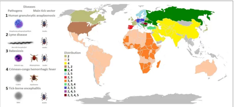
FIGURE 1 | Model organisms: tick-borne pathogens that constitute a growing burden for human and animal health.The pathogens covered in this review include bacteria (A. phagocytophilumandB. burgdorferi), viruses (Crimean-Congo hemorrhagic fever virus, tick-borne encephalitis virus), and protozoa (Babesiaspp.) transmitted by hard ticks (Ixodidae). The most prevalent diseases caused by these pathogens, main tick vectors, and disease distribution worldwide is shown in the figure.
MODEL MICROORGANISMS
In this review, we used different tick-borne microorganisms
including bacteria (
A. phagocytophilum
and
B. burgdorferi
),
viruses (Crimean-Congo hemorrhagic fever virus, tick-borne
encephalitis virus, and louping ill virus), and protozoa (
Babesia
spp.) to illustrate their impact on vector competence, behavior
and transmission (
Figure 1
).
Bacteria
Anaplasma
phagocytophilum
is an obligate intracellular
rickettsial pathogen vectored primarily by
Ixodes
spp. and causes
human granulocytic anaplasmosis (HGA), equine, and canine
granulocytic anaplasmosis, and tick-borne fever (TBF) (
de la
Fuente et al., 2008
). In the vertebrate host,
A. phagocytophilum
infects neutrophils where the pathogen multiplies within a
parasitophorous vacuole or morula (
Ayllón et al., 2015a; Severo
et al., 2015
). In the absence of transovarial passage, ticks must
acquire infection in each generation during a bloodmeal.
A. phagocytophilum
initially infects tick midgut cells and then
subsequently develops in the salivary glands for transmission
to susceptible hosts during tick feeding. Bacteria from the
B. burgdorferi
sensu lato complex are transmitted by Ixodid
ticks and cause various symptoms associated with Lyme disease
(
Radolf et al., 2012
).
B. burgdorferi
s.l. are acquired by larvae
or nymphs from an infected host as they are not transovarially
transmitted (
Rollend et al., 2013
). In the tick, spirochetes
colonize the midgut and then traverse into the hemocoel and
migrate to salivary glands for transmission during tick feeding
(
Pal et al., 2004; Ramamoorthi et al., 2005; Zhang L. et al., 2011;
Coumou et al., 2016
).
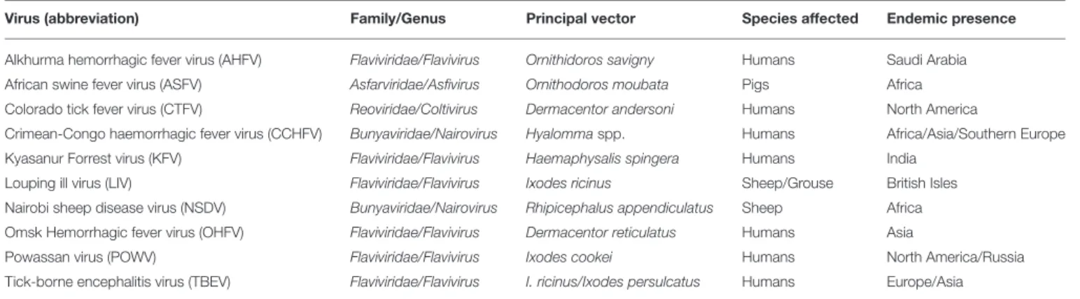
Viruses
Ticks transmit a range of viruses that are of significant public
and veterinary health concern (
Table 1
). It is estimated that
these viruses spend over 95% of their life cycle within the tick
vector. Tick-borne encephalitis virus (TBEV) causes neurological
disease in humans, whereas louping ill virus (LIV) causes
neurological disease in sheep (
Labuda and Nuttall, 2003
). Ixodid
ticks transmit these viruses to particular host species through a
bite (
Doherty and Reid, 1971; Mansfield et al., 2016
).
Crimean-Congo hemorrhagic fever virus (CCHFV) is transmitted to
humans by the bite of infected ticks (
Hyalomma
spp. are the most
competent vectors) or by direct contact with blood or tissues
of viremic patients or animals, causing a disease characterized
by fever, headache, myalgia, and hemorrhagic manifestations
(
Papa, 2010
). If the appropriate receptors are present in the tick,
following a blood meal TBEV and CCHFV enter vector host cells
by endocytosis (
Labuda and Nuttall, 2003; Simon et al., 2009;
Garrison et al., 2013; Shtanko et al., 2014; Suda et al., 2016
).
These viruses replicate in the lining of the tick midgut where they
disseminate to the hemolymph and subsequently infect different
tissues reaching the highest titers in the salivary glands and
reproductive organs to exit the cell via exocytosis (
Dickson and
Turell, 1992
).
Protozoa
Babesia
spp. are tick-borne Apicomplexan protozoans which
invade vertebrate host erythrocytes, where all hemoparasite
phases occur (
Yokoyama et al., 2006; Chauvin et al., 2009;
Florin-Christensen and Schnittger, 2009
).
Babesia bovis
and
Babesia
bigemina
, transmitted mainly by
Rhipicephalus microplus
and
TABLE 1 | Viruses transmitted by ticks of medical or veterinary importance.
Virus (abbreviation) Family/Genus Principal vector Species affected Endemic presence
Alkhurma hemorrhagic fever virus (AHFV) Flaviviridae/Flavivirus Ornithidoros savigny Humans Saudi Arabia African swine fever virus (ASFV) Asfarviridae/Asfivirus Ornithodoros moubata Pigs Africa Colorado tick fever virus (CTFV) Reoviridae/Coltivirus Dermacentor andersoni Humans North America
Crimean-Congo haemorrhagic fever virus (CCHFV) Bunyaviridae/Nairovirus Hyalommaspp. Humans Africa/Asia/Southern Europe Kyasanur Forrest virus (KFV) Flaviviridae/Flavivirus Haemaphysalis spingera Humans India
Louping ill virus (LIV) Flaviviridae/Flavivirus Ixodes ricinus Sheep/Grouse British Isles Nairobi sheep disease virus (NSDV) Bunyaviridae/Nairovirus Rhipicephalus appendiculatus Sheep Africa Omsk Hemorrhagic fever virus (OHFV) Flaviviridae/Flavivirus Dermacentor reticulatus Humans Asia
Powassan virus (POWV) Flaviviridae/Flavivirus Ixodes cookei Humans North America/Russia Tick-borne encephalitis virus (TBEV) Flaviviridae/Flavivirus I. ricinus/Ixodes persulcatus Humans Europe/Asia
Table adapted fromLabuda and Nuttall (2003)andJohnson et al. (2012).
species for their great economic impact on the cattle industry.
Humans are accidental hosts, but human babesiosis caused
by
Babesia microti
is now considered an emerging zoonosis
as cases are increasing yearly (
Schnittger et al., 2012
). Ticks
become infected with
Babesia
parasites when ingesting blood
cells containing piroplasms, which develop into male and female
gametes in the tick midgut (
Uilenberg, 2006
). The zygotes then
multiply and invade numerous tick organs including the ovaries,
which results in transovarial passage for some species such as
B. bovis
and
B. bigemina
but not
B. microti
(
Uilenberg, 2006
).
When ticks attach to a new host, the sporozoites mature and the
parasites are transmitted with tick saliva and infect red blood cells
(
Uilenberg, 2006
).
BIOLOGICAL PROCESSES INVOLVED IN
TICK-PATHOGEN INTERACTIONS
The objective of this paper is to review the information
available on tick-pathogen molecular interactions and their role
in vector competence. To address this objective, we discussed the
main biological processes involved in tick-pathogen interactions.
Additionally, the impact of tick microbiome on these interactions
was considered. Although host-tick and host-pathogen molecular
interactions also affect vector competence, this review focuses
on tick-pathogen interactions for the identification of molecular
drivers affecting vector competence that may result in the
identification of tick-derived and pathogen-derived antigens for
the development of novel control and prevention strategies for
tick-borne diseases.
Role of Bacterial Proteins in Tick-Pathogen
Interactions
Tick-pathogen protein-protein interactions play a crucial role
during pathogen infection, persistence and transmission. The
analysis of
A. phagocytophilum
proteins differentially represented
during infection in ticks demonstrated that heat shock protein 70
(HSP70) and major surface protein 4 (MSP4) interact and bind
to tick cells, thus playing a role in tick-pathogen interactions
(
Villar et al., 2015b
). The type IV secretion system (T4SS) was
proposed to be involved in the secretion of HSP70 and the
MSP4 interaction with tick cells may induce the secretion of
vesicles at the phagocytic cup to aid in adhesin secretion for
rickettsial infection of tick cells (
Villar et al., 2015b
). Recent
results have advanced our understanding of the molecular factors
that are involved in the acquisition, persistence and transmission
of
B. burgdorferi
in ticks (
Rosa et al., 2005; Kung et al., 2013
).
An important protein involved in spirochete colonization of
the tick midgut is the outer surface protein A (OspA), which
binds to the tick receptor for OspA (TROSPA) (
Pal et al.,
2004
). An
I. scapularis
dystroglycan like protein (ISDLP) as
well as a tick receptor for the
B. burgdorferi
protein BBE31
(TRE31) help spirochetes traverse from the tick midgut into
the hemocoel (
Zhang L. et al., 2011; Coumou et al., 2016
).
B. burgdorferi
outer surface protein C (OspC), produced when
bacteria leave the tick midgut, binds to tick salivary protein 15
(Salp15) (
Ramamoorthi et al., 2005
), providing protection against
mammalian antibody/complement-mediated immune response
during bacterial transmission (
Garg et al., 2006; Schuijt et al.,
2011a
). The TROSPA homolog in the
B. bigemina
vectors,
R. microplus,
and
R. annulatus
was proposed to be a putative
receptor for
Babesia
ligands based on the decrease in infection
after RNAi and vaccination experiments targeting this protein
(
Antunes et al., 2012; Merino et al., 2013
). Flaviviruses and
CCHFV enter vertebrate and vector host cells by attachment
of viral envelope proteins to host receptors, which activates the
actin-dependent clathrin-mediated endocytic pathway (
Labuda
and Nuttall, 2003; Simon et al., 2009; Garrison et al., 2013
).
Tick Cytoskeleton
Intracellular
bacteria
induce
cytoskeletal
rearrangement
to establish infection (
Ireton, 2013
). In
I.
scapularis
,
A. phagocytophilum
remodels tick cytoskeleton by altering
the ratio between monomeric globular G actin and filamentous
F actin to facilitate infection through selective regulation of gene
transcription in association with the RNA polymerase II and
the TATA-binding protein (
Sultana et al., 2010
). In
I. scapularis
FIGURE 2 | Tick-pathogen molecular interactions. (A)A. phagocytophilum(B)B. burgdorferis.l.,(C)TBEV, and(D)B. bovis/B. bigeminaactivate mechanisms (panel 1) and manipulate tick protective responses and other biological processes in order to facilitate infection (panel 2), while ticks respond to limit pathogen infection and preserve feeding fitness and vector competence for survival of both ticks and pathogens (panel 3). MG, midgut; HE, hemocyte; SG, salivary gland; MSPs, major surface proteins; HSPs, heat shock proteins; ER, endoplasmic reticulum.
the under-representation of cytoskeleton proteins in response
to
Borrelia
infection, suggesting that some
Borrelia
strains
promote a cytoskeleton rearrangement in ticks (
Cotté et al.,
2014
,
Figure 2B
).
Tick Cell Apoptosis
Apoptosis is an intrinsic immune defense mechanism in
response to microbial infection that results in reduction of
infected cells, but several pathogens have developed different
strategies to inhibit cell apoptosis in order to enhance their
infection, replication and survival (
Ashida et al., 2011
). Infection
of tick salivary glands with
A. phagocytophilum
results in
inhibition of the intrinsic apoptosis pathway through porin
down-regulation, favoring bacterial infection (
Ayllón et al.,
2015a
). Tick cells respond to infection via activation of the
extrinsic apoptosis pathway, which limits
A. phagocytophilum
infection and promotes tick survival (
Ayllón et al., 2015a
). In tick
midguts,
A. phagocytophilum
infection results in activation of the
Janus kinase/signal transducers and activators of transcription
(JAK/STAT) pathway, which inhibits apoptosis and promotes
pathogen infection (
Ayllón et al., 2015a
). The ISE6 cultured cells,
derived from embryonic
I. scapularis
, have provided a model
for tick hemocyte responses to pathogen infection. In this cell
line,
A. phagocytophilum
infection promotes protein misfolding
in the endoplasmic reticulum (ER), counteracting the tick cell
response to infection. However, tick cells respond by activating
protein targeting and degradation, which reduces ER stress and
apoptosis, thus favoring
A. phagocytophilum
infection (
Villar
et al., 2015a
). Additionally,
A. phagocytophilum
may benefit
from the tick cells ability to limit pathogen infection through
phosphoenolpyruvate carboxykinase (PEPCK) inhibition that
results in lower glucose metabolism and the reduction in the
availability of essential metabolites for bacterial growth, which
leads to the inhibition of cell apoptosis that increases infection
in tick cells (
Villar et al., 2015a
). These results show that the
inhibition of tick cell apoptosis is a physiologically relevant
mechanism used by
A. phagocytophilum
to facilitate infection
and multiplication in both tick and vertebrate host cells (
de la
Fuente et al., 2016
,
Figure 4
). Infection of
I. ricinus
cells with
flaviviruses leads to the differential expression of a large number
of genes involved in a variety of cellular functions, including
up-regulation of genes such as
cytochrome c
associated with
cellular stress and apoptosis (
Mansfield et al., 2017
). However,
the lack of detection of
caspase
genes, and the up-regulation
of genes that inhibit apoptosis (including
hsp70
) suggest that
flavivirus infection inhibits tick cell apoptosis in order to
promote cell survival during infection as previously shown for
A. phagocytophilum
(
Ayllón et al., 2015a; Alberdi et al., 2016
).
Tick Innate Immune Response
role in defense to
Anaplasma
,
Borrelia
, flavivirus, and
Babesia
infection or are manipulated by pathogens to facilitate infection
(
Turell, 2007; Hajdušek et al., 2013; Mansfield et al., 2017
,
Figure 2
). With respect to the tick innate immune response,
A. phagocytophilum
subverts tick RNAi by mechanisms other
than reduction of Tudor staphylococcal nuclease (Tudor-SN)
levels to preserve a protein that is important for tick feeding
(
Ayllón et al., 2015b
). In contrast, Subolesin (SUB), also involved
in tick innate immune response for limiting pathogen infection
(
Naranjo et al., 2013; de la Fuente and Contreras, 2015
), is not
manipulated by
A. phagocytophilum
. SUB has been shown to
be required for tick feeding and reproduction and for pathogen
infection, and therefore the preservation of this protein is
important for both tick and pathogen survival (
de la Fuente
and Contreras, 2015
). In
I. scapularis
, the x-linked inhibitor
of apoptosis protein (XIAP) interacts with the E2 conjugating
enzyme Bendless affecting positive and negative regulators of
the immune deficiency (IMD) pathway resulting in protection
against infection by
A. phagocytophilum
(
Severo et al., 2013
).
After molting, tick nymphs attach and start feeding,
displaying an altered midgut transcriptome when infected with
B. burgdorferi
(
Rudenko et al., 2005
). Some of the genes affected
by infection include innate immune factors (defensin and
thioredoxin peroxidase) that possibly limit tick
Borrelia
infection.
Tick salivary protein 20 (Salp20) belongs to a protein family with
complement-inhibitory activity that blocks the host alternative
complement pathway and assists in
Borrelia
transmission
(
Hourcade et al., 2016
). Tick salivary lectin pathway inhibitor
(TSLPI) inhibits the human lectin complement pathway by
interfering with the mannose binding lectin activity and enables
transmission of
Borrelia
by protecting it from
complement-mediated killing (
Schuijt et al., 2011b; Wagemakers et al., 2016
).
Recently,
Smith et al. (2016)
showed that
I. scapularis
respond to
interferon gamma acquired in the blood meal when parasitizing
on
B. burgdorferi
-infected mice, leading to the up-regulation of
the Rho-like GTPase and induction of antimicrobial peptides to
inhibit pathogen infection.
Preliminary studies focusing on transcriptomic changes
induced by TBEV infection of
I. scapularis
and
I. ricinus
cells
have revealed the role of particular proteins within tick innate
immune pathways that act to control infection (
Weisheit et al.,
2015
). A similar approach has identified this response in tick
cells infected with LIV and TBEV, with a range of transcripts
being up and down-regulated (
Weisheit et al., 2015; Mansfield
et al., 2017
). Flavivirus infection also induced transcripts
associated with activation of innate immune pathways in tick
cells, including JAK/STAT and Mitogen-activated protein kinase
(MAPK) pathways (
Mansfield et al., 2017
), with additional
up-regulation of genes with host resistance functions, including
FK506 binding protein (FKBP) and the antiviral helicase Slh1
(
Mansfield et al., 2017
,
Figure 2C
). CCHFV is capable of evading
the tick innate immune response. Following intracoelomic
CCHFV inoculation, virus titers in male and female ticks are the
same and infection rates and titers in salivary glands, ovaries, and
testes increase upon blood feeding (
Dickson and Turell, 1992
).
Therefore, viral replication in tissues associated with possible
CCHFV transmission in infected ticks may be stimulated by
attachment and feeding on susceptible hosts. This might reduce
the stress induced by viral replication while ticks are waiting
to find a vertebrate host, but increase the potential for viral
transmission once the host is infested (
Turell, 2007
).
Using different methodologies, some molecules have been
identified as being implicated in tick-
Babesia
interactions
(
Hajdušek et al., 2013
). Genes involved in immunity, stress,
and defense responses showed up-regulation in response to
B. bovis
infection (
Heekin et al., 2012
), while genes encoding
for calreticulin, kunitz-type serine protease inhibitors and
microplusin which exhibits antimicrobial activity, were
differentially expressed in
B. bovis/B. bigemina
infected
Rhipicephalus
ticks (
Rachinsky et al., 2007; Antunes et al., 2012;
Heekin et al., 2013; Lu et al., 2016
). Tick SUB (
Almazán et al.,
2005
) was shown to be up-regulated in
B. microti
inoculated
intrahemocoelically into
Rhipicephalus haemaphysaloides
(
Lu
et al., 2016
) and
B. bigemina
-infected
R. microplus
(
Merino et al.,
2013
) (
Figure 2D
). The putative role of SUB in
B. bigemina
infection in ticks was supported by showing a decrease
in pathogen levels in ticks fed on cattle immunized with
recombinant SUB (
Merino et al., 2013
).
Tick Cell Epigenetics
Intracellular pathogens manipulate the transcriptional programs
of their host cells via epigenetic mechanisms, leading to
stress, and inflammatory responses (
Gómez-Díaz et al., 2012
).
Recently,
A. phagocytophilum
was shown to manipulate tick
cell epigenetics to increase the levels of the histone modifying
enzymes (HMEs), histone acetyltransferases (HATs; 300/CBP),
and histone deacetylases (HDACs and Sirtuins) resulting in
the inhibition of cell apoptosis to facilitate pathogen infection
and multiplication (
Cabezas-Cruz et al., 2016
). The results of
this study suggested that a compensatory mechanism might
exist by which
A. phagocytophilum
differentially manipulates
tick HMEs to regulate transcription and apoptosis in a
tissue-specific manner to facilitate infection but preserving tick fitness
to guarantee survival of both pathogens and ticks (
Cabezas-Cruz et al., 2016
). As previously discussed (
Cabezas-Cruz et al.,
2016
), the mechanisms by which
A. phagocytophilum
affects
tick cell epigenetics is unknown but effector proteins such as
AnkA, secreted through T4SS or other secretion mechanisms
probably control it (
Garcia-Garcia et al., 2009a,b;
Rennoll-Bankert et al., 2015
). It has been previously demonstrated that
A. phagocytophilum
AnkA recruits host histone deacetylase 1
(HDAC1) and modifies neutrophils gene expression (
Garcia-Garcia et al., 2009a,b; Rennoll-Bankert et al., 2015
). Interestingly,
the homolog of HDAC1 in
I. scapularis
was overrepresented
upon
A. phagocytophilum
infection in tick salivary glands
(
Cabezas-Cruz et al., 2016
). It remains to be tested whether
A. phagocytophilum
AnkA plays the same role in ticks as in
vertebrate neutrophils.
Effect of Pathogen Infection on Tick
Fitness
The characterization of
I. scapularis
-
A. phagocytophilum
while ensuring robust vector capacity (
Ayllón et al., 2015a; Villar
et al., 2015a; Gulia-Nuss et al., 2016; de la Fuente et al., 2016
).
Several lines of evidence suggest that tick-pathogen associations
evolved to form “
intimate epigenetic relationships
” that have the
potential to increase tick fitness (
Cabezas-Cruz et al., 2017
).
At the tick-pathogen interface,
A. phagocytophilum
induces an
antifreeze glycoprotein (IAFGP) and heat shock proteins (HSPs)
to increase tick survival and feeding fitness (
Neelakanta et al.,
2010; Busby et al., 2012
).
Neelakanta et al. (2010)
demonstrated
that
I. scapularis
ticks infected with
A. phagocytophilum
show
enhanced fitness against freezing injury due to the induced
expression of IAFGP. They further showed that improved
survival of infected ticks correlated with higher bacterial
infection, therefore providing a direct link between pathogen
infection and tick fitness in unfavorable ecological conditions.
The fact that
Borrelia
and TBEV-infected ticks choose a higher
questing height suggests that these pathogens help ticks to
survive under dry conditions. In agreement with this hypothesis,
I. ricinus
infected by
B. burgdorferi
move less toward a humid
environment and their survival is higher in highly desiccating
conditions (
Hermann and Gern, 2010; Herrmann and Gern,
2012
). The tick histamine release factor (tHRF), up-regulated
in
B. burgdorferi
-infected
I. scapularis
during feeding, facilitates
tick engorgement and
B. burgdorferi
infection by increasing
the blood flow to the tick-bite site and modulating vascular
permeability (
Dai et al., 2010
).
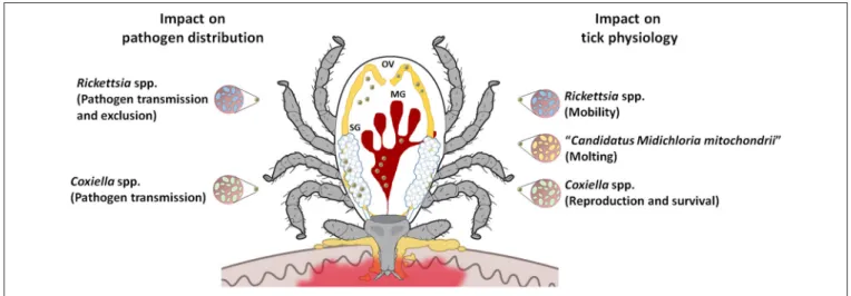
TICK-MICROBIOME INTERACTIONS
The recent development of high-throughput next generation
sequencing technologies has highlighted the complexity of the
tick microbiome that includes both pathogens and potential
symbionts (
Vayssier-Taussat et al., 2015
). It is readily apparent
that interactions frequently occur among tick microbial
communities, as relationships between microorganisms existing
in one environment can be competitive, exclusive, facilitating,
or absent, with many potential implications for human
and animal health that remain to be elucidated (
Ahantarig
et al., 2013; Vayssier-Taussat et al., 2015
). Both positive
and negative associations have been reported for pathogens
(
Mather et al., 1987; de la Fuente et al., 2003
). However,
the role of tick endosymbionts in pathogen transmission
has only been studied in a few selected bacterial and tick
species.
Symbionts may confer crucial and diverse benefits to
their hosts, playing nutritional roles, or affecting fitness,
development, reproduction, defense against environmental
stress, and immunity (
Ahantarig et al., 2013
).
Coxiella
-like
endosymbionts are believed to be the most common vertically
transmitted agents in hard ticks (
Bernasconi et al., 2002; Lee
et al., 2004; Clay et al., 2008; Bonnet et al., 2013; Cooper et al.,
2013
). In
Amblyomma americanum,
the removal of
Coxiella
symbionts following antibiotic treatment reduced tick offspring
production and increased time to oviposition (
Zhong et al.,
2007
). In
I. ricinus
(
Lo et al., 2006; Sassera et al., 2006; Montagna
et al., 2013
),
Candidatus
Midichloria mitochondrii is an
intra-mitochondrial bacterium that has also been detected in other
tick genera (
Harrus et al., 2011; Williams-Newkirk et al., 2012
).
It has been ascribed a possible helper role in tick molting
processes (
Zchori-Fein and Bourtzis, 2011
,
Figure 3
).
Rickettsia
-like symbionts have also been reported to infect hard ticks from
several genera (
Baldridge et al., 2004; Clay et al., 2008; Liu et al.,
2013
). One study reported that
Rickettsia
-infected
Dermacentor
variabilis
have slightly greater motility than uninfected ticks,
indirectly influencing disease risk (
Kagemann and Clay, 2013
).
Francisella
-like symbionts have been reported in several hard
tick genera (
Venzal et al., 2008; Ivanov et al., 2011; Michelet
et al., 2013
), but their effect on tick fitness and biology remains
unknown. Being able to manipulate host reproduction and then
to affect vector populations,
Wolbachia
spp. have also been
identified in several hard tick genera (
Engelstadter and Hurst,
2007; Andreotti et al., 2011; Reis et al., 2011; Zhang X. et al.,
2011
). Their role in pathogen transmission requires further
attention, as reports suggest that this bacterium can protect
some arthropods against microbial infections (
Martinez et al.,
2014
). In
I. ricinus
,
Wolbachia pipientis
is known to be associated
with the hymenoptera tick endoparasitoid
Ixodiphagus hookeri
(
Plantard et al., 2012; Bohacsova et al., 2016
), and
Arsenophonus
spp. symbionts (
Dergousoff and Chilton, 2010
). The latter,
detected in several tick species (
Clay et al., 2008; Dergousoff
and Chilton, 2010; Reis et al., 2011
), are responsible for
sex-ratio distortion in arthropods, and some studies suggest that
they can affect host-seeking success by decreasing tick motility
in
A. americanum
and
D. variabilis
(
Kagemann and Clay, 2013
).
Lastly, some
Spiroplasma
spp. detected in
Ixodes
spp. such as
Spiroplasma ixodetis
(
Tully et al., 1995
) may cause sex-ratio
distortion in some insect species via male killing (
Tabata et al.,
2011
).
Recently,
Abraham
et
al.
(2017)
showed
how
A. phagocytophilum
manipulates
I. scapularis
tick microbiota
to promote infection. Firstly, they showed that IAFGP,
apart from protecting ticks against cold injury (see above),
has antimicrobial activity against biofilm-forming bacteria,
particularly
Staphylococcus aureus
and
Enterococcus faecalis
.
They further showed that by targeting biofilm-forming bacteria,
A. phagocytophilum
modifies the composition of gut microbiota
and alters tick midguts permeability, which results in higher
A. phagocytophilum
infection in the vector (
Abraham et al.,
2017
). Regarding the relationship between symbionts and
pathogens, exclusion has been reported in Rickettsiales,
which may be due to intra-family bacterial cross-immunity.
Exclusion has been documented in
Dermacentor
ticks infected
with
Rickettsia peacockii
or
Rickettsia montana
that limits
Rickettsia rickettsii
and
Rickettsia rhipicephali
distribution,
respectively (
Burgdorfer et al., 1981; Macaluso et al., 2002
,
Figure 3
). It has also been reported that
I. scapularis
male
ticks infected by a rickettsial endosymbiont had significantly
lower rates of infection by
B. burgdorferi
than
symbiont-free males, thus evidencing interactions among microbial
species (
Steiner et al., 2008
). Further research showed that
perturbation of the midgut microbiome in
I. scapularis
influences
B. burgdorferi
colonization of ticks through a
transcriptional mechanism resulting in lower expression of
peritrophin, which perturbs the integrity of the peritrophic
matrix (
Narasimhan et al., 2014
). In
A. americanum,
the
presence of
Coxiella
-related symbionts seems to influence
Ehrlichia chaffeensis
transmission (
Klyachko et al., 2007
), and
infection with
Arsenophonus
appears to be negatively correlated
with the frequency of
Rickettsia
sp. infection (
Clay et al., 2008
,
Figure 3
).
CONCLUSIONS AND FUTURE
DIRECTIONS FOR THE CONTROL OF
TICK-BORNE DISEASES
Over millions of years, arthropod vectors have co-evolved with
a variety of microorganisms including bacteria, viruses, and
protozoa to the point where they appear to co-exist with little
impact on the vector (
Beerntsen et al., 2000; Estrada-Peña et al.,
2015; de la Fuente et al., 2015
). These arthropods have become
efficient vectors of pathogens to humans and other vertebrate
hosts that are susceptible to infection and disease.
Present results show that different pathogens have developed
similar strategies such as manipulation of the immune response
to infect ticks and facilitate multiplication and transmission.
Some of these strategies may be used by pathogens to infect
both ticks and mammalian hosts (
de la Fuente et al., 2016
).
Additionally, recent evidence demonstrates that the microbiome
has an effect on tick fitness and pathogen infection and
transmission, highlighting the importance of tick-microbiome
interactions for vector competence. Overall, these results
illustrate how pathogens activate mechanisms and manipulate
tick protective responses and other biological processes in order
to facilitate infection, while ticks respond to limit pathogen
infection and preserve feeding fitness and vector competence for
survival of both ticks and pathogens. However, how different
molecular mechanisms make certain tick species suitable vectors
for certain pathogens is still not fully characterized. The
presence of tick receptors that are pathogen-specific affects
vector competence for these pathogens, but other mechanisms
are probably also involved in this process. Furthermore, the
biological processes involved in tick-pathogen interactions are
also affected in other arthropod vectors (
Box 2
).
The identification of the molecular drivers that promote
tick survival, spread, and pathogen transmission provides the
opportunity to disrupt these processes and lead to a reduction
in tick burden and prevalence of tick-borne diseases. Targeting
some of the similar mechanisms used by the pathogens for
infection and transmission by ticks may be used to develop
strategies against multiple tick-borne diseases. As shown for
B. burgdorferi
OspA (
Gomes-Solecki, 2014
), pathogen-derived
BOX 2 | Are the biological processes involved in tick-pathogen interactions unique for ticks?
The answer to this question is that several of the processes involved in tick-pathogen interactions have also been identified in other vector-pathogen interactions (see for example,Beerntsen et al., 2000; Vlachou et al., 2005; Wang et al., 2010; Gómez-Díaz et al., 2012; Sabin et al., 2013; Ramphul et al., 2015; Eng et al., 2016; Shaw et al., 2017). For example, as described in ticks, receptor-ligand-like interactions mediate pathogen recognition and infection in mosquitoes (Beerntsen et al., 2000). Remodeling of the cytoskeleton seems to be a general mechanism for tick pathogen infection (Cotté et al., 2014; de la Fuente et al., 2016). Pathogens such as Dengue virus (DENV), West Nile virus (WNV), andPlasmodiumparasites also affect mosquito cytoskeleton during infection (Vlachou et al., 2005; Wang et al., 2010). The finding that some pathogens manipulate tick immune response to facilitate infection has been also reported in mosquitoes infected withPlasmodium falciparum(Beerntsen et al., 2000). Similarly, the expression of immune response genes such as those involved in the JAK/STAT pathway may serve to limit bacterial and fungal proliferation in fruit fly and mosquitoes (Beerntsen et al., 2000). Apoptosis plays an important role in tick-pathogen interactions (de la Fuente et al., 2016). While inhibition of cell apoptosis by pathogens facilitates infection, host cell response may activate alternative apoptotic pathways to limit infection (de la Fuente et al., 2016). These findings have been also described in for exampleAedes aegyptiandAnopheles gambiaemosquitoes infected with DENV andP. falciparum, respectively (Ramphul et al., 2015; Eng et al., 2016). The control of tick cell epigenetics byA. phagocytophilumhas been proposed as a mechanism used by the pathogen to facilitate infection and multiplication (Cabezas-Cruz et al., 2016). Similar mechanisms have been described to operate at the mosquito-Plasmodium
interface (Gómez-Díaz et al., 2012).
However, the functional mechanisms by which these processes are affected at the vector-pathogen interface may vary between pathogen and vector species (Figure 4). The limited information available on the functional characterization of these processes in ticks and other arthropods limits the scope of the comparative analysis between different vectors. Nevertheless, recent results support that in some cases the protein function described in model insect species may be different in the evolutionarily distant ticks. Differences in vector competence may be genetically encoded by differences in the immune response pathways operating at each vector-pathogen interaction (Baxter et al., 2017). For example, Tudor-SN, a conserved component of the basic RNAi machinery with a variety of functions including immune response and gene regulation, is involved in defense against infection inDrosophila(Sabin et al., 2013) but not in ticks (Ayllón et al., 2015b). The IMD pathway is involved in protection against infection in arthropods, but recent results support the existence of two functionally distinct IMD circuits in insects and ticks (Shaw et al., 2017). Future comparative analyses between different vector species will provide additional information on the functional implication of the different biological processes in vector-pathogen interactions and vector competence (Gerold et al., 2017).
FIGURE 4 | Pathogens inhibit vector cell apoptosis by different mechanisms.After infection of tick salivary glands,A. phagocytophiluminhibit apoptosis by decreasing the expression of the pro-apoptotic genes coding for proteins such as ASK1 and Porin. Porin down-regulation is associated with the inhibition of mitochondrial Cyt c release (Ayllón et al., 2015a). In contrast,A. phagocytophiluminfection does not affect Bcl-2 levels, probably because this protein but not Porin is essential for tick feeding (Ayllón et al., 2015a).A. phagocytophilumalso induces ER stress in tick cells which play a role in reducing the levels of MKK that inhibits apoptosis (Villar et al., 2015a). Another interesting mechanism ofA. phagocytophilumto inhibit apoptosis is the manipulation of glucose metabolism by reducing the levels of PEPCK (Villar et al., 2015a). The capacity ofA. phagocytophilumto downregulate gene expression in neutrophils was associated with HDAC1 recruitment to the promoters of target genes by the ankyrin repeat protein AnkA (Garcia-Garcia et al., 2009a,b; Rennoll-Bankert et al., 2015). Tick HDAC1 is overrepresented in
AUTHOR CONTRIBUTIONS
JF, SA, SB, AD, AE, NJ, KM, AN, AP, NR, AF, ROMR conducted
the literature research and wrote the paper. JF, AC, AP, SB, AN,
NJ prepared the figures and tables. All authors provided critical
review and revisions.
FUNDING
Part of the research included in this review was supported by
the Ministerio de Economia y Competitividad (Spain) grant
BFU2016-79892-P and the European Union (EU) Seventh
Framework Programme (FP7) ANTIGONE project number
278976. SA and AD would like to acknowledge FCT for funds
to GHTM - UID/Multi/04413/2013. MV was supported by the
Research Plan of the University of Castilla-La Mancha (UCLM),
Spain. The funders had no role in study design, data collection
and interpretation, or the decision to submit the work for
publication.
ACKNOWLEDGMENTS
We thank members of our laboratories for fruitful
discussions.
REFERENCES
Abraham, N. M., Liu, L., Jutras, B. L., Yadav, A. K., Narasimhan, S., Gopalakrishnan, V., et al. (2017). Pathogen-mediated manipulation of arthropod microbiota to promote infection.Proc. Natl. Acad. Sci. U.S.A.114, E781–E790. doi: 10.1073/pnas.1613422114
Ahantarig, A., Trinachartvanit, W., Baimai, V., and Grubhoffer, L. (2013). Hard ticks and their bacterial endosymbionts (or would be pathogens). Folia. Microbiol.58, 419–428. doi: 10.1007/s12223-013-0222-1
Alberdi, P., Mansfield, K. L., Manzano-Román, R., Cook, C., Ayllón, N., Villar, M., et al. (2016). Tissue-specific signatures in the transcriptional response to Anaplasma phagocytophiluminfection ofIxodes scapularisandIxodes ricinus tick cell lines.Front. Cell. Infect. Microbiol.6:20. doi: 10.3389/fcimb.2016.00020 Almazán, C., Kocan, K. M., Blouin, E. F., and de la Fuente, J. (2005). Vaccination with recombinant tick antigens for the control of Ixodes scapularisadult infestations.Vaccine23, 5294–5298. doi: 10.1016/j.vaccine.2005.08.004 Andreotti, R., Perez de Leon, A. A., Dowd, S. E., Guerrero, F. D., Bendele, K.
G., and Scoles, G. A. (2011). Assessment of bacterial diversity in the cattle tickRhipicephalus (Boophilus) microplusthrough tag-encoded pyrosequencing. BMC. Microbiol.11:6. doi: 10.1186/1471-2180-11-6
Antunes, S., Galindo, R. C., Almazán, C., Rudenko, N., Golovchenko, M., Grubhoffer, L., et al. (2012). Functional genomics studies of Rhipicephalus (Boophilus) annulatus ticks in response to infection with the cattle protozoan parasite, Babesia bigemina. Int. J. Parasitol. 42, 187–195. doi: 10.1016/j.ijpara.2011.12.003
Ashida, H., Mimuro, H., Ogawa, M., Kobayashi, T., Sanada, T., Kim, M., et al. (2011). Host-pathogen interactions cell death and infection: a double-edged sword for host and pathogen survival. J. Cell. Biol.195, 931–942. doi: 10.1083/jcb.201108081
Ayllón, N., Naranjo, V., Hajdušek, O., Villar, M., Galindo, R. C., Kocan, K. M., et al. (2015b). Nuclease Tudor-SN is involved in tick dsRNA-mediated RNA interference and feeding but not in defense against flaviviral or Anaplasma phagocytophilum rickettsial infection. PLoS ONE 10:e0133038. doi: 10.1371/journal.pone.0133038
Ayllón, N., Villar, M., Busby, A. T., Kocan, K. M., Blouin, E. Bonzón-Kulichenko, E., F., et al. (2013). Anaplasma phagocytophilum inhibits apoptosis and promotes cytoskeleton rearrangement for infection of tick cells.Infect. Immun. 81, 2415–2425. doi: 10.1128/IAI.00194-13
Ayllón, N., Villar, M., Galindo, R. C., Kocan, K. M., Šíma, R., López, J. A., et al. (2015a). Systems biology of tissue-specific response to Anaplasma phagocytophilum reveals differentiated apoptosis in the tick vector Ixodes scapularis.PLoS Genet.11:e1005120. doi: 10.1371/journal.pgen.1005120 Baldridge, G. D., Burkhardt, N. Y., Simser, J. A., Kurtti, T. J., and Munderloh,
U. G. (2004). Sequence and expression analysis of the ompA gene of Rickettsia peacockii, an endosymbiont of the Rocky Mountain wood tick, Dermacentor andersoni. Appl. Environ. Microbiol. 70, 6628–6636. doi: 10.1128/AEM.70.11.6628-6636.2004
Baxter, R. H., Contet, A., and Krueger, K. (2017). Arthropod innate immune systems and vector-borne diseases. Biochemistry. 56, 907–918. doi: 10.1021/acs.biochem.6b00870
Beerntsen, B. T., James, A. A., and Christensen, B. M. (2000). Genetics of mosquito vector competence. Microbiol. Mol. Biol. Rev. 64, 115–137. doi: 10.1128/MMBR.64.1.115-137.2000
Bell-Sakyi, L., Zweygarth, E., Blouin, E. F., Gould, E. A., and Jongejan, F. (2007). Tick cell lines: tools for tick and tick-borne disease research.Trends. Parasitol. 23, 450–457. doi: 10.1016/j.pt.2007.07.009
Bernasconi, M. V., Casati, S., Peter, O., and Piffaretti, J. C. (2002).Rhipicephalus ticks infected withRickettsiaandCoxiellain Southern Switzerland (Canton Ticino).Infect. Genet. Evol.2, 111–120. doi: 10.1016/S1567-1348(02)00092-8 Bohacsova, M., Mediannikov, O., Kazimirova, M., Raoult, D., and Sekeyova,
Z. (2016). Arsenophonus nasoniae and Rickettsiae infection of Ixodes ricinusdue to parasitic waspIxodiphagus hookeri.PLoS ONE11:e0149950. doi: 10.1371/journal.pone.0149950
Bonnet, S., de la Fuente, J., Nicollet, P., Liu, X., Madani, N., Blanchard, B., et al. (2013). Prevalence of tick-borne pathogens in adultDermacentorspp. ticks from nine collection sites in France.Vector Borne Zoonotic Dis.13, 226–236. doi: 10.1089/vbz.2011.0933
Burgdorfer, W., Hayes, S., and Mavros, A. (1981). “Non-pathogenic rickettsiae inDermacentor andersoni: a limiting factor for the distribution ofRickettsia rickettsia,” inRickettsia and Rickettsial Disease, ed W. Burgdorfer (New York, NY: Academic Press), 585–594.
Busby, A. T., Ayllón, N., Kocan, K. M., Blouin, E. F., de la Fuente, G., Galindo, R. C., et al. (2012). Expression of heat-shock proteins and subolesin affects stress responses,Anaplasma phagocytophiluminfection and questing behavior in the tick, Ixodes scapularis. Med. Vet. Entomol. 26, 92–102. doi: 10.1111/j.1365-2915.2011.00973.x
Cabezas-Cruz, A., Alberdi, P., Ayllón, N., Valdés, J. J., Pierce, R., Villar, M., et al. (2016). Anaplasma phagocytophilum increases the levels of histone modifying enzymes to inhibit cell apoptosis and facilitate pathogen infection in the tick vector, Ixodes scapularis. Epigenetics 11, 303–319. doi: 10.1080/15592294.2016.1163460
Cabezas-Cruz, A., Estrada-Peña, A., Rego, R. O. M., and De la Fuente, J. (2017). Tick-pathogen ensembles: do molecular interactions lead ecological innovation?Front. Cell. Infect. Microbiol.7:74. doi: 10.3389/fcimb.2017.00074 Chauvin, A., Moreau, E., Bonnet, S., Plantard, O., and Malandrin, L. (2009).
Babesia and its hosts: adaptation to long-lasting interactions as a way to achieve efficient transmission.Vet. Res.40, 37. doi: 10.1051/vetres/2009020
Clay, K., Klyachko, O., Grindle, N., Civitello, D., Oleske, D., and Fuqua, C. (2008). Microbial communities and interactions in the lone star tick,Amblyomma americanum. Mol. Ecol. 17, 4371–4381. doi: 10.1111/j.1365-294X.2008. 03914.x
Cooper, A., Stephens, J., Ketheesan, N., and Govan, B. (2013). Detection ofCoxiella burnetiiDNA in wildlife and ticks in northern Queensland, Australia.Vector Borne Zoonotic Dis.13, 12–16. doi: 10.1089/vbz.2011.0853
Cotté, V., Sabatier, L., Schnell, G., Carmi-Leroy, A., Rousselle, J. C., Arsène-Ploetze, F., et al. (2014). Differential expression ofIxodes ricinussalivary gland proteins in the presence of theBorrelia burgdorferisensu lato complex.J. Proteomics.96, 29–43. doi: 10.1016/j.jprot.2013.10.033
Borrelia burgdorferi migration from the gut. J. Mol. Med. 94, 361–370. doi: 10.1007/s00109-015-1365-0
Cramaro, W. J., Revets, D., Hunewald, O. E., Sinner, R., Reye, A. L., and Muller, C. P. (2015). Integration ofIxodes ricinusgenome sequencing with transcriptome and proteome annotation of the naive midgut.BMC Genomics 16:871. doi: 10.1186/s12864-015-1981-7
Dai, J., Narasimhan, S., Zhang, L., Liu, L., Wang, P., and Fikrig, E. (2010). Tick histamine release factor is critical for Ixodes scapularisengorgement and transmission of the Lyme disease agent. PLoS Pathog. 6:e1001205. doi: 10.1371/journal.ppat.1001205
de Castro, M. H., de Klerk, D., Pienaar, R., Latif, A. A., Rees, D. J., and Mans, B. J. (2016).De novoassembly and annotation of the salivary gland transcriptome of Rhipicephalus appendiculatusmale and female ticks during blood feeding.Ticks Tick Borne Dis.7, 536–548. doi: 10.1016/j.ttbdis.2016.01.014
de la Fuente, J., Blouin, E. F., and Kocan, K. M. (2003). Infection exclusion of the rickettsial pathogenAnaplasma marginalein the tick vectorDermacentor variabilis. Clin. Diagn. Lab. Immunol. 10, 182–184. doi: 10.1128/cdli. 10.1.182-184.2003
de la Fuente, J., and Contreras, M. (2015). Tick vaccines: current status and future directions. Expert Rev. Vaccines 14, 1367–1376. doi: 10.1586/14760584.2015.1076339
de la Fuente, J., Estrada-Peña, A., Cabezas-Cruz, A., and Brey, R. (2015). Flying ticks: anciently evolved associations that constitute a risk of infectious disease spread.Parasit Vectors8, 538. doi: 10.1186/s13071-015-1154-1
de la Fuente, J., Estrada-Peña, A., Cabezas-Cruz, A., and Kocan, K. M. (2016). Anaplasma phagocytophilum uses common strategies for infection of ticks and vertebrate hosts. Trends Microbiol. 24, 173–180. doi: 10.1186/s13071-015-1154-1
de la Fuente, J., Estrada-Peña, A., Venzal, J. M., Kocan, K. M., and Sonenshine, D. E. (2008). Overview: ticks as vectors of pathogens that cause disease in humans and animals.Front. Biosci.13, 6938–6946. doi: 10.2741/3200
de la Fuente, J., Garcia-Garcia, J. C., Blouin, E. F., McEwen, B. R., Clawson, D., and Kocan, K. M. (2001). Major surface protein 1a effects tick infection and transmission ofAnaplasma marginale.Int. J. Parasitol.31, 1705–1714. doi: 10.1016/S0020-7519(01)00287-9
de la Fuente, J., and Kocan, K. M. (2014). “Development of vaccines for control of tick infestations and interruption of pathogen transmission,” inBiology of Ticks, 2nd Edn., ed D. Sonenshine and M Roe (New York, NY: Oxford University Press), 333–352.
de la Fuente, J., Kocan, K. M., Almazán, C., and Blouin, E. F. (2007). RNA interference for the study and genetic manipulation of ticks.Trends Parasitol. 23, 427–433. doi: 10.1016/j.pt.2007.07.002
Dergousoff, S. J., and Chilton, N. B. (2010). Detection of a new Arsenophonus-type bacterium in Canadian populations of the Rocky Mountain wood tick, Dermacentor andersoni. Exp. Appl. Acarol. 52, 85–91. doi: 10.1007/s10493-010-9340-5
Dickson, D. L., and Turell, M. J. (1992). Replication and tissue tropisms of Crimean-Congo hemorrhagic fever virus in experimentally infected adult Hyalomma truncatum (Acari: Ixodidae). J. Med. Entomol. 29, 767–773. doi: 10.1093/jmedent/29.5.767
Doherty, P. C., and Reid, H. W. (1971). Experimental louping ill in the sheep. II, Neuropathology.J. Comp. Pathol.81, 331–337. doi: 10.1016/0021-9975(71) 90020-X
Eng, M. W., van Zuylen, M. N., and Severson, D. W. (2016). Apoptosis-related genes control autophagy and influence DENV-2 infection in the mosquito vector, Aedes aegypti. Insect Biochem. Mol. Biol. 76, 70–83. doi: 10.1016/j.ibmb.2016.07.004
Engelstadter, J., and Hurst, G. D. (2007). The impact of male-killing bacteria on host evolutionary processes. Genetics 175, 245–254. doi: 10.1534/genetics.106.060921
Estrada-Peña, A., de la Fuente, J., Ostfeld, R. S., and Cabezas-Cruz, A. (2015). Interactions between tick and transmitted pathogens evolved to minimise competition through nested and coherent networks. Sci. Rep. 5:10361. doi: 10.1038/srep10361
Estrada-Peña, A., Ortega, C., Sánchez, N., Desimone, L., Sudre, B., Suk, J. E., et al. (2011). Correlation ofBorrelia burgdorferisensu lato prevalence in questing Ixodes ricinusticks with specific abiotic traits in the western Palearctic.Appl. Environ. Microbiol.77, 3838–3845. doi: 10.1128/AEM.00067-11
Florin-Christensen, M., and Schnittger, L. (2009). Piroplasmids and ticks: a long-lasting intimate relationship.Front. Biosci. 14, 3064–3073. doi: 10.2741/3435 Garcia-Garcia, J. C., Barat, N. C., Trembley, S. J., and Dumler, J. S. (2009a).
Epigenetic silencing of host cell defense genes enhances intracellular survival of the rickettsial pathogenAnaplasma phagocytophilum.PLoS Pathog.5:e1000488. doi: 10.1371/journal.ppat.1000488
Garcia-Garcia, J. C., Rennoll-Bankert, K. E., Pelly, S., Milstone, A. M., and Dumler, J. S. (2009b). Silencing of host cell CYBB gene expression by the nuclear effector AnkA of the intracellular pathogenAnaplasma phagocytophilum.Infect Immun. 77, 2385–2391. doi: 10.1128/IAI.00023-09
Garg, R., Juncadella, I. J., Ramamoorthi, N., Ananthanarayanan, S. K., Thomas, V., Rincón, M., et al. (2006). Cutting edge: CD4 is the receptor for the tick saliva immunosuppressor, Salp15. J. Immunol. 177, 6579–6583. doi: 10.4049/jimmunol.177.10.6579
Garrison, A. R., Radoshitzky, S. R., Kota, K. P., Pegoraro, G., Ruthel, G., et al. (2013). Crimean-Congo hemorrhagic fever virus utilizes a clathrin- and early endosome-dependent entry pathway.Virology444, 45–54. doi: 10.1016/j.virol.2013.05.030
Gerold, G., Bruening, J., Weigel, B., and Pietschmann, T. (2017). Protein interactions during the flavivirus and hepacivirus life cycle. Mol. Cell. Proteomics16(4 Suppl. 1), S75–S91. doi: 10.1074/mcp.r116.065649
Gomes-Solecki, M. (2014). Blocking pathogen transmission at the source: reservoir targeted OspA-based vaccines againstBorrelia burgdorferi.Front. Cell Infect Microbiol.4:136. doi: 10.3389/fcimb.2014.00136
Gómez-Díaz, E., Jordà, M., Peinado, M. A., and Rivero, A. (2012). Epigenetics of host-pathogen interactions: the road ahead and the road behind.PLoS Pathog. 8:e1003007. doi: 10.1371/journal.ppat.1003007
Gulia-Nuss, M., Nuss, A. B., Meyer, J. M., Sonenshine, D. E., Roe, R. M., Waterhouse, R. M., et al. (2016). Genomic insights into the Ixodes scapularis tick vector of Lyme disease. Nat. Commun. 7:10507. doi: 10.1038/ncomms10507
Hajdušek, O., Síma, R., Ayllón, N., Jalovecká, M., Perner, J., de la Fuente, J., et al. (2013). Interaction of the tick immune system with transmitted pathogens. Front. Cell Infect Microbiol.3:26. doi: 10.3389/fcimb.2013.00026
Harrus, S., Perlman-Avrahami, A., Mumcuoglu, K. Y., Morick, D., Eyal, O., and Baneth, G. (2011). Molecular detection ofEhrlichia canis,Anaplasma bovis,Anaplasma platys,CandidatusMidichloria mitochondrii andBabesia canis vogeli in ticks from Israel. Clin. Microbiol. Infect 17, 459–463. doi: 10.1111/j.1469-0691.2010.03316.x
Heekin, A. M., Guerrero, F. D., Bendele, K. G., Saldivar, L., Scoles, G. A., Dowd, S. E., et al. (2013). The ovarian transcriptome of the cattle tick,Rhipicephalus (Boophilus) microplus, feeding upon a bovine host infected withBabesia bovis. Parasit. Vectors6:276. doi: 10.1186/1756-3305-6-276
Heekin, A. M., Guerrero, F. D., Bendele, K. G., Saldivar, L., Scoles, G. A., Gondro, C., et al. (2012). Analysis ofBabesia bovisinfection-induced gene expression changes in larvae from the cattle tick,Rhipicephalus (Boophilus) microplus. Parasit. Vectors5:162. doi: 10.1186/1756-3305-5-162
Hermann, C., and Gern, L. (2010). Survival ofIxodes ricinus(Acri: Ixodidae) under challenging conditions of temperature and humidity is influenced by Borrelia burgdorferisensu laro infection. J. Med. Entomol.47, 1196–1204. doi: 10.1603/ME10111
Herrmann, C., and Gern, L. (2012). Do the level of energy reserves, hydration status and Borrelia infection influence walking by Ixodes ricinus (Acari: Ixodidae) ticks?Parasitology139, 330–337. doi: 10.1017/S0031182011002095 Hourcade, D. E., Akk, A. M., Mitchell, L. M., Zhou, H. F., Hauhart, R., et al. (2016).
Anti-complement activity of theIxodes scapularissalivary protein Salp20.Mol. Immunol.69, 62–69. doi: 10.1016/j.molimm.2015.11.008
Ireton, K. (2013). Molecular mechanisms of cell-cell spread of intracellular bacterial pathogens.Open Biol.3:130079. doi: 10.1098/rsob.130079
Ivanov, I. N., Mitkova, N., Reye, A. L., Hübschen, J. M., Vatcheva-Dobrevska, R. S., Dobreva, E. G., et al. (2011). Detection of new Francisella-like tick endosymbionts in Hyalomma spp. and Rhipicephalus spp. (Acari: Ixodidae) from Bulgaria. Appl. Environ. Microbiol. 77, 5562–5565. doi: 10.1128/AEM.02934-10