Imaging findings of myocardial revascularization
at multidetector computed tomography – where are
the bypass grafts?: an iconographic essay*
Achados de imagem da revascularização do miocárdio pela tomografia computadorizada por múltiplos detectores – onde estão os enxertos?: ensaio iconográfico
Marcelo Targa Ripari1, Rogério Santaniello1, Roberto Sasdelli Neto2, César Higa Nomura2, Rodrigo Caruso Chate2, Rodrigo Bastos Duarte Passos2, Fernando Uliana Kay2, Marcelo Buarque de Gusmão Funari3
Multidetector coronary computed tomography angiography is a precise method for evaluating post-coronary revascularization arterial and venous bypass grafts, and is somehow superseding coronary catheterization that is an invasive and more expensive technique. The present iconographic essay is aimed at anatomically demonstrating the bypass grafts most frequently utilized, how to differentiate between arterial and venous grafts and how to find them. The studies were performed in 64-row multidetector computed tomography equipment, with breath hold, controlled heart rate and appropriate protocols with later MPR, MIP and 3D reconstructions according to electrocardiogram. The localization of the bypass grafts and anastomoses at computed tomography studies focused on chest and coronary arteries may represent a difficulty in the images analysis by the radiologist who is not familiar with the matter, so the knowledge of the surgical techniques adopted and possible courses of the saphenous bypass grafts and arterial grafts can aid in the analysis of both studies, avoiding diagnostic errors.
Keywords: Multidetector coronary computed tomography angiography; Saphenous bypass graft; Coronary artery bypass graft.
A angiotomografia por multidetectores de coronárias constitui um método preciso para avaliação dos enxer-tos venosos e arteriais pós-revascularização coronariana e vem substituindo em parte o cateterismo, o qual é um método invasivo e de maior custo. Este ensaio iconográfico tem como objetivo a demonstração ana-tômica dos enxertos mais comumente utilizados, como diferenciar enxertos venosos e arteriais e como loca-lizá-los. Os exames foram realizados em aparelhos de tomografia computadorizada multislice de 64 fileiras
de detectores, com apneia, frequência cardíaca controlada e protocolos adequados com posterior reconstru-ções MPR, MIP e 3D, de acordo com o eletrocardiograma. A localização dos enxertos e anastomoses em tomografias computadorizadas direcionadas para as artérias coronárias e para o tórax pode dificultar a aná-lise do exame pelo radiologista não familiarizado, sendo que o conhecimento das técnicas cirúrgicas utiliza-das e dos possíveis trajetos utiliza-das pontes de safena e enxertos arteriais ajuda na análise de ambos os exames, evitando também erros diagnósticos.
Unitermos: Angiotomografia das coronárias por multidetectores; Ponte de safena; Enxerto arterial coronariano. Abstract
Resumo
* Study developed at Department of Imaging – Hospital Israe-lita Albert Einstein, São Paulo, SP, Brazil.
1. MDs, Radiologists, Fellows, Department of Imaging – Hos-pital Israelita Albert Einstein, São Paulo, SP, Brazil.
2. MDs, Radiologists, Department of Imaging – Hospital Israe-lita Albert Einstein, São Paulo, SP, Brazil.
3. PhD, MD, Radiologist, Manager of Department of Imaging – Hospital Israelita Albert Einstein, São Paulo, SP, Brazil.
Mailing address: Dr. Marcelo Targa Ripari. Avenida Albert Eins-tein, 627, 4º andar, Bloco D, Sala de Laudos, Departamento de
grafts. However, the assessment of patency of arterial and saphenous grafts utilized in the management of CAD has been per-formed by means of coronary computed tomography (CT) angiography since the introduction of multidetector CT equip-ment. Recently, a meta-analysis demon-strated the high accuracy of this method, with 98% sensitivity, 97% specificity, 93% positive predictive value, and 99% negative predictive value(3).
Notwithstanding, one of the difficulties faced by the investigator during the
analy-Ripari MT, Santaniello R, Sasdelli Neto R, Nomura CH, Chate RC, Passos RBD, Kay FU, Funari MBG. Imaging findings of myocardial revascularization at multidetector computed tomography – where are the bypass grafts?: an iconographic es-say. Radiol Bras. 2009;42(5):327–331.
lar bypass grafts is the technique of choice, particularly in cases of diffuse or multivas-cular disease. Annually, recurrence of symptoms of ischemia is observed in 4% to 8% of patients, and is closely related to the grafts patency(1,2).
Currently, coronary cineangiography still remains as the gold standard in the postoperative follow-up of vascular bypass
INTRODUCTION
Coronary artery disease (CAD) is one of the main causes of death in the world and surgical management by means of
vascu-Imagem. São Paulo, SP, Brazil, 05652-901,. E-mail: mtripari@ uol.com.br
sis of chest images either focused on the evaluation of myocardial revascularization or not, is the identification and localization of the bypass graft course as well as the respective anastomoses that may vary among patients and according to the car-diac surgeon’s experience and technique.
The present essay includes a description of the main types of arterial grafts (right and left internal thoracic artery graft – RITA and LITA) and venous grafts (saphe-nous vein graft – SVG) through images reconstruction and coronary CT angiogra-phy images.
The present iconographic essay is aimed at demonstrating the most frequent sites of arterial and venous bypass grafts for coro-nary arteries by reviewing corocoro-nary CT angiography studies performed in 64-row multidetector CT equipment in the postop-erative follow-up of patients with previous history of myocardial revascularization.
All the studies were performed in an Aquilion 64-row multidetector CT scanner (Toshiba Medical Systems; Tokyo, Japan) with ECG-gating and inspiratory breath-holding(4). The heart rate was previously controlled with beta-blocker, in accordance with the standard protocol of the institu-tion. The images were acquired after 100– 120 ml nonionic iodinated contrast (350 mg/ml) injection into antecubital vein at a rate of 5.0 ml/s. Later, the images were processed on a Vitrea workstation (Vital Images; Minnesota, USA), with multipla-nar and 3D reconstructions.
DESCRIPTION OF THE GRAFTS
CAD can be surgically managed with either venous or arterial bypass grafts.
Venous grafts
Among the venous grafts, the most fre-quently utilized is the SVG, because of its availability, easy harvesting, favorable and widely utilized surgical technique(5) (Fig-ure 1).
Arterial grafts
Internal thoracic artery (ITA) – The LITA is most frequently utilized because of its anatomic proximity, surgical technique, improved survival and extent of the graft patency. Generally, the RITA requires more time to be displaced and anastomosed than
Figure 1.A: Axial images and 3D reconstruction of coronary CT angiography demonstrating SVG (arrows) from the origin of the aorta to the anastomosis in left marginal branch. B: Axial images and 3D recon-struction of coronary CT angiography demonstrating SVG (arrows) from the origin of the aorta to the anastomosis in the anterior descending artery. C: Axial images and 3D reconstruction of coronary CT angiography demonstrating SVG (arrows) from the origin of the aorta to the anastomosis in the distal third of the right coronary artery.
A
B
the LITA does, and seems to be most fre-quently associated with mediastinal infec-tion(5) (Figures 2, 3, 4 and 5).
ITA grafts can be anastomosed with more than one vessel, with sequential, latero-lateral or latero-terminal techniques (Figure 6).
Radial artery – Some authors consider the radial artery graft as the second most relevant in myocardial revascularization. Thrombosis resulting from flow competi-tion and spasm constitute complicacompeti-tions that must be considered by the surgeon in the selection of the graft(5).
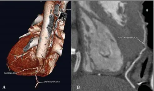
Gastroepiploic artery – The right gas-troepiploic artery is very useful as in situ
graft combined with other grafts in proce-dures without extracorporeal circulation where clamping of aorta is not required (Figure 7). Limitations include: variable extent, vulnerability to flow competition
Figure 4.A,B,C: Axial images demonstrating the absence of RITA and LITA in their respective native beds, but found in oblique routes in the mediastinum (arrows) D: Note the distal anastomosis of the RITA with diagonal branch (arrow).
Figure 3. 3D reconstruction and curved MIP demonstrating LITA anastomosis with the normal anterior descending artery (DA) with more than 20 years after surgery.
Figure 7. 3D reconstruction and curved MIP demonstrating right gastroepiploic artery anastomosis with marginal branch displaced from the respective native bed.
Figure 6. Axial images and 3D reconstruction of coronary CT angiography showing LITA (straight arrows) with sequential anastomosis (curved arrows) in diagonal branch and in the anterior descending artery.
and more extensive involvement of its ori-gin by atherosclerosis, as compared with the ITA(5).
DISCUSSION
Studies demonstrate rates of coronary artery graft occlusion ranging from 10% to 30% within 1 to 2 years, and from 45% to 55% within 10 to 12 years(6).
time required for the analysis of the images, since axial images provide all the medias-tinal fields required for the search for grafts in a single acquisition.
Thus, the localization of grafts and re-spective anastomoses on a study focused on coronary arteries and on general stud-ies of the chest by computed tomography may represent a difficulty in the analysis of the images by a radiologist who is not fa-miliar with the matter, since the knowledge on the surgical techniques utilized and the possible (usual and unusual) routes of the saphenous vein grafts and arterial anasto-moses are useful in the analysis of both imaging methods, besides avoiding wrong diagnoses.
The present iconographic essay also demonstrates some differences that allow the differentiation between arterial and venous grafts: arterial grafts generally present smaller caliber than venous grafts and frequently are associated with the pres-ence of metal clips placed in the origin of their small branches. Venous grafts gener-ally are calibrous and may present gross
parietal calcifications associated with natu-ral degeneration, besides fibrolipidic or “mixed” plaques(3,10).
Therefore, the evaluation of the native bed of the internal thoracic arteries is rel-evant as a first step in this differentiation.
CONCLUSION
Technical developments of CT equip-ment allow the identification of coronary artery and vein grafts on studies with and without ECG-gating so the knowledge on coronary grafts by radiologists is increas-ingly relevant.
REFERENCES
1. Cameron AAC, Davis KB, Rogers WJ. Recur-rence of angina after coronary artery bypass sur-gery: predictors and prognosis (CASS registry). J Am Coll Cardiol. 1995;26:895–9.
2. Eagle KA, Guyton RA, Davidoff R, et al. ACC/ AHA guidelines for coronary artery bypass graft surgery. A report of the American College of Car-diology/American Heart Association Task Force on Practice Guidelines (Committee to revise the 1991 guidelines for coronary artery bypass graft surgery). J Am Coll Cardiol. 1999;34:1262–347. 3. Hamon M, Lepage O, Malagutti P, et al.
Diagnos-tic performance of 16- and 64-section spiral CT for coronary artery bypass graft assessment: meta-analysis. Radiology. 2008;247:679–86.
4. Mahesh M, Cody DD. Physics of cardiac imag-ing with multiple-row detector CT. Radiograph-ics. 2007;27:1495–509.
5. Frazier AA, Qureshi F, Read KM, et al. Coronary artery bypass grafts: assessment with multidetec-tor CT in the early and late postoperative settings. Radiographics. 2005;25:881–96.
6. Ha JW, Cho SY, Shim WH, et al. Noninvasive evaluation of coronary artery bypass graft patency using three-dimensional angiography obtained with contrast-enhanced electron beam CT. AJR Am J Roentgenol. 1999;172:1055–9. 7. Yoo KJ, Choi D, Choi BW, et al. The comparison
of the graft patency after coronary artery bypass grafting using coronary angiography and multi-slice computed tomography. Eur J Cardiothorac Surg. 2003;24:86–91.
8. Burgstahler C, Kuettner A, Kopp AF, et al. Non-invasive evaluation of coronary artery bypass grafts using multi-slice computed tomography: initial clinical experience. Int J Cardiol. 2003;90: 275–80.
9. Ko YG, Choi DH, Jang YS, et al. Assessment of coronary artery bypass graft patency by multislice computed tomography. Yonsei Med J. 2003;44: 438–44.