Clinicoradiological Session
Case 5/2008 – Eight-Month-Old Male Infant, with Down’s Syndrome,
Interventricular Communication and Pulmonary Artery Hypertension
Edmar Atik
Hospital Sírio-Libanês de São Paulo, São Paulo, SP - Brazil
Mailling address: Edmar Atik •
InCor - Av. Dr. Enéas Carvalho de Aguiar, 44 - 05403-000 - São Paulo, SP - Brazil
E-mail: conatik@incor.usp.br
Key Words
Heart defects, congenital; heart septal defects, atrial; hypertension, pulmonary.
Clinical data
Accentuated fatigue was noticed from birth, with no significant alterations throughout time, in addition to low weight gain. Medication use, represented by digoxin, furosemide, captopril and potassium chloride did not alter the clinical evolution.
At physical examination, the patient was dyspneic +++, acyanotic, had normal skin color and pulses were normally palpated in the lower and upper limbs. Weight was 5,330 g, BP = 90/60 mmHg, HR=126 bpm. The aorta was not palpated at the suprasternal notch.
The precordium showed concavity and slight impulsions and the ictus cordis was not palpated. The heart sounds were hyperphonetic, with the second sound being high-pitched in the pulmonary area. No heart murmur had been ausculted at several examinations. Sometimes, a systolic murmur was heard, with a +intensity, limited to the 3rd and 4th left intercostal spaces.
The lungs were clean and the liver was palpated at 4 cm from the right costal border.
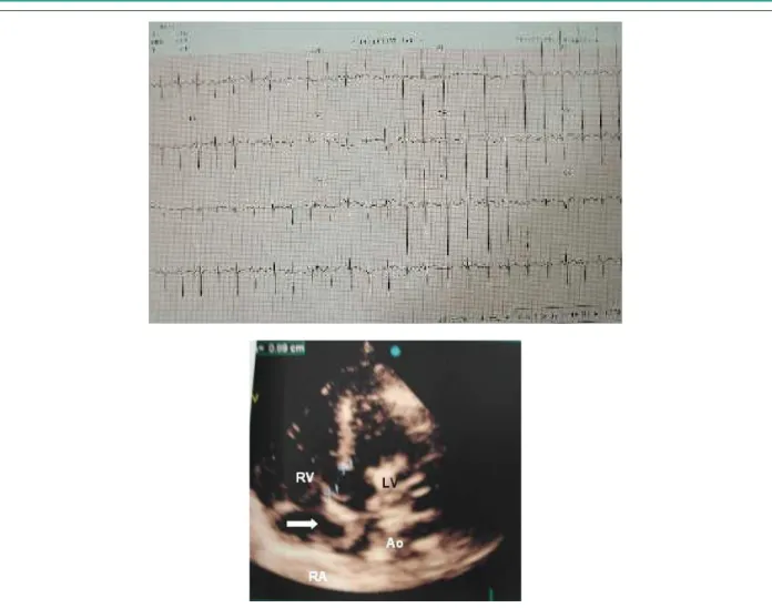
The electrocardiogram (Figure 1) showed a sinus rhythm and signs of biventricular overload, with an RS complex in V2 to V5 and Rs in V1. ÂQRS:+180o, ÂP: +30o and ÂT:+50o.
Radiographic imaging
The radiographic imaging showed moderately increased cardiac area and pulmonary vascular network. The atrial and ventricular contours were noticeable, with a rectified middle arch (Figure 2).
Diagnostic impression
This image is compatible with a diagnosis of acyanogenic congenital cardiopathy with interventricular-communication type pulmonary hyperflow.
Differential diagnosis
Other cardiopathies with atrioventricular septum defect can be considered, as well as arteriovenous fistulae.
Diagnostic confirmation
The clinical elements were decisive to attain the diagnosis of interventricular communication with pulmonary hypertension. The echocardiogram (Figure 1) confirmed the existence of a 7-mm diameter defect in a perimembranous region. There was a gradient between the two ventricles of 20 mmHg and the right ventricle systolic pressure was estimated at 70 mmHg, with a detected left-to-right shunt.
Procedure
At surgery, an 8-mm interventricular communication was closed through the tricuspid valve. The right ventricle and the pulmonary trunk were greatly augmented and the interventricular pressure ratio was 0.5 after the correction of the defect. The postoperative evolution was favorable and the patient was discharged in good clinical condition under the usual anticongestive medication (furosemide and captopril).
Considerations
This case presented great difficulty and raised some concerns at the surgical indication considering the absence of heart murmur. However, the set of all the other elements was decisive to define the adopted procedure, especially considering the presence of signs of heart failure (dyspnea, hepatomegaly, cardiomegaly), in addition to the biventricular overload at the ECG, the left-to-right shunt demonstrated at the echocardiogram, even in the absence of hemodynamic data.
It is worth mentioning that the timing for the operation in this patient was inadequate, as he was exposed to evolution risks, considering the severe consequences of the defect from birth to eight months of age, when the surgical correction was carried out.
As a result of that, the patient already showed signs of pulmonary hypertension (high-pitched second heart sound and absence of heart murmur).
Clinico Radiological Session
Edmar AtikArq Bras Cardiol 2008;91(4):265-266
Figure 1 – Electrocardiogram; the upper part shows signs of biventricular overload, with a predominance of the right ventricle. The lower part shows, in the 5-chamber
apical view, the large perimembranous interventricular communication; RV - Right ventricle; LV - Left ventricle; Ao – Aorta; RA – Right atrium
Figure 2 – Chest x-ray showing the augmented cardiac area and pulmonary vascular network. The atrial and ventricular contours are evident and a rectiied middle
arch is seen.
