Arq Bras Cardiol volume 72, (nº 2), 1999
Clinocopathologic Session
205 Editor: Alfredo José Mansur
Associate Editors: Desiderio Favarato; Vera Demarchi Aiello
Mailing address: Alfredo José Mansur Incor Av. Dr. Enéas C. Aguiar, 44 -05403-000 - São Paulo, SP
(Case report 1/99 - Instituto do Coração do Hospital das Clínicas - FMUSP)
Clinicopathologic Session
Clinocopathologic Session
The patient is a 65-year-old male with a history of dys-pnea during slight exertion(12/17/96).
Six months prior to admission, he developed edema of the lower limbs and, in the last15 days, progressive nea during exertion, which eventually progressed to dysp-nea at rest. Two days prior to admission, he was given intra-venous furosemide for severe dyspnea.
His past medical history is notable forgeneralized to-nic-clonic seizures in the last 10 years, for which he was being treated with phenobarbital. He was born in Con-quista, in the state of Minas Gerais. He reported smoking (2 cigarettes a day) since the age of 38 years. There is no family history of heart disease. His mother and one of his brothers died of stroke. The year before, he had two episodes of pneumonia.
On physical examination (12/17/96) the patient was dyspneic, his blood pressure (BP) was 160/70mmHg, his heart rate (HR) 130bpm and he had a temperature of 38.2oC.
His venous jugular pressure was elevated (+++/4+). Lung examination showed globally diminished breath sounds, with no rales, wheezing or rhonchi. His heart examination revealed a nonpalpable ictus, arrhythmic heart sounds and a systolic murmur grade 2 in the tricuspid area, which increa-sed during inspiration. There was edema of the abdominal wall and ascites. His liver was felt 5cm below the right costal margin. Edema of the lower limbs (+++/4+), varicose veins and stasisdermatitis of the lower limbs were also noted.
Chest X-ray showed an enlarged cardiacsilhouette, (+++/4+), bilateral pleural effusion and mild signs of lung congestion.
The electrocardiogram (ECG) showed atrial fibrillation with frequent ventricular premature depolarizations, low-voltage QRS complexes in all leads, right axis deviation, and diffuse abnormalities of ventricular repolarization.
He was diagnosed with dilated cardiomyopathy com-plicated with atrial fibrillation and a suspected infectious process. He was treated with digoxin, 0.25mg; furosemide, 40mg; ramipril, 2.5mg; and amoxicillin, 1.5g daily, for 10 days. At follow-up 11 days later, the signs and symptoms of heart failure had improved, his BP was 120/60 mm Hg and his HR was 88bpm.
The echocardiogram showed septum and left
ventri-cular posterior wall thickness of 9mm, a ventriventri-cular diastolic diameter of 48mm, and a LV ejection fraction of 73%. The diameter of the aorta was 35mm, left atrium (LA) was 57mm, and right ventricle (RV) was 42mm. The RV was diffusely hypokineticand the right atrium (RA) markedly enlarged. Pulmonary arterial pressure estimated by the Doppler effect was 57mmHg. Marked tricuspid regurgitation and pericar-dial effusion were observed.
Laboratory test results (Jan/97) documented a creati-nine of 0.7mg/dL, a glucose of 104mg/dL, total cholesterol of 120mg/dL (LDL=86mg/dL, HDL=26mg/dL, and VLDL=8mg/ dL) and triglyceridesof 44mg/dL.
The diagnosis of restrictive syndrome wassuspected. Cardiac catheterization (11/4/97) showed moderate hyper-tension of the right chambers and of the pulmonary artery, and slight diffuse hypocinesia of the LV and RV. No coronary lesions were found. Pressures are shown in table I.
The clinical diagnosis was chronic constrictive peri-carditis requiring surgical therapy.
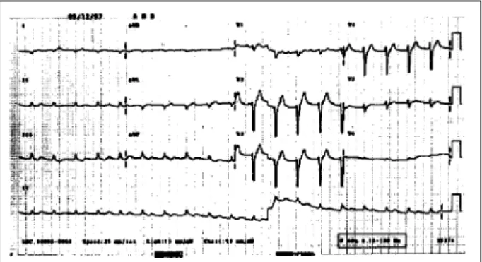
Preoperative ECG (12/1/97) revealed atrial fibrillation, mean heart rate of 110bpm, low-voltage QRS complexes in the frontal plane, QRS axis of + 120o , indirect signs of RA
hypertrophy (Penaloza-Tranchesi) and decreased left ventricular forces. There was no increase in the voltage of the R wave from V1 to V6, suggesting RV hypertrophy (fig. 1).
Additional laboratory test results are shown in table II. The patient underwent surgery (12/9/97), which revealed the following findings: a slightly thickened and adhered pericardium, constrictive epicarditis and a large right serosanguineous pleural effusion. Pericardiectomy from phrenic to phrenic, epicardiectomy and bilateral pleural drainage were performed. The patient had several episodes of ventricular fibrillation easily resolved intraoperatively. His outcome was eventful. At his arrival to the intensive care unit, he was in cardiogenic shock, requiring high doses of vasoactive drugs (dopamine followed by norepi-nephrine)and using intraaorticballoon counter pulsation. These procedures were unsuccessful.
Table I - Cardiac catheterization, pressure measurements (mmHg)
Chamber Systole Protodiastole Telediastole Mean
Right Atrium 12
Right ventricle 50 0 15
Pulmonary artery 50 20 30
Wedge 12
Aorta 95 50 65
206
Clinocopathologic Session Arq Bras Cardiol
volume 72, (nº 2), 1999
The echocardiogram (12/9/97), which was performed with some technical problems, showed a markedly increa-sed RA and a dilated RV, even though contraction was nor-mal, and a LV with normal systolic function.
In the early hours of the day following the surgery, the patient experienced several cardiopulmonary arrests and died (12/10/97).
Discussion
Clinical aspects - Considering the symptoms of heart failure mainly on the right side and the imaging tests showing pulmonary hypertension and involvement of the right cardiac chambers,two diagnostic hypotheses based
on two syndromic pictures can be made: pulmonary hyper-tension with cor pulmonale or restrictive syndrome.
Among the causes of secondary pulmonary hyper-tension, the most common are those caused by chronic obstructive pulmonary disease and autoimmune diseases (systemic lupus erythematosus, rheumatoid arthritis, scleroderma); however, the clinical findings are not suggestive of these diseases. Pulmonary hypertension secondary to pulmonary thromboembolism presenting as chronic microembolisms could be suspected in this case; however, this condition is more related to deficiencies in antithrombotic protective systems, which is not suggested in this case. Finally, pulmonary hypertension caused by parasitosis, stressingin this case pulmonary hypertension caused by S. mansoni, would be a strong diagnostic
hypothesis, since the patient came from an endemic area -Minas Gerais - and this disease is a well defined cause of pulmonary hypertension.
Barbosa et al 1 found that, in addition to cor pulmonale
and pulmonary hypertension, schistosomiasis can rarely cause myocarditis, cardiomyopathy and endomyocardial fibrosis; this may explain the epicarditis found in this patient1.
According to the World Health Organization (WHO), the chronic form of cor pulmonale comprises a combination of RV hypertrophy and dilation secondary to pulmonary hypertension, whichis in turn caused by a disorder of the parenchyma, the pulmonaryvasculature, or both.
It is believed that this severe form of cor pulmonale is associated with the hepatosplenic form of schistosomiasis and that the eggs would reach the lungs through secondary portacaval anastomosesand preexistentliver involvement.
Clinically, these patients show dyspnea as their main symptom. In severe cases, peripheral edema, right ventri-cular failure and portal hypertension can be found.
It is difficult to establish the diagnosis of pulmonary involvement, as the clinical features can only be found long after the infection. Thus, in most studies, the causative diagnosis is based on a strong clinical suspicion.
ECG shows abnormalities that range from inversion of the T wave in the right precordial leads,in mild forms, to the presence of Q waves in the right precordial leads,in cases of severe pulmonary hypertension.
X-rays may show enlargement of the RV and the pul-monary artery. In regard to pressure measurements, pulmo-nary pressure is often increased and pulmopulmo-nary capillary pressure and LA pressure are within the normal range; this finding indicates precapillary pulmonary hypertension, a clinical feature consistent with the one described here.
In respect to the rarer forms or to the yet undefined forms of heart involvement, schistosomiasis can lead to the direct involvement of the myocardium or pericardium due to the presence of erraticeggs, and true myocarditis can ensue; however, this is considered an extremely rare fin-ding. Moreover, some reports suggest an association of schistosomiasis with endomyocardial fibrosis, but there are no conclusive data and further studies are necessary.
Fig. 1 – ECG - Atrial fibrillation; low-voltage QRS complex in the frontal plane; right chambers’ hypertrophy.
Table II - Laboratory test results during hospitalization
12/2/97 12/8/97 12/10/97 Red blood cells/mm3 3.500.000 3.500.000
Hematocrit (%) 33 36 36
Hemoglobin (g/dL) 11,8 12 12
MCV (m3) 100 103
MCH (pg) 34 34
White blood cells/mm3 8.500 10.300
Band forms (%) 1 7
Segmented neutrophils 80 72
Eosinophils (%) 1 2
Lymphocytes (%) 8 8
Monocytes (%) 10 11
Platelets/mm3 67.000 160.000
Prothrombin time (s) 15,2 (11,7) 14,7 (11,7)
INR 1,7 1,59
APTT (s) 40,9 (27) 35,3 (27) Urea nitrogen (mg/dL) 92 172 Creatinine (mg/dL) 0,9 1,6
Sodium (mEq/L) 138 127 128
Potassium (mEq/L) 4 5 6,9
ESR (mm) 48 (20)
Mucoproteins (mg% tyrosine) 3,3 (<4,6) C-reactive protein (g/mL) 6,6 (<5) AFB [acid-fast bacilli] (sputum) Absent AFB [acid-fast bacilli] (pleura) Absent Blood gases
p H 7,21
pCO2 (mmHg) 31
pO
2 (mmHg) 199
Oxygen saturation (%) 99,5
Bicarbonate(mEq/L) 12,4
Arq Bras Cardiol volume 72, (nº 2), 1999
Clinocopathologic Session
207
Therefore, one of the possible diagnoses in this case is pulmonary hypertension caused by schistosomiasis.
In regard tothe features of restrictive syndrome, it is assumed that the restriction involves an area extending from the endocardium to the pericardium. However, peri-cardial involvement might be excluded in this case due to the different LV and RV pressures during cardiac cathete-rization and the lack of a characteristic finding during surgery.
Among the restrictive heart diseases, there are more frequent conditions such as endomyocardialfibrosis, whose clinical, radiological and electrocardiographic fin-dings can be similar to the case presented here. Endomyo-cardial fibrosis usually shows abnormalities in the ventri-cular shape incontrast ventriculography, which is the gold standard for the diagnosis of that entity 2,3. These findings
were not shown in the present case.
There is also amyloidosis, a disease where heart invol-vement is a common feature and represents the most fre-quent cause of death in these patients 4,5. The most frequent
clinical feature of this disease is a restrictive cardiomyopa-thy whose presentation is dominated by right ventricular failure. Chest X-ray may show a normal cardiac silhouette in the restrictive forms or an enlarged heart silhouette when systolic dysfunction and pleural effusions are present.
ECG findings are consistent with those observed in the case presented here, with low-voltage QRS complexes, atrial fibrillation and absenceofR waves or small R waves in the right precordial leads.
The echocardiogram of the presented case is not very suggestive of amyloidosis, considering the fact that in amyloidosis it usually reveals a thickened ventricular wall with granular texture, diminished ventricular cavities, and markeddilationof the atrial chambers.
Biopsy is the most valuable diagnostic method. It is performed through a simple technique consisting of aspi-ration of fat from the peritoneum, rectum, medulla, kidneys or heart, according to the clinical manifestations.
Other less common causes of restrictive heart diseases are not consistent with the current case report.
(Dra. Luciana Diniz Nagem)
Diagnostic hypotheses - Pulmonary hypertension secondary to schistosomiasis; restrictive syndrome caused by cardiac amyloidosis.
Autopsy
Grossly, there was fibrinous epicarditis, a common fin-ding after any heart surgery. The heart was enlarged, with di-lation of all chambers - slight didi-lation of the LV, moderate dila-tionof the LA, and marked dilationof the right chambers. The valves, the muralendocardium,and the coronary vasculature were within the normal range. The liver exhibited extensive fibrotic zones, in the centrolobular areas - as a result of
passive chronic congestion - and in the portal areas, as well. The lungs showed a brownish color due to passive chronic congestion. Additionally, an adenomatous goiter was found. There were no significant abnormalities in other organs.
Based on the clinical data - the patient suffered from heart failure with a restrictive syndrome - and on the ma-croscopic findings (from surgery and autopsy), three main diagnostic possibilities were raised. First, chronic pericar-ditis per se, although pericardial involvement, especially of the parietal leaflet, did not seem exuberant. Second, restric-tive cardiomyopathy, although there was dilation of the chambers, it was slight for the LV and more marked for the right chambers. In this case, a storage disease should be considered, either extracellular, such as amyloidosis, or intracellular, such as glycogenosis. Finally, as the third diagnostic possibility, primary pulmonary hypertension should be considered, because the dilation of the right chambers was more marked (one should remember, howe-ver, that this finding could be due to superimposed subse-quent thromboembolism). Hemodynamic data, however, did not support this last hypothesis. If present, pulmonary hypertension could be primary or secondary to schistoso-miasis (which would then be the cause of the liver fibrosis found at autopsy) or to chronic thromboembolism.
The microscopic study of the heart, liver and lungs would indicate the most likely hypotheses for this case. It was observed that: the liver condition corresponded to cirrhosis, possibly caused by viral hepatitis; and the pulmo-nary microcirculation impairment was due to abnormalities secondary to stasis, with recent foci of thrombosis and intra-alveolar hemorrhage and no signs of chronic throm-boembolism. Myocardial findings were nonspecific and amyloidosis or other deposits were not found in this loca-tion. Venous dilation, possibly secondary to impaired heart filling, was found.
The surgical specimen revealed chronic pericarditis with fibrosis and fibrin deposits. Tests for bacteria or fungi were negative.
208
Clinocopathologic Session Arq Bras Cardiol
volume 72, (nº 2), 1999
1. Barbosa MM, Lamounier JA, Lambertucci J. Cardiopulmonary involvement in schistosomiasis. Arq Bras Cardiol 1995; 65: 343-8.
2. Freers J, Amandua J, Mugerwa R. Endomyocardial fibrosis and eosinophilia. Lancet 1993; 342: 1233.
3. Shaper AG. What is new in endomyocardial fibrosis? Lancet 1993; 342: 255-6.
References
After excluding other diagnoses, chronic pericarditis was considered the main cause of the pathophysiological abnormalities. As frequently occurs in most cases, the cau-se of chronic pericarditis was not determined. Although it may seem natural to think that there is a direct correlation between the intensity of the inflammatory process and fibrosis and the degree of heart restriction, there are no data in the literature supporting this view. On the other hand,
cases like the one presented here, where restriction caused by pericarditis only results in left side impairment, have been described 6.
(Dr. Paulo Sampaio Gutierrez)
Anatomical diagnoses: Constrictive pericarditis; liver cirrhosis of viral origin.
4. Hackel AJ, Wagner GS, Reimer K, Hackel AB. Amyloid disease of the heart. Clin Cardiol 1994; 17: 619-22.