Clinical Efficacy of Percutaneous Renal Revascularization with Stent
Placement in Atherosclerotic Renovascular Disease
Janaína Andréa Altemar Gonçalves, Jorge Eduardo Amorim, Milton Macedo Soares Neto, Artur Beltrame Ribeiro,
Valter Correa Lima
Hospital do Rim e Hipertensão da Fundação Oswaldo Ramos (FOR)/ Universidade Federal de São Paulo – Escola Paulista de Medicina - São Paulo, SP, Brazil
Objective: To evaluate the clinical efficacy of percutaneous renal revascularization with stenting to control hypertension and preserve/restore renal function in patients with atherosclerotic renovascular disease.
Methods: From May/1999 to October/2003, 46 patients with atherosclerotic renal artery stenosis (ARAS) underwent revascularization with stenting. The indication for the procedure was hypertension control and/or renal function preservation/ restoration. Clinical characteristics: age range: 33-84 years (median = 58.5 ±10.7), males: 26 (56.5%), 4 (10%) patients with dia betes mellitus, 21 (46%) patients with coronary artery disease, creatinine <2.0mg/dl: 39 (64%), 6 patients (14%) with congestive heart failure, 20 (43%) patients with ostial stenosis and 15 (33%) patients with bilateral stenosis. Hypertension control was evaluated by the number of drugs used before the procedure and at follow-up (FU) and by blood pressure (BP) measurement.
Results: The minimum follow-up was 7 months (range of 7-52, median: 23, mean: 24.2 ± 15.2). There were no major complications. No patient experience any cardiovascular event. There was only one non-cadiac death (2%) and one technical failure in the treatment(2%). There was no serious complication in the procedure. None of the patients presented cardiovascular events. The renal function improved or stabilized in 32 patients (82%) and worsened in 4 (10%). The BP control improved in 19/44 (43,8%) patients and worsened / stabilized in 6 patients (14%).
Conclusion: Angioplasty with renal artery stenting for ARAS showed to be an effective treatment strategy to restore and preserve renal function and to control blood pressure.
Key words: Renovascular hypertension, renal artery, angioplasty.
Mailing Address: Janaína A. A. Gonçalves •
Rua Dr. José de Andrade Figueira, 540/52 - 05709-010 – São Paulo, SP, Brazil
Atherosclerotic renal artery stenosis (ARAS) is one of the causes of systemic hypertension (SH) and renal failure1-3. Its real prevalence is unknown, but it is estimated that up to 5% of hypertensive patients have ARAS as the etiology of SH. The clinical relevance of ARAS has increased due to global longer life expectancy and higher prevalence of SH and diabetes mellitus4,5. In this group of patients, the simultaneous occurrence of atherosclerosis in different sites is very common6.
A study carried out by Baboolal, in 1998, described the mortality, the rate of renal function decrease and the incidence of end-stage renal disease in 51 patients with bilateral ARAS, who were followed up during a mean of 52 months7. Such patients did not undergo percutaneous or surgical intervention. The evaluation of renal function showed impairment at the time of angiography and a variable, non-uniform decline during the observation period. After 5 years, there was a progressive increase in the incidence of end-stage renal disease (ESRD). Six out of 51 patients who were evaluated
with angiography presented ESRD.
Between 3 and 30% of patients with ARAS showed progressive loss of renal mass within 3 to 5 years8. Another study of patients with established or recently diagnosed ESRD identified a ARAS prevalence of 10-22%1,2.
The therapeutic resources for ARAS are the control of SH and other risk factors for cardiovascular events and renal revascularization, which can be achieved by surgery or angioplasty (with or without stent placement).
Percutaneous revascularization techniques are alternatives that are less invasive than surgery and have shown good results when they are appropriately indicated. These considerations have led clinicians to recommend percutaneous revascularization more frequently for treating ARAS.
period, by means of measurements of arterial blood pressure (BP) in conformity with the guidelines for blood pressure measurement included in the IV Brazilian Guidelines on the treatment of systemic arterial hypertension)12.
Patients considered to be diabetic were those with three FRQVHFXWLYH EORRG JOXFRVH WHVWV PJG/ REWDLQHG LQ different days.
Renal function was evaluated through serum creatinine levels, in mg/dL, both before and after the intervention. Pre-stent serum creatinine and later measurement available during the follow-up period (days and months after intervention) were considered for analysis.
Criteria for creatinine evaluation - Renal function was evaluated by serum creatinine levels, both before the procedure and later on during the follow-up period. Normalization: serum creatinine <1.2 mg/dL; improvement: decrease of (at least) 15% compared with baseline values; stabilization: serum creatinine levels are the same as pre-intervention levels, with no deterioration during the study period; worsening: increase in serum creatinine levels by over 10% as compared with baseline values.
Criteria for blood pressure evaluation - Cure: diastolic blood pressure (DBP) <90 mmHg and systolic blood pressure (SBP) <140 mmHg without medication; improvement: DBP <90 mmHg and/or SBP <140 mmHg with the same or lower number of medications, or reduction of DBP by at least 15 mmHg with the same or lower number of medications. Worsening: unchanged BP or with no criteria for inclusion in any of the other categories described above.
The statistical analyses used were Student´s t test and Mann-Whitney test (for creatinine values) and quantitative and qualitative descriptive analyses.
Inclusion criteria - Patients were followed up at the
Outpatient Clinic of Renovascular Hypertension of the
Hospital do Rim e Hipertensão da Fundação Oswaldo Ramos
(FOR)/ Universidade Federal de São Paulo – Escola Paulista de Medicina.
The main inclusion criterion was the presence of an atherosclerotic stenosis of one or both renal arteries SUHVHQWLQJ RFFOXVLRQ DQGRU V\VWROLF JUDGLHQW >20 mmHg in the lesion, associated with high blood pressure of difficult management (or refractory hypertension) and presenting recent deterioration (< 6 months) of renal function. After the initial approach, all patients underwent renal arteriography that confirmed the diagnosis of renal artery stenosis before stenting.
As to measurement of blood pressure, all patients were considered hypertensive. This classification followed the guidelines of the World Health Organization, i.e., according WRWKHPHDQYDOXHVRIEORRGSUHVVXUHOHYHOVPP+J on three consecutive visits, with an interval of at least one week between visits (WHO, 1996). They were also classified according to the grade of congestive heart failure according to the New York Heart Association.
Among the criteria used, we clarify that refractory hypertension was defined as high blood pressure >140/90 mmHg , while using three or more antihypertensive in order to obtain the clinical control equivalent to balloon
angioplasty, about half patients randomized for medical treatment were submitted to angioplasty as per physician’s decision. Additionally, angioplasty was not performed with stenting, which resulted in restenosis in 50% of patients treated in this protocol9.
A meta-analysis of seven studies with surgical intervention and four studies with percutaneous renal angioplasty with stent placement showed that 25% to 30% of patients presented improvement of the glomerular filtration rate (GFR) and 45% to 50% evolved with stabilization of GFR; only 20 to 25% presented a decrease in GFR10,11.
In this paper we prospectively evaluated the clinical efficacy of percutaneous renal revascularization with stent placement in consecutive cases.
Methods
We evaluated patients diagnosed with renal artery stenosis, which was an additional finding during coronary angiography performed in patients very likely to have renovascular disease concomitant with coronary heart disease. Furthermore, we evaluated patients from the Outpatient Clinic of Renovascular Hypertension of the Hospital do Rim e Hipertensão da Fundação Oswaldo Ramos (FOR)/ Universidade Federal de Sao Paulo – Escola Paulista de Medicina, between May/1999 and October/2003. At that time, the patients had clinical and/or laboratory indications of renovascular hypertension (according to the IV Brazilian guidelines on treatment of systemic arterial hypertension).
The criteria for tracking stenosis during coronary angiography were at the discretion of the physician performing the catheterization procedure. In general, such an evaluation was performed when the patient presented one or more conditions indicating a high likelihood of renovascular disease, namely diffuse or multiple vessel coronary artery disease, coronary artery disease and hypertension of difficult management or associated with mild or moderate deterioration of renal function, systemic atherosclerosis (cerebrovascular and/or peripheral vascular), especially in patients aged over 70 years.
All patients were followed up with outpatient visits which included physical examination, history-taking and complementary exams (laboratory and imaging exams), before and after the intervention, during the follow-up and whenever necessary in the emergence of any other complaints.
Forty-six patients with atherosclerotic renal artery stenosis underwent placement of a stent in the affected artery. The indications for the procedure included control of hypertension and/or preservation/restoration of renal function.
Baseline clinical features: ages between 33-84 years (median = 58.5 ± 10.7), 26 (56.5%) men, four (10%) patients with diabetes mellitus, 39 (64%) patients with serum creatinine <2.0mg/dl, 6 (13.6%) patients with congestive heart failure ( NYHA grades I-III), 20 (43%) patients with ostial stenosis and 15 (33%) patients with bilateral stenosis.
medications. Recent loss of renal function was related to serum creatinine levels >1.2 mg/dL that were present for a period PRQWKVEHIRUHGLDJQRVLV
The study included patients with type I and type II diabetes, comprising Afro-Brazilian, Caucasian and Asian individuals.
Exclusion criteria - The study excluded patients with valve diseases, neoplastic diseases, degenerative diseases (diseases of the connective tissue), patients with chronic parenchymatous nephropathy (of any etiology) with severe renal atrophy (kidney size <7 cm in its longer axis measured by ultrasound), patients with renovascular disease of any etiology other than atherosclerosis (fibromuscular dysplasia, arteritis), stenosis of renal artery < 50% identifiable with renal arteriography, or lesion <50% and gradient <20 mmHg13, and patients on dialysis.
Study design -The patients selected for renal angioplasty with stenting were evaluated through physical examination, serial measurements of creatinine and creatinine clearance, blood glucose level, serum urea, electrocardiogram (ECG) at rest, Doppler ultrasound of the kidneys with renal arteries (US-Doppler), diagnostic renal arteriography and renal angioplasty with stent placement.
Results
Out of 46 patients studied, 20 were women (43.5%) and 26 were men (56.5%). The mean age was 58.5 years and age ranged from 33 to 84 years (SD: 10.7 years). Most patients were white (80%). All patients were receiving drug treatment for hypertension.
The patients were followed up for 7 to 52 months, with a median of 23 months (mean: 24.5 ± 15.2 months).
Blood pressure - Blood pressure (BP) was controlled
(cure) in 43.8% of patients in the analysis comparing the pre-intervention and late follow-up values. BP was stabilized in 9.1% of patients, while 4.5% of patients showed worsening of BP levels.
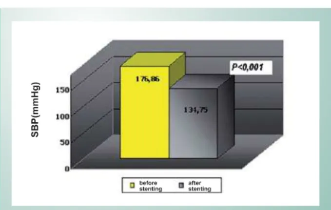
Although decrease in diastolic (DBP) and systolic (SBP) blood pressures was statistically significant (p<0.001), when SBP was analyzed separately from DBP, a greater impact of this reduction was seen with SBP.
The overall results of the main measurements of BP are presented in Table 1 and Figures 1 and 2.
Diastolic blood pressure (DBP) presented a mean reduction by approximately 15 mmHg (p<0.001) (Figure 1).
According to evaluation of systolic blood pressure values, there was a mean decrease of SBP by 42.11 mmHg after intervention (SD ± 37.85); this reduction was statistically significant (p<0.001) (Figure 2).
Renal function - Following the intervention, serum
creatinine was evaluated on the next day and months after the procedure; the statistical analysis compared the late results with the baseline results.
The values found both in the pre-intervention and late evaluations are listed in Table 2.
The mean value of creatinine before the intervention compared with the mean value after the procedure
demonstrated a reduction greater than 17%, which was significant in terms of clinical improvement.
The (mean) decrease in creatinine levels was from 2.33 mg/ dL to 1.94 mg/dL (p<0.001) (Mann-Whitney test) (Figure 3).
SBP (before)*
SBP (after)*
DBP (before)*
DBP (after)*
Mean 176.86 134.75 97.95 83.10
Standard
deviation 30.34 28.13 16.57 8.25
Minimum value 124 100 80 70
Maximum value 248 180 170 100
N 44 44 39 39
SBP - systolic blood pressure; DBP - diastolic blood pressure; * BP measurement unit: mmHg.
Table 1 - Blood pressure variables measured before and after intervention in patients followed up for arterial hypertension at
outpatient clinics.
Fig. 1 -PAd média antes e após a intervenção. 98
83
m
m
H
g
Impact of renal stenting on diastolic BP – mean
before stenting after stenting
p<0,0001
Fig. 2 -Mean SBP (mmHg) before and after intervention.
SB
P(m
m
H
g
)
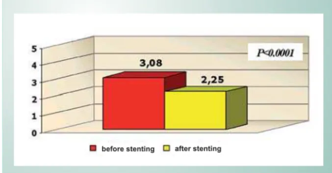
before
Number of drugs - According to the number of drugs taken before and after the procedure, there was a significant reduction by one drug in average (p<0.0001). This information is shown in Figure 4 (above) and in Table 3.
late follow-up, the patient evolved with worsening of renal function (creatinine reached 9.0 mg/dL) and hemodialysis was indicated.
There was no severe complication associated with the procedure. No patient presented cardiovascular events during the study follow-up.
Discussion
All 46 patients evaluated in this study underwent renal artery stenting. All presented significant atherosclerotic OHVLRQVLQUHQDODUWHULHVRFFOXVLRQZLWKKHPRG\QDPLF UHSHUFXVVLRQHYLGHQFHGE\DJUDGLHQWRIEORRGSUHVVXUH mmHg in the lesion). Patients were referred to renal artery angioplasty with placement of a stent according to the clinical history and physical examination analyzed in conjunction with stenosis of renal artery suggested by the renal US-Doppler and with the results of renal arteriography, similarly to procedures recommended and performed in other previous studies14-17.
In the late follow-up, the patients were evaluated both clinically (physical examination and laboratory tests) and by renal US to investigate restenosis of renal artery. Renal US-Doppler was used since it is sensitive to detect post-stenting renal artery stenosis18,19. The literature reports post-stenting renal artery stenosis rates of 15%20. In the sample studied, the patients did not present restenosis in the follow-up period and there was only one case of technical failure (2%) characterized by dissection of the renal artery during the procedure. In this patient, the renal artery originated from the aorta and formed an acute angle that hindered stent access. Later on, the patient evolved with a worsening of the renal function (creatinine reached 9.0 mg/dL) and hemodialysis was indicated. Despite the small number of patients involved in this study, the sample tried to select the cases with a clear clinical indication for percutaneous renal revascularization, either to control systemic hypertension or to preserve or restore renal function3,12,20,21. In fact, several studies have evaluated the renal function after renal angioplasty with stent placement10,11,16-18,20-23.
A study carried out by Jensen24 evaluated 107 patients with ARAS in a one-year follow-up after stenting in renal artery and found that this group showed decreased BP and increased glomerular filtration rate. Watson25 also noticed the repercussion of stent angioplasty in the renal function of 33 patients with azotemia for 20 months (mean follow-up) and reported that the deteriorating renal function had stabilized or improved after the procedure.
Creatinine (before)
Creatinine (after)
Mean 2.33 1.94
Standard deviation 1.27 1.43
Minimum value 1.0 1.0
Maximum value 6.1 9.0*
N 39 39
*only one patient presented this increase (technical failure); the second highest creatinine level was 4.4 mg/dl.
Table 2 - Renal function assessed per serum creatinine level (mg/ dl) measured before and after intervention in patients followed up
at outpatient clinics
Fig. 3 -Evaluation of the impact of stenting on renal function Creatinine (mean)
before after
Drugs (before) Drugs (after)
Mean 3.075 2.25
Standard
deviation 1.28 1.08
Minimum value 0 0
Maximum value 6 4
N 40 40
Table 3 - Evaluation of the number of drugs taken before and after intervention
Fig. 4 -Evaluation of the mean number of drugs taken before and after intervention.
before stenting after stenting
The number of daily doses of each drug were not evaluated.
In the current study about renal function, according to serum creatinine measured before the procedure and compared with the last measurement, we were able to notice a significant reduction in its levels. The detailed results related with the repercussion on the renal function and blood pressure (systolic and diastolic) were exposed in the previous section. The data found in this study show that stent placement in the treatment of atherosclerotic renal artery stenosis is an effective and safe therapy, and it should be appropriately indicated for a group of patients who can benefit from renal control and
preservation, as well as from blood pressure control.
Conclusions
The use of a stent in atherosclerotic renal artery stenosis was efficient and effective to restore and/or preserve renal function. The use of a stent in ARAS was effective to attain the control of blood pressure (reduction in the number of antihypertensive drugs). Treatment of the atherosclerotic renal artery lesion proved to be a safe and effective method, with low mortality rates during hospitalization and follow-up periods.
References
1. Uzu T, Inoue T, Fujii T, Nakamura S, Inenaga T, Yutani C, et al. Prevalence and predictors or renal artery stenosis in patients with myocardial infarction. Am J Kidney Dis. 1997; 29: 733-8.
2. Maiolloux LU, Napolitano B, Bellucci AG, Vernace M, Wilkes BM, Mossey RT. Renal vascular disease causing end-stage renal disease, incidence, clinical correlates, and outcomes: A 20-year clinical experience. Am J Kidney Dis. 1994; 24: 622-9.
3. Pohl MA, Kaplan N. Renal artery stenosis, renal vascular hypertension, and ischemic nephropathy. In: Schrier RW, Gottschalk CW (eds). Diseases of the kidney and urinary tract. 7th ed. Philadelphia: Lippincott Williams & Wilkins; 2001. p. 1399-457.
4. Detection, evaluation, and treatment of renovascular hypertension. Final report. Working Group on Renovascular Hypertension. Arch Intern Med. 1987;147: 820-9.
5. Hansen KJ, Edwards MS, Craven, TE, Cherr GS, Jackson AS, Appel RG, et al. Prevalence of renovascular disease in the elderly: a population-based study. J Vasc Surg. 2002; 36: 443-51.
6. Alcazar JM, Rodicio, JL. Ischemic nephropathy: clinical characteristics and treatment. Am J Kidney Dis. 2000; 36: 88-93.
7. Baboolal, K, Evans, C, Moore, RH. Incidence of end-stage renal disease in medically treated patients with severe bilateral atherosclerotic renovascular disease. Am J Kidney Dis. 1998; 31: 971-7.
8. Caps MT, Zierler RE, Polissar NL, Bergelin RO, Beach KW, Cantwell-Gab K, et al. Risk of atrophy in kidneys with atherosclerotic renal artery stenosis. Kidney Int. 1998; 53: 735-42.
9. Van Jaarsveld BC, Krijnen P, Pieterman H, Derkx FH, Deinum J, Postma CT, et al. The effect of ballon angioplasty on hypertension in atherosclerotic renal-artery stenosis. Dutch Renal Artery Stenosis Intervention Cooperative Study Group. N Engl J Med. 2000; 342: 1007-14.
10. Textor SC, Wilcox CS. Renal artery stenosis: A commom, treatable cause or renal failure? Annu Rev Med. 2001; 52: 421-42.
11. Textor SC. Revascularization in atherosclerotic renal artery disease. Kidney Int. 1998; 53: 799-811.
12. IV Diretrizes Brasileiras de Hipertensão Arterial. Sociedade Brasileira de Cardiologia, Sociedade Brasileira de Nefrologia e Sociedade Brasileira de Hipertensão. Arq Bras Cardiol. 2004; 82 (supl. 4): 1.
13. Cooper CJ, Murphy TP, Matsumoto A, Steffes M, Cohen DJ, Jaff M, et al. Stent revascularization for the prevention of cardiovascular and renal events among patients with renal artery stenosis and systolic hypertension: rationale and design of the CORAL trial. Am Heart J. 2006; 152: 59-66.
14. Chábová V, Schrirger A, Stanson AW, Mckusick MA, Textor SC.Outcomes of atherosclerotic renal artery stenosis managed without revascularization. Mayo Clin Proc. 2000; 75: 437-44.
15. Campo A, Boero R, Stratta P, Quarello F. Selective stenting and the course of atherosclerotic renovascular nephropathy. J Nephrol. 2002; 15: 525-9.
16. Tuttle KR, Raabe RD. Endovascular stents for renal artery revascularization. Curr Opin Nephrol Hypertens. 1998; 7: 695-701.
17. Leertouwer TC, Derkx FHM, Pattynama PM, Deinum J, van Dijk LC. Functional effects of renal artery stent placement on treated and contralateral kidneys. Kidney Int. 2002; 62: 574-9.
18. Ramos F, Kotliar C, Alvarez D, Baglivo H, Rafaelle P, Londero H, et al. Renal function and outcome of PTRA and stenting for atherosclerotic renal artery stenosis. Kidney Int. 2003; 63: 276-82.
19. Bakker J, Beutler JJ, Elgersma OE, de Lange EE, de Kort GA, Beek FJ, et al. Duplex ultrasonography in assessing restenosis of renal artery stents. Cardiovasc Intervent Radiol. 1999; 22: 475-80.
20. van de Ven PJ, Beutler JJ, Kaatee R, Beek FJ, Mali WP, Geysker GG, et al. Transluminal vascular stent for ostial atherosclerotic renal artery stenosis. Lancet. 1995; 346: 672-4.
21. Isles CG, Robertson S, Hill D. Management of renovascular disease: a review of renal artery stenting in tem studies. QJM. 1999; 92: 159-67.
22. Rees CR. Stents for atherosclerotic renovascular disease. J Vasc Interv Radiol. 1999; 10: 689-705.
23. Bloch MJ, Pickering TG: Renal vascular disease: medical management, angioplasty and stenting. Semin Nephrol. 2000; 20: 474-88.
24. Jensen G, Zachrisson BF, Delin K, Volkmann R, Aurell M. Treatment of renovascular hypertension: one year results of renal angioplasty. Kidney Int. 1995; 48: 1936-45.