Original Article
Prevalence of and Risk Factors for Combined Coronary Artery
Disease and Aortic Aneurysm
Carlos Romério Costa Ferro, Dinaldo Cavalcanti de Oliveira, Fábio de Freitas Guimarães Guerra, Alexandre Jorge
de Lucena, Fabiana Piech Nunes, Sergio Tranchesi Ortiz, Enilton Sergio Tabosa Egito, Luis Carlos Bento de Sousa,
Adib Domingos Jatene, Leopoldo Soares Piegas
Hospital do Coração and Associação do Sanatório Sírio - São Paulo, SP, Brazil
Objective: To evaluate coronary artery disease (CAD) prevalence in patients with aortic aneurysm, as well as differences related to aneurysm topographies. To describe the primary risk factors for CAD related to this association and their occasional differences according to AA topographies.
Methods: This was an open, prospective, nonrandomized study that evaluated 95 patients (62 men and 33 women, mean age 63 ± 11.8). All patients, asymptomatic for CAD, had undergone aortic CT and coronary angiography. According to the AA topography, they were classified into three groups: 1) Patients with thoracic aortic aneurysm (TAA); 2) thoracoabdominal aortic aneurysm (TAAA); and 3) abdominal aortic aneurysm (AAA). A database was created to store information from clinical data and complementary examinations. Statistical analysis was performed using the Student’s t test or analysis of variance (ANOVA) for continuous variables and chi-square test for categorical variables. P values < 0.05 were considered statistically significant.
Results: CAD prevalence was 63.1%, and AAA was more prevalent than TAA and TAAA (76% vs. 70% vs.30%, p = 0.001). The comparative analysis of CAD risk factors based on the aortic aneurysm topography revealed that smoking and dyslipidemia were more prevalent among AAA patients (74.5% vs. 42.3% vs.60%, p = 0.01 and (54.2% vs. 19.9% vs. 60%, p = 0.007, UHVSHFWLYHO\$VIRUFRURQDU\OHVLRQVHYHULW\LQWKHSRSXODWLRQRI$$SDWLHQWVKDGDWOHDVWRQHOHVLRQDQG )LIWHHQSDWLHQWVKDGVLQJOHYHVVHOGLVHDVHKDGWZRYHVVHOGLVHDVHDQGKDGWKUHH vessel disease.
Conclusion: Asymptomatic CAD is highly prevalent in AA patients, particularly among those with AAA. Study results suggest the need for diagnostic stratification for CAD in patients with AA, especially those with AAA.
Key words: Coronary artery disease, aortic aneurysm, risk factors.
Mailing Address: Dinaldo Cavalcanti de Oliveira •
Rua Desembargador Eliseu Guilherme, 123 – 04040-030 – São Paulo, SP – Brasil E-mail: [email protected]
Manuscript received November 24, 2005; revised manuscript received January 17, 2006; accepted February 04, 2006.
Aortic aneurysm (AA) is defined as a localized dilation greater than 50% the expected normal luminal diameter of that given aortic segmenta. Its incidence has been rising in the last decades, owing both to the increase in the mean age of the population and improvement in diagnostic methods.
The incidence of thoracic AA (TAA) is estimated at six cases per 100,000 patients/year, while Abdominal AA (AAA) is estimated at 25 per 100,000 patients/yearb,c. Around 10% of the patients diagnosed with AA have multiple aneurysms and in different segments of the aorta. Approximately 20 to 25% of the subjects with TAA have concomitant AAA. Abdominal aortic aneurysms are more common than thoracic aortic aneurysms and thoracoabdominal aortic aneurysmsd,e.
According to population-based studies using ultrasonography as their screening tool, AAA prevalence ranged from 4% to 9% in men and 1% in women6. The major risk factors for AAA include: age, male gender, smoking, systemic arterial hypertension, family history of AAA, and atherosclerotic disease7,8,9,10.
The presence of common risk factors and pathophysiological substrates has established a strongly correlation between
AAA and coronary artery disease (CAD)11. Some series have reported a CAD prevalence ranging from 40% to 60% in AAA patients12,13.Hertzer et al noted that acute myocardial infarction accounted for half of the deaths (intra- and postoperative) in the surgical correction of AAA12.
The aim of this study was to evaluate the relationship between AA and CAD, as well as the prevalence of and risk factors for this association.
Methods
From July 2003 to July 2005 we conducted an open, prospective, nonrandomized trial involving 95 patients (66 men and 33 women, mean age 63 ± 11.8). Inclusion criteria were the following: diagnosis of AA confirmed by computed tomography and coronary angiography examination, but presenting neither CAD symptoms nor known CAD. Among the excluded patients were those younger than 18 or 80 years or older, those with traumatic, dissected or ruptured aneurysms, and those who refused to participate in the study. The
Original Article
Ferro et alPREVALENCE OF AND RISK FACTORS FOR COMBINED CORONARY ARTERY DISEASE AND AORTIC ANEURYSM
Arq Bras Cardiol 2007; 88(1) : 37-40
following risk factors were analyzed: age, gender, systemic arterial hypertension, diabetes mellitus, dyslipidemia, and smoking.
The presence of AA was defined as an aortic segment exceeding 1.5 times the expected diameter of that area. Based on angiographic visual analysis, CAD was defined DVWKHSUHVHQFHRIDFRURQDU\REVWUXFWLRQDQGZDV classified as mild (30% – 49%), moderate (50% – 69%), and VHYHUH
Patients included were divided into three groups according to AA topography: 1) TAA, aneurysm located in the ascending aorta, aortic arch or descending aorta above the diaphragm; 2) TAAA, aneurysm involving most part of the descending aorta and upper portion of the abdominal aorta (Crawford I), the entire descending and abdominal aorta (Crawford II) or the lower portion of the thoracic and abdominal aorta (Crawford III), and 3) AAA, aneurysm located in the abdominal aorta).
A database was created (SPSS) containing patients’ information from clinical data and supplementary examinations. Results are expressed as mean ± standard deviation and percentages. Statistical analysis was performed using the Student’s t test or analysis of variance (ANOVA) for continuous variables and chi-square test for categorical variables. P values < 0.05 were considered statistically significant.
Results
Of the 95 patients studied, 26 had TAA (21 in the ascending aorta, 1 in the aortic arch and 4 in the descending aorta), 10 had one TAAA (5 Crawford I, 1 Crawford II and 4 Crawford
III), and 59 had one AAA (56 infrarenal), (Fig. 1).
AA patients age 50 or older (n = 83 patients) showed higher prevalence of CAD when compared with those younger than 50 (n = 12 patients) [58 patients (69%) vs. 2 (16.6%), p = 0.01).
CAD prevalence was 63.1%, and was higher in the AAA group than in the TAA and TAAA groups [45 patients (76%)
vs. 7 (70%) vs.8 (30%), p = 0.001; Fig. 2].
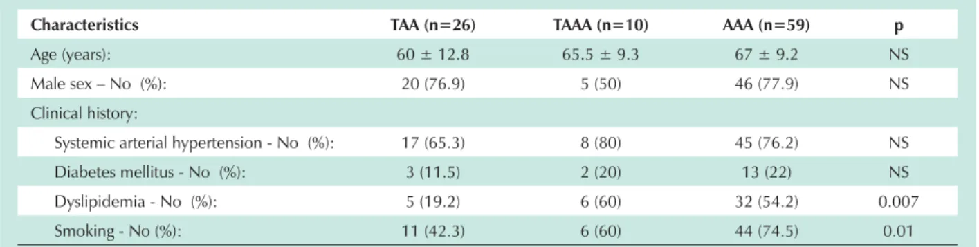
Risk factors distribution is shown in Table 1. A comparative analysis based on AA topography revealed a greater number of smokers and dyslipidemic patients in the AAA group (Tab. 2). With respect to the number and importance of the coronary lesions, it was found that: 12 (20%) patients had at least one OHVLRQ DQG )LIWHHQ SDWLHQWV DQG )LIWHHQ SDWLHQWVDQG )LIWHHQ SDWLHQWV )LIWHHQ SDWLHQWV 50%. Fifteen (25%) patients had single-vessel disease, 11 (18%) had two-vessel disease, and 34 (57%) had three-vessel disease (Fig. 3).
In the AAA group, 18 patients (40%) had at least one lesion DQGKDGRQHOHVLRQ1LQHKDG single-vessel disease, 14 (31.1%) had two-vessel disease, and 22 (48.8%) had three-vessel disease.
CAD patients were older (66.7 ± 8.1 vs. 61.9 ± 12.7, p = 0.02), dyslipidemic (51.6% vs. 28.5 %, p = 0.04), hypertensive (80%vs. 60%, p = 0.03) and smokers (75% vs. 45.7%, p = 0.06) (Fig. 4).
Figure 5 depicts two patients of this series with TAA and AAA and their respective computed tomography and coronary angiography.
Risk factors for CAD AA (n=95)
Age (years): 66 ± 11.8
Males No (%) 72 (75.7)
Clinical history:
Systemic arterial hypertension - No (%): 70 (73.6)
Diabetes mellitus - No (%): 18 (18.9)
Dyslipidemia - No (%): 41 (43.1)
Smoking - No (%): 61 (64.2)
AI/IAM sem supra ST31 (44,3%)
Table 1 - Risk factors in the population studied
Fig. 1 -AA distribution according to the aortic segment involved.
AA topography
thoracoabdominal aneurysm
n = 95 patients
TAA
Fig. 2 -Prevalence of CAD in patients according to AA topography.
Thoracoabdominal TAA
Original Article
Ferro et al PREVALENCE OF AND RISK FACTORS FOR COMBINED CORONARY ARTERY DISEASE AND AORTIC ANEURYSMArq Bras Cardiol 2007; 88(1) : 37-40
Discussion
When the AA etiology is related to atherosclerotic disease and its risk factors, the latter become manifestations of the vascular disease, which affects both medium and large arteries, making patients more likely to develop cerebrovascular, renal, and coronary diseases, all of which hike mortality rates.
The high CAD prevalence found in this study is similar to that of other series reported in the literature, such as that of Hetzer et al, in which 1000 patients with AA underwent coronary angiography prior to surgical repair of the aneurysm, and 60% of them were found to have CAD14.
In our series, 75% of the CAD patients had abdominal aortic aneurysm, more precisely infrarenal aneurysm,
Characteristics TAA (n=26) TAAA (n=10) AAA (n=59) p
Age (years): 60 ± 12.8 65.5 ± 9.3 67 ± 9.2 NS
Male sex – No (%): 20 (76.9) 5 (50) 46 (77.9) NS
Clinical history:
Systemic arterial hypertension - No (%): 17 (65.3) 8 (80) 45 (76.2) NS
Diabetes mellitus - No (%): 3 (11.5) 2 (20) 13 (22) NS
Dyslipidemia - No (%): 5 (19.2) 6 (60) 32 (54.2) 0.007
Smoking - No (%): 11 (42.3) 6 (60) 44 (74.5) 0.01
Table 2 - Comparative analysis of risk factors for CAD between groups
Fig. 5 -A: Tomographic image of ascending aorta aneurysm and coronary angiography demonstrating moderate lesion in the middle third and severe lesion in the distal third of the right coronary artery, and mild lesion in the proximal third and severe lesion in the middle third of the anterior descending artery. B: Tomographic image of abdominal aorta aneurysm and coronary angiography demonstrating severe lesion in the middle and distal thirds of the right coronary artery, and severe lesion in the anterior descending artery after the first diagonal branch presenting severe ostial lesion.
MAC, 59 years, TAA. RAS, 65 years, AAA.
followed by 13.3% of thoracic aortic aneurysm and 11.6% of thoracoabdominal aortic aneurysm, suggesting a greater association between AAA and CAD.
This association is determined by common etiology and pathophysiological substrates. It is known that oxygen and nutrient supply to the outer half of the aortic wall, including part of the media layer, is provided by the vasa vasorum. However, in humans, the media layer of the infrarenal aorta does not have vasa vasorum and, therefore, the inner part of the media should receive nutrients and oxygen through the blood diffusion mechanism of the aortic lumen15.
Atherosclerosis causes a thickening of the intima, which impairs the nutrient diffusion to the media layer. Thus, hypoxemia occurs, predisposing to the development of ischemic lesions in the media and eventually causing the degeneration of this layer and its elastic components. This degeneration weakens the aortic wall and makes it prone to aneurysmsf.
Ascending thoracic aortic aneurysms result from cystic medial degeneration (CMD), leading to the weakening of the aortic wall16. Cystic medial degeneration is related to age and systemic arterial hypertension (SAH), known risk factors for CAD. Fig. 3 -Severity and size of CAD in patients with AA and AAA.
One-vessel disease
Two-vessel disease
Three-vessel disease Aortic aneurysms Abdominal AA
Fig. 4 -Comparison of risk factors among patients with and without CAD.
no CAD
Risk factors
Male sex
SAH
DM
Dyslipidemia
Dmoking
CAD
Original Article
Ferro et alPREVALENCE OF AND RISK FACTORS FOR COMBINED CORONARY ARTERY DISEASE AND AORTIC ANEURYSM
Arq Bras Cardiol 2007; 88(1) : 37-40
Descending thoracic aortic aneurysms are associated with atherosclerosis and its risk factors17. Aneurysms that extend to the thoracic and abdominal aorta have etiopathogenic characteristics of both segments.
In this study, AAA patients tended to be older, dyslipidemic and smokers, compared with the TAA and TAAA, thus presenting a clinical profile of higher risk of atherosclerotic disease and its manifestations.
When patients with and without CAD were compared, risk factors were found to be more frequent in the coronary disease group; yet only SAH and dyslipidemia were statistically significant. A trend towards statistical significance was noted for smoking.
Both the prevalence and quantity of risk factors for CAD in the population studied were high, and AAA patients showed more risk factors for CAD, as well as a higher prevalence of this disease. This might be explained by the similar etiology and pathophysiology of these diseases.
The five-year survival after AAA surgical repair is around 70%, and CAD accounts for about one-third of the deaths18.
Non-invasive tests, considered screening tools for myocardial ischemia, have some limitations in this population. Some authors recommend routine coronary angiography to AAA patients scheduled for elective surgery or stenting. There is a direct relationship between aortic aneurysm size and CAD severity19,20.
7KH KLJK LQFLGHQFH RI OHVLRQV D DQG WKH number of patients with two-or three-vessel disease in our population suggest that a careful preoperative evaluation is warranted regarding CAD risk in patients with aortic aneurysm, especially those with abdominal aortic aneurysm. Patients with risk factors should be considered for invasive stratification of CAD.
Conclusions
CAD prevalence in patients with aortic aneurysm was high, especially those with abdominal aortic aneurysms. In our opinion, in patients with aneurysms involving the abdominal aorta, preinterventional coronary angiography is highly recommended.
References
1. Johnston KW, Rutherford RB, Wilson MD, et al. Suggested standards for reporting on arterial aneurysms. Subcommittee on Reporting Standards for Arterial Aneurysms, Ad Hoc Committee on Reporting Standards, Society for Vascular Surgery and North American Chapter, International Society for Cardiovascular Surgery. J Vasc Surg 1991; 13:452-260.
2. Bickerstaff LK, Pairolero PC, Hollier LH, et al. Thoracic aortic aneurysms: A population-based study. Surgery 1982; 92:1103-08.
3. Eickhoff JH. Incidence of diagnosis, operation and death from abdominal aortic aneurysms in Danish hospitals: results from a nation-wide survey,
1977-1990. Eur J Surg 1993;159:619-23.
4. Crawford ES, Cohen ES. Aortic aneurysm: A multifocal disease. Arch Surg 1982; 117(11):1393-400.
5. Pressler V, McNamara JJ. Aneurysms of the thoracic aorta: Review of 260 cases. J Thorac Cardiovasc Surg 1985; 89(1):50-4.
6. Holmes DR, Liao S, Parks WC, Thompson RW. Medial neovascularization in abdominal aortic aneurysms: a histopathologic marker of aneurysm degeneration with pathophysiologic implications. J Vasc Surg 1995;21:761-71.