365
Acute pericarditis is a syndrome characterized by pericardial inflammation and clinical manifestations like thoracic pain, peri-cardial friction, and electrocardiographic and echocardiographic abnormalities 1. The clinical history frequently reveals thoracic pain and dyspnea 2. Pericardial friction, when present, is a pathog-nomonic signal of acute pericarditis. Fever, muscle pain, weakness, fatigue, and prostration may already be present 3.
All causes of acute pericarditis can evolve to pericardial effusion. Signs of pericardial effusion can range from minimal to no clinical symptoms to compression leading to symptoms of cardiac tampo-nade 1 (fig. 1).
Several diseases can evolve in the pericardium. The main causes of pericarditis are infections, myocardial infarction (MI), heart failure (HF), renal failure, cancer, and systemic and metabolic diseases 2.
The viral and idiopathic causes are often confused. The clinical findings do not always distinguish the viral and idiopathic forms, and, probably, many cases of idiopathic pericarditis are unrecog-nized viral infections. In general, it is not productive to try to isolate or identify the potential virus for this disease 4.
Acute idiopathic pericarditis, in general, is a self-limiting disease lasting 1 to 3 weeks with the potential for complications like myo-carditis, cardiac tamponade, and late constrictive pericarditis 5.
M ethods
From January 1999 to December 2001, 1.656 patients from a heart clinic sought treatment for suspected or diagnosed cardio-logic disease. In this period, 84 patients had clinically and echocar-diographically diagnosed pericarditis. Of this population, 61 (Group A) had known causes for the disease; on the other hand, 23 (Group B) individuals did not have an explanation for the disease and therefore were labeled as idiopathic. Because of this high incidence of idiopathic pericarditis compared with that in previous years, the authors reevaluated data on anamnesis, clinical exami-nations, and complementary examinations to clarify the causes.
Of these patients, known causes of pericarditis like infections, myocardial infarction, heart failure, cancer, renal failure, and other systemic and metabolic diseases were considered and investigated through clinical history and/or specific complementary exami-nations.
The effusion was identified by visualization of spaces around the heart free of echoes, which increased through the level of visceral and parietal pericardial separation, being restricted to the posterior wall where mild effusion was present and around the entire heart where the most significant effusion occurred 6.
Original Article
Pericardit is. Series of 84 Consecut ive Cases
M arco Tulio Zanet t ini, João Ot avio Zanet t ini, Jacira Pisani Zanet t ini
Caxias do Sul, RS - Brazil
Eletrocor Laboratório Cárdio Diagnóstico e Universidade de Caxias do Sul Correspondência: Marco Tulio Zanettini - Rua Bento Gonçalves, 2048 - 2º andar - Cep 95020-412 - Caxias do Sul, RS, Brazil
E-mail: [email protected] Received: 11/19/02
Accepted: 10/20/03
Objective
To identify differential clinical, laboratory, and echocardio-graphic characteristics in persons with diagnosed idiopathic and secondary pericarditis.
M ethods
From January 1999 to December 2001, 84 patients with clinically and echocardiographically diagnosed pericarditis were identified in a heart clinic. These patients were analyzed according to age, sex, anthropometric measurements, body habitus, casual blood pressure (BP), signs and symptoms, morbid history, me-dicines and complications. The individuals were divided into 2 groups: group A comprised 61 patients with known causes of pericarditis and group B comprised 23 patients with idiopathic causes. The groups were compared with chi-square test. P≤ 0.05 was considered statistically significant.
Results
The population of these 2 groups was similar in age, sex, anthropometric measures, body habitus, and casual BP. In group B (idiopathic), 23 (100%) cases were diagnosed between April and August versus 24 (39.4%) in the same period for group A (P<0.01). Twenty-three (100%) group B patients received anti-influenza vaccine versus none in group A. Breathlessness (P=0.02) and swelling (P=0.01) were more frequent in group A, but fatigue was more common in group B (P=0.01). For treatment, non-steroidal anti-inflammatory drugs (NSAID) were prescribed to 5 (8.2%) patients in group A and 19 (82.6%) in group B (P=0.01).
Conclusion
In this series, patients labeled as having idiopathic pericar-ditis who had previously taken the influenza vaccine had seasonal distribution, a lower prevalence of previous disease, less exuberant signs and symptoms, and clinical regression with NSAID use.
Key w ords
366
The statistical analysis was performed for age, sex, smoking, casual blood pressure based on the VI Report of the JNC classifi-cation 7, and body mass index was measured (BMI) in kg/m2. Normal BMI was considered to be from 18.5 to 24.9 kg/m2, overweight from 25.0 to 29.9 kg/m2, obesity level I from 30 to 34.9 kg/m2, obesity level II from 35.0 to 39.9 kg/m2, and morbid obesity above 40 kg/m2 8. Also considered were the month of the diagnosis, signs and symptoms, morbid past, medicines, previous vaccinations and complications of the disease. Specific viral sero-logic measures were not obtained; in our center, this diagnostic tool is not available.
The groups were compared with chi-square test, with P≤0.05 being considered significant.
Results
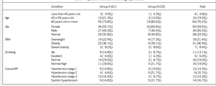
The population in the 2 groups was similar for age, sex, anthro-pometric measures, body habitus, and casual BP. Of the 84 individuals, 64 (76.2%) were 60 years old or more, 50 (59.5%) were women, 56 (66.7%) had BMI ≥ 25 kg/m2, 11 (13.1%) were smokers, and 47 (56.0%) had casual BP ≥ 140/90 mmHg (tab. I). The clinical manifestations occurred, predominantly, for up to 3 weeks in both groups [38 (62.3%) in group A versus 16 (69.5%) in group B]. Ten (16.4%) group A patients were asymptomatic compared with 2 (8.7%) in group B. In 23 (100%) group B patients, the diagnoses were made between April and August
compared with 24 (39.4%) at the same time in group A (P<0.01) (fig. 2) (tab. II).
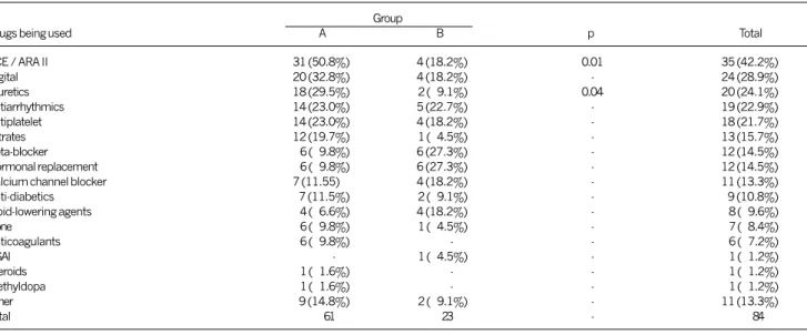
Twenty-three (100%) group B patients were vaccinated against the influenza virus previously and in the same year of the pericarditis versus none on group A. Dyspnea (P=0.02) and swelling (P=0.01) were more frequent in group A, while fatigue was more commonly reported in group B (P=0.01) (tab. III). More group A patients took diuretics (P=0.04) and angiotensin-converting enzyme inhi-bitors/angiotensin II receptor antagonists (ACE/ARA II) (P=0.01) compared with group B patients (tab IV). Analysis of comorbid conditions showed that ischemic cardiopathy was more frequent in group A (19 cases) compared with only 2 cases in group B (P=0.03) (tab. V). Heart failure was the main cause of pericarditis in group A, occurring in 27 (44.3%) patients versus none in group B (P=0.01) (tab. VI). With regard to therapeutics, non-steroidal anti-inflammatory drugs (NSAID) were prescribed to 5 (8.2%) patients in group A and 19 (82.6%) in group B (P=0.01) (tab. VII). On follow-up, 17 (73.9%) patients in group B did not have any complications, but 3 (13.0%) developed heart failure (P=0.05) and 3 (13.0%) respiratory infection. In group A, 31 (50.8%) did not have any complications; 20 (32.8%) evolved to HF. Of the other 14 (16.4%) patients in group A with complications, 5 (8.2%) required cardiac surgery.
Fig. 1 - A large pericardial effusion visualized with echocardiography.
Table I - Population characteristics for groups A and B
Condition Group A (61) Group B (23) Total
Less than 40 years old 3 (04.9%) 1 (04.3%) 4 (04.8%) Age 40 a 59 years old 13 (21.3%) 3 (13.0%) 16 (19.0%) 60 years old or more 45 (73.8%) 19 (82.6%) 64 (76.2%)
Sex Female 34 (55.7%) 16 (69.6%) 50 (59.5%)
Male 27 (44.3%) 7 (30.4%) 34 (40.5%)
Normal 20 (32.8%) 8 (34.8%) 28 (33.3%)
BMI Overweight 14 (22.9%) 4 (17.3%) 18 (21.4%)
Obesity 22 (36.1%) 9 (39.1%) 31 (36.9%)
Severe obesity 5 (08.2%) 2 (08.6%) 7 (08.4%)
Smoking Yes 9 (14.8%) 2 (08.7%) 11 (13.1%)
Excellent 4 (06.6%) 1 (04.3%) 5 (06.0%) Normal 14 (23.0%) 2 (08.7%) 16 (19.0%) Normal High 11 (18.0%) 5 (21.7%) 16 (19.0%) Casual BP Hipertension stage 1 9 (14.8%) 3 (13.0%) 12 (14.3%) Hipertension stage 2 4 (06.6%) 5 (21.7%) 9 (10.7%) Hipertension stage 3 10 (16.4%) 2 (08.7%) 12 (14.3%) Systolic hypertension 9 (14.8%) 5 (21.7%) 14 (16.7%)
% 100
90
80 70
60 50 40 30
20 10 0
Group A Group B
42.6
8.7
29.5 91.3
27.9
0
January to April
May to August
September to December
Trimestrality (p < 0.01)
367
Table II - Classification by month of diagnosis
Group
Month Diagnosed A B Total
January 9 (14.8%) - 9 (10.7%) February 4 (06.6%) - 4 (04.8%) March 7 (11.5%) - 7 (08.3%) April 6 (09.8%) 2 (08.7%) 8 (09.5%) May 2 (03.3%) 11 (47.8%) 13 (15.5%) June 2 (03.3%) 6 (26.1%) 8 (09.5%) July 5 (08.2%) 2 (08.7%) 7 (08.3%) August 9 (14.8%) 2 (08.7%) 11 (13.1%) September 4 (06.6%) - 4 (04.8%) October 5 (08.2%) - 5 (06.0%) November 7 (11.5%) - 7 (08.3%) December 1 (01.6%) - 1 (01.2%)
Total 61 23 84
* p < 0.01. Comparison of groups A and B by pericarditis cases between April and August.
Table III - Group classification by signs and symptoms
Group
Signs and Symptoms A B p Total
Dyspnea 30 (49.2%) 5 (21.7%) 0.02 35 (42.2%)
Fatigue 12 (19.7%) 14 (60.9%) 0.01 26 (31.0%)
Precordial / retrosternal 17 (27.9%) 5 (21.7%) - 22 (26.2%)
Swelling 17 (27.9%) - 0.01 17 (20.2%)
Cough 11 (18.0%) 5 (21.7%) - 16 (19.0%)
Palpitations 11 (18.0%) 2 (08.7%) - 13 (15.5%)
Non symptomatic 11 (18.0%) 2 (08.7%) - 13 (15.5%)
Muscle pain 3 (04.9%) 4 (17.4%) - 7 (08.3%)
Fever 3 (04.9%) 2 (08.7%) - 5 (06.0%)
Pericardial friction 1 (01.6%) - - 1 (01.2%)
Syncope / pre-syncope 5 (08.2%) - - 5 (06.0%)
Total 61 23 - 84
Obs.: Multiple answer questions.
Discussion
The Coxsackie B virus, Echo type 8, mumps virus, influenza, mononucleosis virus, poliomyelitis virus, zoster, and the hepatitis vaccine are some of main causal agents of acute pericarditis 9.
Meester et al 10, Streifler et al 11, and Desson et al 12 reported, consecutively, 2, 1, and 1 cases of acute pericarditis after anti-influenza vaccine administration. In these cases, the diagnosis was confirmed by serologic, electrocardiographic, and echocar-diographic means.
Zanettini et al 13,14 reported on a series of cases of pericarditis after influenza vaccination at the XII and XIII Congress of Cardiology of the Rio Grande do Sul.
The incidence of influenza increases during the winter, leading to massive vaccination during the autumn and winter months. The immunity provided by vaccination varies from 60 to 90%, being lower in the elderly and persons with compromised immune systems 15.
During influenza infection, the incubation period depends on the viral dose and the immunology host stage 16. It is known that the target population in vaccination campaigns is composed, in large part, of the elderly and those with comorbidities.
Table IV - Group classification by drugs being used by patients in the groups before of the pericarditis diagnosis
Group
Drugs being used A B p Total
ACE / ARA II 31 (50.8%) 4 (18.2%) 0.01 35 (42.2%)
Digital 20 (32.8%) 4 (18.2%) - 24 (28.9%)
Diuretics 18 (29.5%) 2 (09.1%) 0.04 20 (24.1%)
Antiarrhythmics 14 (23.0%) 5 (22.7%) - 19 (22.9%)
Antiplatelet 14 (23.0%) 4 (18.2%) - 18 (21.7%)
Nitrates 12 (19.7%) 1 (04.5%) - 13 (15.7%)
Beta-blocker 6 (09.8%) 6 (27.3%) - 12 (14.5%)
Hormonal replacement 6 (09.8%) 6 (27.3%) - 12 (14.5%) Calcium channel blocker 7 (11.55) 4 (18.2%) - 11 (13.3%)
Anti-diabetics 7 (11.5%) 2 (09.1%) - 9 (10.8%)
Lipid-lowering agents 4 (06.6%) 4 (18.2%) - 8 (09.6%)
None 6 (09.8%) 1 (04.5%) - 7 (08.4%)
Anticoagulants 6 (09.8%) - - 6 (07.2%)
NSAI - 1 (04.5%) - 1 (01.2%)
Steroids 1 (01.6%) - - 1 (01.2%)
Methyldopa 1 (01.6%) - - 1 (01.2%)
Other 9 (14.8%) 2 (09.1%) - 11 (13.3%)
Total 61 23 - 84
Obs.: Multiple answer questions.
368
Table V - Group classification by morbid past
Group
Morbid past A B p Total
Mitral valvopathy 25 (41.0%) 13 (56.5%) - 38 (45.2%) Hypertensive cardiopathy 19 (31.1%) 8 (34.8%) - 27 (32.1%)
Other valvopathy 20 (32.8%) 6 (26.1%) - 26 (31.0%)
Ischemic cardiopathy 19 (31.1%) 2 (08.7%) 0.03 21 (25.0%)
Arrhythmia 13 (21.3%) 7 (30.4%) - 20 (23.8%)
Hypercholesterolemia 9 (14.8%) 6 (26.1%) - 15 (17.9%)
Cancer 11 (18.0%) - - 11 (13.1%)
Hypertension 8 (13.1%) 3 (13.0%) - 11 (13.1%)
Hypothyroidism 8 (13.1%) 1 (04.3%) - 9 (10.7%)
Heart failure 7 (11.5%) - - 7 (08.3%)
Cardiomyopathy 7 (11.5%) - - 7 (08.3%)
Chronic obstructive pulmonary disease 4 (06.6%) 3 (13.0%) - 7 (08.3%) Myocardial infarction 5 (08.2%) 1 (04.3%) - 6 (07.1%)
Diabetes 5 (08.2%) - - 5 (06.0%)
None 2 (03.3%) 2 (08.7%) - 4 (04.8%)
Myocardial revascularization 2 (03.3%) - - 2 (02.4%)
Renal failure 1 (01.6%) - - 1 (01.2%)
Other heart surgeries 1 (01.6%) - - 1 (01.2%)
Marfan´s syndrome 1 (01.6%) - - 1 (01.2%)
Congenital cardiopathy 1 (01.6%) - - 1 (01.2%)
Other 5 (08.2%) 8 (34.8%) - 13 (15.5%)
Total 61 23 - 84
Obs.: Multiple answer questions.
Table VI - Pericardial effusion causes
Causes Group A Group B p
Heart failure 27 (44.3%) - 0.01
Cancer 11 (18.0%) -
-After cardiac surgery 8 (13.1%) -
-Ischemic cardiopathy 7 (11.5%) -
-Hypothyroidism 7 (11.5%) -
-Valvopathy 5 (08.2%) -
-Cardiomyopathy 4 (06.6%) -
-Hypertensive cardiopathy 4 (06.6%) -
-Viral 2 (03.3%) -
-Collagenous disease 1 (01.6%) -
-Idiopathic 1 (01.6%) 23 (100%) 0.01
After myocardial infarction 1 (01.6%) -
-Total 61 23 84
Obs.: Multiple answer questions.
Table VII - Group classification by drugs prescribed before the diagnosis of pericarditis
Group
Prescribed Drugs A B p Total
Piroxicam SL 5 (08.2%) 19 (90.5%) 0.01 24 (29.3%)
Other NSAI 1 (01.6%) - - 1 (01.2%)
Steroids 2 (03.3%) 1 (04.8%) - 3 (03.7%)
ACE / ARA II 12 (19.7%) 5 (23.8%) - 17 (20.7%)
Antiarrhythmics 11 (18.0%) 5 (23.8%) - 16 (19.5%)
Digoxin 13 (21.3%) 1 (04.8%) - 14 (17.1%)
Diuretics 11 (18.0%) 1 (04.8%) - 12 (14.6%)
Antibiotics 5 (08.2%) 3 (14.3%) - 8 (09.8%)
Nitrates 8 (13.1%) - - 8 (09.8%)
Antiplatelet 7 (11.5%) - - 7 (08.5%)
Beta-blocker 6 (09.8%) - - 6 (07.3%)
Calcium channel blockers 3 (04.9%) - - 3 (03.7%) Hormonal replacement therapy 3 (04.9%) - - 3 (03.7%)
Lipid-lowering agents 1 (01.6%) - - 1 (01.2%)
Anti-diabetics 1 (01.6%) - - 1 (01.2%)
Other 1 (01.6%) - - 1 (01.2%)
None 13 (21.3%) - - 13 (15.9%)
Total 61 23 - 84
369
described by the laboratories include pain, rush, local swelling, low fever, malaise, muscle pain, anaphylactic and hypersensitivity reactions such as asthma and Guillain Barré syndrome 16.
The literature teaches that idiopathic pericarditis treatment is determined by clinical manifestations. When pericardial pain is present, NSAID should be used. When evaluation of a large effusion or cardiac tamponade is performed, pericardiocentesis is indicated associated or not with pericardioscopy with an epicardial biopsy. Specific antivirals are indicated for treating viral or idiopathic peri-carditis in persons with compromised immunity 17.
Analysis of the group B population showed that the patients were mainly elderly, with controlled heart disease in which the basic disease was not related to pericarditis. In group B, 13 (56.5%) patients had mitral valvopathy, 1 (4.3%) previous myo-cardial infarction, and 1 (4.3%) hypothyroidism; all, however, were under specific treatment and their base disease was com-pensated (tab V).
The clinical findings, in group B, were predominantly mild, benign, and prodromics of viral disease; three individuals, however, required hospitalization due to heart failure.
All group B individuals received anti-influenza vaccine in the same year and before the onset of pericarditis; all either took the
1. Fragata Filho A. Pericardites. In: Timerman A, César LAM. Manual de Cardiologia. 1a ed. São Paulo: Atheneu, 2000: 242-51.
2. Shabetai R. Doenças do Pericárdio. In: Bennett JC, Plum F Cecil. Tratado de Me-dicina Interna. 20a, Rio de Janeiro: Guanabara Koogan, 1997: 372-8.
3. Lorell BH. Pericardial Disease. In: Braunwald E. Heart Disease: A Textbook of Car-diovascular Medicine. 5a, Philadelphia: W.B Saunders Company, 1997: 1478-534.
4. Sagrista-Sauleda J, Almenar Bonet L, Angel Ferrer J et al. The clinical practice of the Sociedad Española de Cardiología on pericardial pathology. Rev Esp Cardiol 2000; 53: 394-412.
5. Muir P, Nicholson F, Tilzey AJ et al. Cronich relapsing pericarditis and delated cardio-myopathy: Serologic evidence of persistent enterovirus infection. Lancet 1989; 1: 804. 6. Andrade JL, Campos Filho O. Ecocardiografia nas Pericardiopatias e Cardiomio-patias. In: Timerman A, César LAM. Manual de Cardiologia. 1a, São Paulo:
Athe-neu, 2000: 339-46.
7. The Sixth Report of the Joint National Committee on Prevention, Detection, Eva-luation, and Treatment of High Blood Pressure - National Institutes of Health, NIH. Publication Nº. 98-4080, November 1997.
8. National Heart, Lung and Blood Institute / National Institutes of Diabetes and Di-gestive and Kidney Disease. Clinical guidelines on the identification, evaluation and treatment of overweight and obsesity in adults: the evidence report. Bethesda: National Institutes of Health, 1988: 1-228.
References
9. Bensaid J, Denis F. Benign acute pericarditis after vaccination against hepatitis B. Press Med 1993; 22: 269.
10. Meester A, Luwaert R, Chaudron JM. Symptomatic pericarditis after influenza vac-cination: report of two cases. Chest 2000; 117: 1803-5.
11. Streifler J, Rosenfeld J, Dux S, Garty M. Recurrent pericarditis: a rare complica-tion of influenza vaccinacomplica-tion. BMJ 1981; 283: 526-7.
12. Desson E, Leprèvast M, Vabret F, Davy A. Péricardite aiguë bénigne après vaccina-tion antigrippale. Press Med 1997; 26: 415.
13. Zanettini JO, Zanettini MT, Zanettini JP. Pericardite pós vacina antiinfluenza: série de casos. In: Anais do XII Congresso de Cardiologia do Rio Grande do Sul. Gra-mado: Sociedade de Cardiologia do Rio Grande do Sul, 2001: 25.
14. Zanettini JO, Zanettini JP, Zanettini MT. Pericardite: série de 84 casos consecuti-vos. In: Anais do XIII Congresso de Cardiologia do Rio Grande do Sul. Gramado: Sociedade de Cardiologia do Rio Grande do Sul, 2002: 46.
15. Melnick JL, Alberg EA. Ortomixovírus (Vírus da Influenza). In: Jawetz E. Microbio-logia Médica. 20a, Rio de Janeiro: Guanabara Koogan, 1998: 356-64.
16. Nichol KL. Side effects associated with influenza vaccination in healthy working adults. Arch Intern Med, 22 July 1996; 156.
17. Maisch B. Treatment of idiopathic pericarditis: viral versus autoreactive disease. In: Seferovic PM, Spodick DH, Maisch B. Pericardiology: Editora Science. Belgra-do 2000: 373-80.
initiative to be vaccinated or the vaccination was indicated by external sources. The vaccines were provided by renowned labo-ratories.
The diagnosis of pericarditis in group B was seasonal and coincided with the period for influenza vaccination.
Concluding, the patients in this study were predominantly elderly women with body mass indexes and blood pressures above normal. The individuals said to have idiopathic pericarditis had compensated heart failure, had previously received an influenza vaccination, and were diagnosed seasonally. These patients had prodromic viral signs and symptoms and experienced clinical regres-sion after taking NSAIDs. They also demonstrated the possibility of developing heart failure as a complication. The persons with secondary pericarditis were diagnosed during all months of the year, had known causes of pericarditis, signs and symptoms related to the base disease with regression of symptoms after the imple-mentation of specific medication.