Application of Whole Exome Sequencing in
Six Families with an Initial Diagnosis of
Autosomal Dominant Retinitis Pigmentosa:
Lessons Learned
Berta Almoguera1☯, Jiankang Li2☯, Patricia Fernandez-San Jose3,4, Yichuan Liu1,
Michael March1, Renata Pellegrino1, Ryan Golhar1, Marta Corton3,4, Fiona Blanco-Kelly3,4, Maria Isabel López-Molina4,5, Blanca García-Sandoval4,5, Yiran Guo1, Lifeng Tian1,
Xuanzhu Liu2, Liping Guan2, Jianguo Zhang2, Brendan Keating1, Xun Xu2, Hakon Hakonarson1☯, Carmen Ayuso3,4☯
*
1Center for Applied Genomics, The Children's Hospital of Philadelphia, Philadelphia, PA, 19104, United States of America,2BGI-Shenzhen, Shenzhen 518083, China,3Department of Genetics and Genomics, IIS-Fundacion Jimenez Diaz, 28040, Madrid, Spain,4Center for Biomedical Network Research on Rare Diseases (CIBERER), ISCIII, Madrid, Spain,5Department of Ophthalmology, Fundacion Jimenez Diaz, 28040, Madrid, Spain
☯These authors contributed equally to this work. *cayuso@fjd.es
Abstract
This study aimed to identify the genetics underlying dominant forms of inherited retinal dys-trophies using whole exome sequencing (WES) in six families extensively screened for known mutations or genes. Thirty-eight individuals were subjected to WES. Causative ants were searched among single nucleotide variants (SNVs) and insertion/deletion vari-ants (indels) and whenever no potential candidate emerged, copy number variant (CNV) analysis was performed. Variants or regions harboring a candidate variant were prioritized and segregation of the variant with the disease was further assessed using Sanger sequencing in case of SNVs andindels, and quantitative PCR (qPCR) for CNVs. SNV and
indelanalysis led to the identification of a previously reported mutation inPRPH2. Two addi-tional mutations linked to different forms of retinal dystrophies were identified in two families: a known frameshift deletion inRPGR, a gene responsible for X-linked retinitis pigmentosa and p.Ser163Arg inC1QTNF5associated with Late-Onset Retinal Degeneration. A novel heterozygous deletion spanning the entire region ofPRPF31was also identified in the affected members of a fourth family, which was confirmed with qPCR. This study allowed the identification of the genetic cause of the retinal dystrophy and the establishment of a cor-rect diagnosis in four families, including a large heterozygous deletion inPRPF31, typically considered one of the pitfalls of this method. Since all findings in this study are restricted to known genes, we propose that targeted sequencing using gene-panel is an optimal first approach for the genetic screening and that once known genetic causes are ruled out, WES might be used to uncover new genes involved in inherited retinal dystrophies.
OPEN ACCESS
Citation:Almoguera B, Li J, Fernandez-San Jose P, Liu Y, March M, Pellegrino R, et al. (2015) Application of Whole Exome Sequencing in Six Families with an Initial Diagnosis of Autosomal Dominant Retinitis Pigmentosa: Lessons Learned. PLoS ONE 10(7): e0133624. doi:10.1371/journal.pone.0133624
Editor:Dror Sharon, Hadassah-Hebrew University Medical Center, ISRAEL
Received:March 3, 2015
Accepted:June 30, 2015
Published:July 21, 2015
Copyright:© 2015 Almoguera et al. This is an open access article distributed under the terms of the
Creative Commons Attribution License, which permits unrestricted use, distribution, and reproduction in any medium, provided the original author and source are credited.
Data Availability Statement:All data underlying the findings in our study are freely available in the paper and supplemental files.
Introduction
Despite the advances over the last decades on the genetics of inherited retinal dystrophies, molecular diagnosis of this heterogeneous group of diseases is still challenging. The inherited retinal dystrophies include a wide spectrum of diseases caused by more than 190 genes
identi-fied so far (RetNet;https://sph.uth.edu/retnet/), and represent the most frequent cause of
genetic blindness in the Western world. With an overall prevalence of up to 1 in 4000
individu-als worldwide [1], retinitis pigmentosa (RP) is the most common form of inherited retinal
dys-trophy and accounts for almost half of the patients [2].
RP is a set of inherited progressive and degenerative retinal diseases that lead to loss of
vision (reviewed in [2] and [3]). All types of Mendelian inheritance patterns have been
described for RP, with autosomal dominant RP (adRP) accounting for 15–25%, autosomal
recessive RP (arRP) for 35–50%, and X-linked RP (xlRP) for up to 15% of the families [4].
One particular characteristic of RP is its extreme genetic and allelic heterogeneity, what makes the diagnosis of patients a complex task. As of the last update of the Retinal
Informa-tion Network (RetNet;https://sph.uth.edu/retnet/), more than 2,800 mutations in 81 genes
had been identified to cause RP: 26 in adRP, 52 in arRP and three causing xlRP. To make it even more complex, large phenotypic heterogeneity is also observed, with mutations in the same genes causing different diseases and the same mutation displaying extensive variation
in clinical expression, if not clinically distinct entities, among individuals [3]. Symptoms and
phenotypes are variable between families and also in different members of the same family,
and several genes display incomplete penetrance [2].
Until the advent of Next Generation Sequencing (NGS), molecular diagnosis of RP was mainly based on a combination of arrayed primer extension (APEX) technology of previously known mutations, and Sanger sequencing, which results unaffordable for the screening of all potentially causative genes. Due to its high-throughput nature, NGS is revolutionizing the way
disease-causing mutations of Mendelian disorders are identified [5], [6] being able to
simulta-neously scan multiple genes in a cost-efficiently manner and has proven very productive in RP,
with high mutation detection and diagnostic rates [7–11], as well as the discovery of new genes
[12–14]. Specifically, targeted capture of known and candidate genes is emerging as the most
optimal diagnostic tool for RP [10,11,15,16] with a mutation detection rate of 20–70%,
depending on the inheritance pattern and selection criteria [3], [7,8]. Notably, approximately
50% of the cases of adRP are estimated to harbor mutations in novel genes [3] that might not
be captured by gene-panels, and in these cases whole exome sequencing (WES), which targets the complete coding part of the genome, could help identify the missing causative genes. In this study, we used WES in six families with a suspected diagnosis of adRP as a part of a larger effort on the identification of the genetic causes of Mendelian disorders. The application of WES led to the characterization of four of the six families, allowing the reappraisal of the
diag-nosis in three, and the identification of a novel large deletion inPRPF31responsible for the
phenotype in a fourth family.
Materials and Methods
Subjects
A total of 66 individuals from six large unrelated Spanish families from the Fundacion Jimenez Diaz University Hospital, with an initial diagnosis of non-syndromic adRP were included in
the current study (S1 Fig) The criteria for the assignment of autosomal dominant inheritance
were based on that previously described by Ayusoet al. [17].
(NHGRI433 sponsored eMERGE Network); and by the Shenzhen Municipal Government of China (No GJHZ20130417140916986).
Thirty-eight members out of the 66 were selected for WES (S1 Fig). Written informed con-sent was obtained from all individuals involved in the study, and the research was performed in accordance with the tenets of the Declaration of Helsinki and further reviews. Protocols were approved by the Bioethics Committee of the IIS-Fundacion Jimenez Diaz.
Genomic DNA was extracted from peripheral blood lymphocytes and/or saliva (Oragene containers, DNA Genotek) using standard methods. Index cases enrolled were previously screened for known causes of adRP using a combined strategy of molecular tools: Single Strand Conformation Polymorphism (SSCP), CG-clamped Denaturing Gradient Gel Electrophoresis (DGGE), genotyping ADRP Chip (Asper Biotech, Tartu, Estonia), Sanger sequencing of
preva-lent adRP genes [18], [19], [20] and a NGS-based approach with a custom panel for 73 genes
related to retinal dystrophies [10,20]. (SeeS1andS2Tables).
Clinical evaluation
The clinical ophthalmic evaluation included the assessment of visual acuity (VA), intraocular pressure, ocular motility, pupillary reaction, biomicroscopic slit-lamp examination, and dilated fundus examination in all members of the six families. Visual function was performed by static perimetry, D15 panel testing, and Ganzfeld electroretinography according to the International
Society for Clinical Electrophysiology of Vision (ISCEV) Standards [21] with a UTAS 2000
sys-tem (LKC Technologies, Gaithersburg, USA) and jet electrodes.
RP diagnosis was made in patients with night blindness, progressive visual field constric-tion, poor VA in advanced stages, and confirmed by altered or abolished electroretinogram
(ERG) responses [22].
Whole exome sequencing
DNA samples were subjected to library construction using Agilent Sure Select Human All Exon kit version 2 covering 46MB of coding region (Agilent Technologies, Santa Clara, CA, USA), and sequenced on HiSeq 2000 instruments (Illumina, San Diego, CA, USA). Default parameters predefined in the Illumina sequencing workflow were applied to call bases from raw images, which produced raw sequencing reads that were mapped against the human
ref-erence genome (UCSC hg19), using the Burrows–Wheeler alignment tool [23]. Genome
Analysis Tool Kit version 1.4 [24] was integrated with own scripts to design a variant calling
pipeline for genomic variant detection, including single nucleotide variants (SNVs) and
small insertions/deletions (indels). ANNOVAR [25] was used for variant functional
annotation.
Variant prioritization
Causative variants were first searched among all SNVs andindelsand whenever no potential
candidate emerged, copy number variant (CNV) analysis was performed using WES data. Var-iants or regions harboring a candidate variant in genes previously associated with retinal dys-trophies or with expression in the retina were prioritized and segregation of the variant with the disease was further assessed. Databases used for such prioritization were the Retinal
Infor-mation Network Database (RetNet;https://sph.uth.edu/retnet/) and the Human Gene
Muta-tion Database (HGMD;www.hgmd.org/).
SNVs andindelsin coding regions and potentially functional (nonsynonymous, splice
acceptor and donor site SNVs, or frameshiftindels) were considered for the analysis. From
those, only novel variants or those with a MAF<1% in a cohort of more than 8,000 control
Sequencing Project-ESP6500SI;http://evs.gs.washington.edu/EVS/), and 669 in-house whole-exomes) were kept for the subsequent analyses.
CNV analysis was performed using the standard Exome Hidden Markov Model (XHMM)
[26]. Briefly, target regions with extreme GC content (<10% or>90%), and low complexity
regions were filtered out. Then, read depths of all targets and samples were calculated with
GATK [24] and normalized using principal component analysis (PCA) to remove inherent
biases in sample preparation and sequencing. Samples with extreme variability in normalized read depth were removed. Finally, per-sample CNV detection with a Hidden Markov Model was performed and quality metrics assigned to all samples for detected CNVs.
Genetic characterization of
ORF15
and molecular validation of the
candidate variants
Sanger sequencing. Sanger sequencing was used to validate the candidate SNVs andindels selected, their segregation in the families and also to sequence the 3' end of a highly repetitive region of exon open reading frame 15 (ORF15) of RPGR in family RP-0502. All primers were designed using Primer3 (frodo.wi.mit.edu/). PCR products were enzymatically purified with ExoSAP-it (USB, Affymetrix), sequenced on both strands using Big Dye Terminator Cycle Sequencing Kit v3.1 Kit (Applied Biosystems) and resolved on an automated sequencer (ABI 3130xl Genetic Analyzer, Applied Biosystems).
For the mutation screening inORF15, 13 primer sets were used for the amplification of
exon 14 (ORF14) and exon 15 (ORF15) of RGPR (RefSeq NM_001034853) (S3 Table) [27].
PCR amplifications were done in 50-μL reactions using FastStart polymerase (Roche)
accord-ing to the recommended protocols. PCR conditions were: 95°C for 5 minutes, followed by 35 cycles of pre-incubation at 95°C for 1 minute, annealing for 1 minute at the indicated
tempera-ture inS3 Tableand extension at 72°C for 1 minute. After amplification, PCR products were
enzymatically purified with ExoSAP-it (USB, Affymetrix) and sequenced on reverse strand using Big Dye Terminator Cycle Sequencing Kit v1.1 Kit (Applied Biosystems) in presence of 10% of betaine (Sigma). PCR products were purified on a 96-well multiscreen filter plate (Mon-tage SEQ96 Sequencing Reaction Cleanup Kit, Millipore, Bedford, MA) and resolved on an automated sequencer (ABI 3130xl Genetic Analyzer, Applied Biosystems).
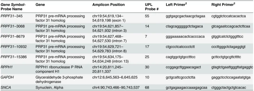
CNV validation. Validation of the large deletion in the genePRPF31was performed in the two affected and six unaffected members of family RP-0777 using quantitative PCR (qPCR) with two different methods: TaqMan assays using the predesigned probes
Hs01877341_cn (chr19:54,618,875) and Hs01993463_cn (chr19:54,619,056) (Applied Biosys-tems TaqMan Copy Number Assays, Life Technologies, Inc.) and the Universal Probe Library (UPL; Roche, Indianapolis, IN) with slight modifications of what was previously described in
[28] and [29]. Briefly, primer and UPL probe combinations were designed againstPRPF31
genomic DNA sequence using the Probe Finder v2.49 software (Roche, Indianapolis, IN). Five assays spanning the length of the gene were selected for validation (genomic coordinates of
each targeted amplicon listed inTable 1).
Quantitative PCR was performed on an ABI Prism 7900HT Sequence Detection System (Applied Biosystems, Foster City, CA). Data were evaluated using the Sequence Detection
Soft-ware v2.4 (Applied Biosystems, Foster City, CA) and further analyzed by theΔΔCT method.
The geometric mean of the CT values for the three control sequences (GAPDH,RPPH1, and
Results
SNV and
indel
analysis with further confirmation of family segregation by
Sanger sequencing allowed the genetic characterization of three
families
Seventeen affected and 21 unaffected members from the six families were subjected to WES. A total of 107.7 GB of data on target genomic regions were generated for the 38 samples, with a mean coverage of target region of 63.67 fold (minimum coverage was 34.85 fold). An average
of 50,562 SNVs and 8,476indelswere called for the 38 exomes, however, for further variant
fil-tering only variants located in the coding regions and splicing boundaries were considered,
thus reducing the number to an average of 18,278 SNVs and 708indels.
Variant filtering was initially performed in the index cases from all six families and then seg-regation with the disease was assessed in the remaining family members analyzed by WES.
SNV andindelanalysis led to the identification of three previously reported mutations
associ-ated with retinal dystrophies in families RP-0107, RP-0858, and RP-0911. A description of the phenotypic features of the affected members of these families and figures with
electroretino-gram and eye fundus are shown inTable 2andS2andS3Figs.
Family RP-0107 harbored a missense change inPRPH2(NM_000322.4:c556G>A; p.
Asp186Asn) [30] and the complete segregation in all five affected and one unaffected members
from RP-0107 was further confirmed by Sanger sequencing. RP-0858 carried a known
frame-shiftdeletion inRPGR(NM_000328.2: c.485_486delTT; p.Phe162Tyrfs4) [31], a gene associ-ated with xlRP. This mutation completely segregassoci-ated in all 25 members from family RP-0858: it was present in eight cases, including six symptomatic and two asymptomatic female carriers and absent in the 17 unaffected members of the family. In RP-0911, a novel nucleotide change inC1QTNF5(NM_015645.3: c.489C>A), leading to the missense mutation p.Ser163Arg
previ-ously associated with Late-Onset Retinal Degeneration (LORD, #605670) [32] was identified in
the index case. Validation and segregation ofC1QTNF5p.Ser163Arg in the family revealed
that, along with the five affected individuals, two of the family members initially considered
unaffected also carried the mutation (S1 Fig, V:1 and IV:15).Table 3summarizes causal
muta-tions identified in the four families characterized.
Table 1. UPL Probes used for validation ofPRPF31heterozygous deletion. The amplicon position is that reported by UCSC genome browser (hg19)in silicoPCR tool. All primers are listed 5’to 3’.
Gene
Symbol-Probe Name
Gene Amplicon Position UPL
Probe #
Left Primer2 Right Primer2
PRPF31–345 PRP31 pre-mRNA processing factor 31 homolog
chr19:54,619,134–
54,619,198 (exon 1)
55 ggtgagcgactaacgctagaa cgtggtctccatcacactca
PRPF31–3068 PRP31 pre-mRNA processing factor 31 homolog
chr19:54,621,857–
54,621,932 (intron 3)
14 ctagcagggggctctagaca gtcagaatccagcactcttcaa
PRPF31–8679 PRP31 pre-mRNA processing factor 31 homolog
chr19:54,627,468–
54,627,530 (intron 7)
7 gggaaaaacactcacccaca gtggtcatctctgggtttcc
PRPF31–10932 PRP31 pre-mRNA processing factor 31 homolog
chr19:54,629,721–
54,629,783 (intron 8)
17 ctgccctcatcccctctt cccttgggctctagaggtgt
PRPF31–15386 PRP31 pre-mRNA processing factor 31 homolog
chr19:54,634,175–
54,634,248 (intron 13)
25 cagtggctgtgcctttcc gcttcctgtgcgttcttttc
RPPH1 RPPH1 ribonuclease P RNA
component H1
chr14:20,811,245–
20,811,337
30 ccggagcttggaacagact gtagtctgaattgggttatgaggtc
GAPDH Glyceraldehyde 3-phosphate
dehydrogenase
chr12:6,645,563–6,645,625 10 gctgcattcgccctctta gaggctcctccagaatatgtga
SNCA Synuclein, Alpha chr4:90,743,466–90,743,537 68 gctgagaagaccaaagagcaa ctgggctactgctgtcacac
Table 2. Clinical features of the four families genetically characterized in the present study.All the ages are expressed in years. adRP = autosomal dominant retinitis pigmentosa, DOB = date of birth, BE = both eyes, ERG = Electroretinogram, HM = High myopia, LE = left eye, LORD = Late Onset Retinal Dystrophy, LP = Light perception, MA = myopic astigmatism, MD = macular degeneration, MM = myopic maculopathy, NA = not available, NB = night blind-ness, NR = Non recordable, RE = right eye, RP = retinitis pigmentosa; RPE = Retinal pigment epithelium, VA = visual acuity, VEP = visual evoked potentials, VF = visual field, xlRP = X-linked retinitis pigmentosa.
Family Subject Revised diagnosis Age at diagnosis Age at onset Age at time of testing
Visualfield Eye fundus ERG Visual acuity
Other
(Gene) (DOB) NB/VF/
VA
RE/LE
RP-0107 III:2 (1915)
adRP NA 12/40/35 NA NA NA NA NA Myopia (18y) and
cataract
(PRPH2) III:6 (1924)
adRP 14 12/14/62 63 Absolute
scotoma
Typical RP with macular alteration
NA NA Cataract (55y)
IV:3 (1950)
adRP NA 40/40/40 NA NA Salt-and-pepper
pigmentation
NA NA
V:3 (1976)
adRP NA NA NA NA Salt-and-pepper
pigmentation
NA NA
V:4 (1980)
adRP 17 13/16/20 17 Diffuse
relative scotoma
Salt-and-pepper pigmentation
NA 0.7/0.8 Dyschromatopsia
RP-0777 II:4 (1966)
adRP NA NA 32 Peripheral
constriction
Typical RP, no macular affectation in BE
Reduced amplitudes typical of bilateral retinopathy 1.0/1.0 (PRPF31) III:1 (1934)
adRP 32 27/32/NA 79 Tubular
field
Normal vessels and papilla, peripapillar atrophy 360°. No pigmentary lessions in BE
Rods: minimum reduced amplitude and increment of latencies; mix: minimum reduced amplitude in a wave and increment latencies in a and b waves; cones: minimum reduced amplitude b wave and minimum increment latencies in waves;flicker: normal in BE
0.7/0.8
RP-0858 III:4 (1947)
xlRP 43 08/08/08 44 Concentric
narrowing Pale papilla, attenuated retinal vessels, peripheral pigment deposits in BE
NR BE FC/LP HM and PSC and altered VEP in BE (56y)
(RPGR) III:6 (1943)
xlRP NA NA NA NA NA NA Glaucoma
IV:6 (1965)
xlRP 31 3/23/3 31 Severe
concentric narrowing in BE
Typical RP NR BE FC/0.4 Strabismus, amblyopia RE, and myopia LE
Table 2. (Continued)
Family Subject Revised diagnosis Age at diagnosis Age at onset Age at time of testing
Visualfield Eye fundus ERG Visual acuity
Other
(Gene) (DOB) NB/VF/
VA
RE/LE
IV:8 (1966)
xlRP 37 NA 45 Severe
concentric narrowing in BE
Typical RP NA 0.2/0.1 MM (BE)
IV:9 (1969)
xlRP 35 Childhood 37 Severe
concentric narrowing in BE Pale papilla, attenuated retinal vessels, macular affectation in BE
Diffuse and severe impairment but not abolished 0.15/ 0.15
HM, MA, MM
IV:10 (1974)
xlRP 35 NA NA NA NA NA 0.8/0.8 HM, MM (BE)
V:2 (1998)
xlRP 14 NA NA Severely
affected
Typical RP NA 0.5/0.5 MA
V:3 (1996)
xlRP 14 NA 14 NA NA NA NA NA
RP-0911 III:7 (1925)
LORD 60 43/43/40 67 NA Macular atrophy
and bone spicules in periphery
NA 0.5/0.2 Cataract (67y)
(C1QTNF5) IV:1 (1946)
LORD 54 54/54/No 66 Central
scotome Normal papilla, RPE macular atrophy, no pigment Rods and cones: abnormal amplitudes 0.8/0.8 IV:6 (1946)
LORD NA 61/61/61 67 Central
scotome nasal superior RPE macular atrophy and hipopigmentary rounded areas Rods: NR, mix: very reduced amplitudes cones and flicker: reduced amplitudes BE
0.1/0.4 Cataract (63y)
IV:10 (1952)
LORD 59 59/No/No 62 Normal Macular drusen Normal 0.9/1
IV:11 (1956)
LORD 60 60/60/60 60 Central
scotome RPE macular atrophy, bone spicules in periphery Rods: very reduced amplitude in b wave; mix and cones: reduced amplitude in a and b waves; cones: reduced amplitude in a and b waves, flicker: reduced amplitude in b wave BE
0.5/0.2 Cataract (56y)
V:1 (NA)
LORD NA NA NA NA NA NA NA NA
A novel large deletion spanning the entire
PRPF31
gene was identified in
family RP-0777
The presence of causative CNVs was investigated in the 3 families not characterized by SNP/ indelanalysis with the XHMM algorithm: RP-0502, RP-0777, and RP-1405 using 663
individu-als from 212 families as controls. A heterozygous deletion in the region chr19: 54,600,186–
54,628,017 (Fig 1) was identified in the two affected members of family RP-0777 with XHMM.
The region identified by this prediction tool included the first exon of the geneOSCAR, the
entire sequence ofNDUFA3andTFPTand exons 1 to 7 ofPRPF31(Fig 1).
For validation of the CNV predicted by XHMM inPRPF31, we used a TaqMan pre-designed
probe in exon 1 and five UPL probes spanning the entire gene region: exon 1, and introns 3, 7, 8
and 13 (Table 2). Both CNV assays with pre-designed TaqMan and UPL probes confirmed that
the coding sequence ofPRPF31from chr19:54,618,875 to chr19:54,634,248 (exons 1 to 13) was
hemizygously deleted in the two affected and present at two copies in the six unaffected
mem-bers of the family. The phenotype of family RP-0777 is summarized inTable 2.
No CNV was identified in families RP-0502 and RP-1405.
Screening of ORF15 in family RP-0502
In view of the results observed in family RP-0858 and due to the absence of male-to-male transmission in family RP-0502, we decided to screen this family for an X-linked inheritance
model. SinceRPGRwas negative in this family (both by WES and gene-panels) and a large
pro-portion of causal mutations ofRPGRoccur in the 3' end of theORF15coding sequence, which
was poorly covered in those sequencing assays, Sanger sequencing was used to scan for muta-tions in this gene. However, no mutation was found and therefore this family and family RP-1405, remain uncharacterized at the molecular level.
Discussion
In the present study, we applied WES to six families with an initial diagnosis of adRP that had been extensively screened for known causative mutations and/or genes. Using this approach, we were able to characterize four out of the six families and, although the four families carried mutations in known genes, the identification of the genetic defect by WES led to the reappraisal of the phenotype from the initial adRP to xlRP in RP-0858, to LORD in RP-0911 and cone-rode dystrophy in RP-0107. This allowed the establishment of a correct diagnosis, estimation of risk recurrence and genetic counseling in these families. adRP was initially considered the
most plausible phenotype based on the mode of inheritance in families, patients’report on
onset of symptoms such as night blindness, or visual acuity loss, and the ophthalmological data regarding fundus and visual field assessments. Clinical information was limited in some cases or exclusively recorded at later stages of the disease. This limitation, which is a common situa-tion when studying this type of diseases, along with the clinical overlap of symptoms in Table 3. Mutations detected in the four families included in the study. adRP: autosomal dominant Retinitis Pigmentosa; AMD: age-related macular degeneration; adCRD: autosomal dominant cone-rod dystrophy; LORD: Late-onset retinal degeneration.
Family ID Suspected diagnosis Final diagnosis Mutation Reference
RP-0107 adRP adCRD PRPH2 c556G>A; p.Asp186Asn (NM_000322.4) Kohlet al. 2012 [33] RP-0777 adRP adRP PRPF31 del 54,619,134 to 54,634,248 (NM_015629.3) This study
RP-0858 adRP + high myopia xlRP RPGR c.485_486delTT; p.Phe162Tyrfs*4 (NM_000328.2) Sharonet al. 2000 [31] RP-0911 adRP + AMD LORD C1QTNF5 c.489C>A; p.Ser163Arg (NM_015645.3) Hayward Cet al. 2003 [32]
Fig 1. Region predicted by XHMM to harbor a heterozygous deletion.The x-axis represents the genome locus, with the genes and exons included in the algorithm, and the y-axis is the computed Z-score of PCA normalized read depth where positive values indicate predicted duplication and negative values deletion. The two affected individuals from family RP-0777 are highlighted with colors red (member II:4) and purple (member III:1) while grey lines represent the 663 control individuals used. Each point indicates a region containing an exon and they are paired with the corresponding exon/gene in a display of the region from the UCSC Genome Browser (http://genome.ucsc.edu/cgi-bin/hgGateway). On the left side of the UCSC Genome Browser track, the genes involved in the deletion are represented. From left to right, the genes/exons illustrated are exons 5 to 1 of theOSCARgene, the entire genesNUDFA3and
TFPT(4 and 6 exons, respectively), and exons 1 to 12 ofPRPF31.
different forms of inherited retinal dystrophies made establishing the precise diagnosis
extremely complex and explains the reclassification of the phenotype in the families upon iden-tification of the genetic cause. Therefore the use of a hypothesis free approach for mutation detection, such as WES, helps minimizing the impact that the availability of patient or family information has on the diagnostic success of retinal dystrophies.
Obligate carrier females of mutations inRPGRmay display manifestations of the disease [8,
34], [35] and even be as severely affected as males [35,36], as observed in family RP-0858, with
all carrier females displaying a severe RP phenotype. What was remarkable in this family is that females affected largely outnumbered affected males (seven females versus three males). Addi-tionally, the onset of symptoms was similar among both female and male carriers and there was no significant intra-familial variability in the symptomatology. These features are not often seen in xlRP and were along with the highly penetrant phenotype females presented the reasons why a dominant rather than an X-linked model was initially considered. Also, although myopia has
been associated with xlRP caused by mutations in bothRPGRandRP2[37–40], it is not
exclu-sively found in this phenotype [41], and therefore was not used as a diagnostic criterion of xlRP.
These results are consistent with the underestimated frequency of xlRP previously reported [35]
and highlight the need of reviewing all adRP families with no male-to-male transmission, as
already reported in [35], regardless of the severity of the symptoms, for testing of X-linked genes.
Family RP-0911 carried the mutation p.Ser163ArgC1QTNF5responsible for LORD, a very
rare fully penetrant autosomal dominant retinal dystrophy with symptoms overlapping with a
number of hereditary retinal conditions [42]. The phenotype of LORD evolves with time and
in early stages can be misdiagnosed as early age-related macular degeneration (AMD), and
later on with RP [42,43], [44],[45], [46]. Due to the limited clinical data available in this family
and the lack of literature about this entity by the time this family was first evaluated, LORD was never considered and different diagnoses were attributed to the three affected members from RP-0911: RP in IV:1, age-related macular degeneration in IV:6, and both in IV:11, ini-tially suspecting of two distinct entities co-segregating in the family. Further clinical re-evalua-tion of the family evidenced that some of the symptoms were consistent with what has been reported in families affected with LORD whereas others, like neovascularization, was not pres-ent in any member of the family at the time of diagnosis. Interestingly, member IV:10, who had referred night blindness as the only symptom when she was 59 years, had evidence of dru-sen in the eye fundus at time of testing (62 years), but the rest of the ophthalmological study was completely normal. Unfortunately, we do not have information on the progression of the phenotype in this family, so a detailed description of the symptomatology and progression of the disease cannot be provided in this study.
WES also helped in the correct diagnosis of family RP-0107, who was found to harbor a
mutation inPRPH2previously identified causing autosomal dominant cone dystrophy [30].
All affected members from family RP-0107 presented eye fundus and visual fields compatible with RP plus macular affectation, except for individual V:4 that had a diffuse retinal degenera-tion. Because the family was studied in the latest stages of the disease, the phenotype at that time was more compatible with RP than cone-rode dystrophy. Remarkably, in our diagnosis
algorithm of adRP,PRPH2is one of the first genes to be screened, however in the index case of
this family this was done by DGGE and possibly due to the sensitivity of the methodology, the mutation was not detected.
A large deletion spanning the entire region ofPRPF31was detected in family RP-0777 using
exome data.PRPF31is one of the most frequently mutated genes in adRP, accounting for 5–10%
of the cases and with large deletions being responsible for almost 3% of the cases [34]. This
dele-tion found in this study is similar to that reported by Kohn et al. [47] that besidesPRPF31also
CNV in intron 11 and in our family, the deletion expands up to intron 13, at least, as evidenced by experimental validation with the UPL assays. The phenotype found in RP-0777, summarized inTable 3, is very similar to that reported by Kohn and colleagues [47] and surprisingly mild
given the size ofPRPF31deleted. Interestingly, despite NGS being widely used in search for
muta-tions in retinal dystrophies, so far only Eisenbergeret al. [48] and Nishiguchiet al. [49] have
reported the discovery of large deletions in genes causing retinal dystrophies using sequencing data. With this study we support the feasibility of detecting CNV in genes responsible for retinal dystrophy using NGS, thus expanding the potential of this tool in the diagnosis of these diseases.
To our knowledge, WES has not been applied systematically to date as diagnostic tool in dominant forms of retinal dystrophies. This study was part of a larger multi-center sequencing effort on Mendelian disorders where WES has been successful identifying the genetic cause of a number of phenotypes, including the discovery of a new genetic cause of a syndromic form of
RP in one of our Spanish families [50]. However, the results from the current study, with only
known genes identified, indicate that WES may be an adequate and efficient tool once all known genetic causes of retinal dystrophy have been ruled out. For that purpose, targeted
sequencing is regarded now as the most optimal approach of candidate gene screening [7,10,
15,51]. Almost simultaneous to this project, our group developed an NGS custom panel with
73 genes related to retinal dystrophies that was applied to 59 index cases of families with adRP,
including the non-characterized families RP-0502 and RP-1405 [10]. The authors found a
detection rate of 27% with 64% of the cases carrying new mutations in known genes, which is
in line with previous results of studies performed in similar conditions [7], [8]. Very recently,
Consugar et al. published a comparative analysis on the performance of panel-based versus
WES [15]. The authors concluded that targeted sequencing was more sensitive for variant
detection than WES, with a superior and more even coverage of genes, and therefore the
pre-ferred method for genetic diagnostic testing [15].
Based on our experience over the past years on the use of NGS technologies and in line with
previous reports [7,15,51], we propose to start the diagnostic testing with targeted sequencing
of candidate genes, due to the methodological and cost advantages over WES. As a second step,
due to the high incidence of xlRP among families initially classified as dominant ([35] and this
study), we propose to screenORF15in any family showing no male-to-male transmission,
regardless of the symptomatology or the number of females affected, to rule out a possible
X-linked inheritance.ORF15is responsible for up to 60% of xlRP disease-causing mutations [52],
and because of its high repetitive nature is not usually adequately covered by NGS methods. Finally, once known causes of inherited retinal dystrophies have been ruled out either WES or WGS may be used in the almost 50% of remaining cases that are estimated to harbor mutations
in rarer novel genes [3].
Supporting Information
S1 Fig. Pedigrees of the six families studied.+ and +/+: wild type genotypes. m: mutation detected in hemizygosis; m/+: mutation detected in heterozygosis. Filled and unfilled symbols represent affected and unaffected individuals respectively. Squares indicate males and circles females. Arrows indicate the index cases. Red circles represent individuals subjected to whole exome sequencing.
(TIFF)
S2 Fig. Eye fundus images from members of families RP-0777 and RP-0911.Each patient presents two fundus images per eye. A) Family RP-0777 and B) Family RP-0911. RE = Right eye. LE = Left eye.
S3 Fig. Electroretinogram recordings from members of families RP-0777 and RP-0911. (TIFF)
S1 Table. Genetic screening performed to the six families prior to whole exome sequencing. SSCP:Single Strand Conformation Polymorphism;DGGE:CG-clamped Denaturing Gradient Gel Electrophoresis. The parentheses indicate the exons targeted by these techniques; otherwise
the entire gene was screened. For ADRP Chip, version 1 includes 355 SNPs inCA4,CRX,
FSCN2,IMPDH1,NR2E3,NRL,PRPF3,PRPF31,PRPF8,PRPH2,RHO,ROM1,RP1,RP9, TOPORS; andversion 2 includes 414 SNPs inCA4, CRX, FSCN2, IMPDH1, KLHL7, NR2E3, NRL, PRPF3, PRPF31, PRPF8, PRPH2, RHO, ROM1, RP1, RP9, TOPORS; Sanger sequencing was used to screen mutations in exons 16 and 25 for SNRNP200, exon 2 for NR2E3 and exon 13 for GUCY2D. For IMPDH1 all exons were sequenced. RD_NGS_Panel refers to the custom Next Generation Sequencing panel fromS2 Table.
(DOCX)
S2 Table. Genes associated with RP and LCA included in the customized RD_NGS_Panel. Macular dystrophy (MD); retinitis pigmentosa (RP); Leber's congenital amaurosis (LCA); con-genital stationary night blindness (CSNB); choroideremia (CHM), cone-rod dystrophy (CORD); autosomal recessive (ar); autosomal dominant (ad); X-linked (xl); McKusick-Kauf-man syndrome (MKKS); Senior Loken Syndrome (SLS); vitreoretinopathy proliferative (VRP); enhanced S-cone syndrome (ESC).
(DOCX)
S3 Table. Primers and conditions used for PCR and sequencing ofORF15.
(DOCX)
Acknowledgments
The authors thank all individuals participating in this study. Patricia Fernandez San Jose’s
work is supported by a Rio Hortega grant (CM12/00013) and Marta Corton by a Miguel Servet grant (CP/03256), both from Instituto de Salud Carlos III. This work was funded in part by FJD-Biobank (RD09/0076/00101), CIBER-ER (06/07/0036), FIS (PI:13/00226), ONCE 2014
and Fundaluce (4019–002); by an Institution Development Award from The Children’s
Hospi-tal of Philadelphia, and U01-HG006830 (NHGRI-sponsored eMERGE Network); and by the Shenzhen Municipal Government of China (No GJHZ20130417140916986).
Author Contributions
Conceived and designed the experiments: BA PFS MM RP YG LT XX HH CA. Performed the experiments: PFS MM RP XL LG JZ. Analyzed the data: BA JL PFS YL MM RP RG MC YG LT. Contributed reagents/materials/analysis tools: JL XL LG JZ XX. Wrote the paper: BA PFS YL MM RP MC YG LT BK HH CA. Clinical evaluation of the patients and the interpretation of the phenotypic analysis: FBK MILM BGS.
References
1. Hamel C. Retinitis pigmentosa. Orphanet journal of rare diseases. 2006; 1:40. Epub 2006/10/13. 1750-1172-1-40 [pii] doi:10.1186/1750-1172-1-40PMID:17032466; PubMed Central PMCID:
PMC1621055.
2. Hartong DT, Berson EL, Dryja TP. Retinitis pigmentosa. Lancet. 2006; 368(9549):1795–809. Epub 2006/11/23. S0140-6736(06)69740-7 [pii] doi:10.1016/S0140-6736(06)69740-7PMID:17113430. 3. Daiger SP, Sullivan LS, Bowne SJ. Genes and mutations causing retinitis pigmentosa. Clin Genet.
4. Ayuso C, Millan JM. Retinitis pigmentosa and allied conditions today: a paradigm of translational research. Genome Med. 2010; 2(5):34. Epub 2010/06/04. gm155 [pii] doi:10.1186/gm155PMID: 20519033; PubMed Central PMCID: PMC2887078.
5. Ng SB, Buckingham KJ, Lee C, Bigham AW, Tabor HK, Dent KM, et al. Exome sequencing identifies the cause of a mendelian disorder. Nature genetics. 2010; 42(1):30–5. Epub 2009/11/17. ng.499 [pii] doi:10.1038/ng.499PMID:19915526; PubMed Central PMCID: PMC2847889.
6. Rope AF, Wang K, Evjenth R, Xing J, Johnston JJ, Swensen JJ, et al. Using VAAST to identify an X-linked disorder resulting in lethality in male infants due to N-terminal acetyltransferase deficiency. Am J Hum Genet. 2011; 89(1):28–43. Epub 2011/06/28. S0002-9297(11)00210-2 [pii] doi:10.1016/j.ajhg. 2011.05.017PMID:21700266; PubMed Central PMCID: PMC3135802.
7. Audo I, Bujakowska KM, Leveillard T, Mohand-Said S, Lancelot ME, Germain A, et al. Development and application of a next-generation-sequencing (NGS) approach to detect known and novel gene defects underlying retinal diseases. Orphanet journal of rare diseases. 2012; 7:8. Epub 2012/01/27. 1750-1172-7-8 [pii] doi:10.1186/1750-1172-7-8PMID:22277662; PubMed Central PMCID: PMC3352121.
8. Bowne SJ, Sullivan LS, Koboldt DC, Ding L, Fulton R, Abbott RM, et al. Identification of disease-caus-ing mutations in autosomal dominant retinitis pigmentosa (adRP) usdisease-caus-ing next-generation DNA sequenc-ing. Investigative ophthalmology & visual science. 2011; 52(1):494–503. Epub 2010/09/24. iovs.10-6180 [pii] doi:10.1167/iovs.10-6180PMID:20861475; PubMed Central PMCID: PMC3053293. 9. Corton M, Nishiguchi KM, Avila-Fernandez A, Nikopoulos K, Riveiro-Alvarez R, Tatu SD, et al. Exome
sequencing of index patients with retinal dystrophies as a tool for molecular diagnosis. PLoS One. 2013; 8(6):e65574. Epub 2013/08/14. doi:10.1371/journal.pone.0065574PONE-D-13-04068 [pii]. PMID:23940504; PubMed Central PMCID: PMC3683009.
10. Fernandez-San Jose P, Corton M, Blanco-Kelly F, Avila-Fernandez A, Lopez-Martinez MA, Sanchez-Navarro I, et al. Targeted next generation sequencing improves the diagnosis of autosomal dominant Retinitis Pigmentosa in Spanish patients. Investigative ophthalmology & visual science. 2015. doi:10. 1167/iovs.14-16178PMID:25698705.
11. Shanks ME, Downes SM, Copley RR, Lise S, Broxholme J, Hudspith KA, et al. Next-generation sequencing (NGS) as a diagnostic tool for retinal degeneration reveals a much higher detection rate in early-onset disease. European journal of human genetics: EJHG. 2013; 21(3):274–80. Epub 2012/09/ 13. ejhg2012172 [pii] doi:10.1038/ejhg.2012.172PMID:22968130; PubMed Central PMCID: PMC3573204.
12. Avila-Fernandez A, Perez-Carro R, Corton M, Lopez-Molina MI, Campello L, Garanto A, et al. Whole-exome sequencing reveals ZNF408 as a new gene associated with autosomal recessive retinitis pig-mentosa with vitreal alterations. Human molecular genetics. 2015. doi:10.1093/hmg/ddv140PMID: 25882705.
13. Ma X, Guan L, Wu W, Zhang Y, Zheng W, Gao YT, et al. Whole-exome sequencing identifies OR2W3 mutation as a cause of autosomal dominant retinitis pigmentosa. Scientific reports. 2015; 5:9236. doi: 10.1038/srep09236PMID:25783483; PubMed Central PMCID: PMC4363838.
14. Wang F, Wang Y, Zhang B, Zhao L, Lyubasyuk V, Wang K, et al. A missense mutation in HK1 leads to autosomal dominant retinitis pigmentosa. Investigative ophthalmology & visual science. 2014; 55 (11):7159–64. doi:10.1167/iovs.14-15520PMID:25316723; PubMed Central PMCID: PMC4224578. 15. Consugar MB, Navarro-Gomez D, Place EM, Bujakowska KM, Sousa ME, Fonseca-Kelly ZD, et al.
Panel-based genetic diagnostic testing for inherited eye diseases is highly accurate and reproducible, and more sensitive for variant detection, than exome sequencing. Genetics in medicine: official journal of the American College of Medical Genetics. 2015; 17(4):253–61. doi:10.1038/gim.2014.172PMID: 25412400.
16. Neveling K, Collin RW, Gilissen C, van Huet RA, Visser L, Kwint MP, et al. Next-generation genetic test-ing for retinitis pigmentosa. Hum Mutat. 2012; 33(6):963–72. Epub 2012/02/16. doi:10.1002/humu. 22045PMID:22334370; PubMed Central PMCID: PMC3490376.
17. Ayuso C, Garcia-Sandoval B, Najera C, Valverde D, Carballo M, Antinolo G. Retinitis pigmentosa in Spain. The Spanish Multicentric and Multidisciplinary Group for Research into Retinitis Pigmentosa. Clin Genet. 1995; 48(3):120–2. Epub 1995/09/01. PMID:8556816.
18. Reig C, Antich J, Gean E, Garcia-Sandoval B, Ramos C, Ayuso C, et al. Identification of a novel rhodop-sin mutation (Met-44-Thr) in a simplex case of retinitis pigmentosa. Hum Genet. 1994; 94(3):283–6. Epub 1994/09/01. PMID:8076945.
20. Blanco-Kelly F, Garcia-Hoyos M, Corton M, Avila-Fernandez A, Riveiro-Alvarez R, Gimenez A, et al. Genotyping microarray: mutation screening in Spanish families with autosomal dominant retinitis pig-mentosa. Mol Vis. 2012; 18:1478–83. Epub 2012/06/28. PMID:22736939; PubMed Central PMCID: PMC3380913.
21. Marmor MF, Zrenner E. Standard for clinical electroretinography (1999 update). International Society for Clinical Electrophysiology of Vision. Documenta ophthalmologica Advances in ophthalmology. 1998; 97(2):143–56. PMID:10765968.
22. Marmor MF, Zrenner E. Standard for clinical electro-oculography. International Society for Clinical Electrophysiology of Vision. Archives of ophthalmology. 1993; 111(5):601–4. PMID:8489436. 23. Li H, Durbin R. Fast and accurate short read alignment with Burrows-Wheeler transform.
Bioinformat-ics. 2009; 25(14):1754–60. Epub 2009/05/20. btp324 [pii] doi:10.1093/bioinformatics/btp324PMID: 19451168; PubMed Central PMCID: PMC2705234.
24. McKenna A, Hanna M, Banks E, Sivachenko A, Cibulskis K, Kernytsky A, et al. The Genome Analysis Toolkit: a MapReduce framework for analyzing next-generation DNA sequencing data. Genome Res. 2010; 20(9):1297–303. Epub 2010/07/21. gr.107524.110 [pii] doi:10.1101/gr.107524.110PMID: 20644199; PubMed Central PMCID: PMC2928508.
25. Wang K, Li M, Hakonarson H. ANNOVAR: functional annotation of genetic variants from high-through-put sequencing data. Nucleic Acids Res. 2010; 38(16):e164. Epub 2010/07/06. gkq603 [pii] doi:10. 1093/nar/gkq603PMID:20601685; PubMed Central PMCID: PMC2938201.
26. Fromer M, Moran JL, Chambert K, Banks E, Bergen SE, Ruderfer DM, et al. Discovery and statistical genotyping of copy-number variation from whole-exome sequencing depth. Am J Hum Genet. 2012; 91 (4):597–607. Epub 2012/10/09. S0002-9297(12)00417-X [pii] doi:10.1016/j.ajhg.2012.08.005PMID: 23040492; PubMed Central PMCID: PMC3484655.
27. Garcia-Hoyos M, Garcia-Sandoval B, Cantalapiedra D, Riveiro R, Lorda-Sanchez I, Trujillo-Tiebas MJ, et al. Mutational screening of the RP2 and RPGR genes in Spanish families with X-linked retinitis pig-mentosa. Investigative ophthalmology & visual science. 2006; 47(9):3777–82. doi: 10.1167/iovs.06-0323PMID:16936086.
28. Edelmann L, Prosnitz A, Pardo S, Bhatt J, Cohen N, Lauriat T, et al. An atypical deletion of the Wil-liams-Beuren syndrome interval implicates genes associated with defective visuospatial processing and autism. J Med Genet. 2007; 44(2):136–43. Epub 2006/09/15. jmg.2006.044537 [pii] doi:10.1136/ jmg.2006.044537PMID:16971481; PubMed Central PMCID: PMC2598069.
29. Glessner JT, Wang K, Cai G, Korvatska O, Kim CE, Wood S, et al. Autism genome-wide copy number variation reveals ubiquitin and neuronal genes. Nature. 2009; 459(7246):569–73. Epub 2009/05/01. nature07953 [pii] doi:10.1038/nature07953PMID:19404257; PubMed Central PMCID: PMC2925224. 30. Kitiratschky VB, Glockner CJ, Kohl S. Mutation screening of the GUCA1B gene in patients with autoso-mal dominant cone and cone rod dystrophy. Ophthalmic Genet. 2011; 32(3):151–5. Epub 2011/03/17. doi:10.3109/13816810.2011.559650PMID:21405999.
31. Sharon D, Bruns GA, McGee TL, Sandberg MA, Berson EL, Dryja TP. X-linked retinitis pigmentosa: mutation spectrum of the RPGR and RP2 genes and correlation with visual function. Investigative oph-thalmology & visual science. 2000; 41(9):2712–21. Epub 2000/08/11. PMID:10937588.
32. Hayward C, Shu X, Cideciyan AV, Lennon A, Barran P, Zareparsi S, et al. Mutation in a short-chain col-lagen gene, CTRP5, results in extracellular deposit formation in late-onset retinal degeneration: a genetic model for age-related macular degeneration. Human molecular genetics. 2003; 12(20):2657–
67. Epub 2003/08/29. doi:10.1093/hmg/ddg289ddg289 [pii]. PMID:12944416.
33. Kohl S, Kitiratschky V, Papke M, Schaich S, Sauer A, Wissinger B. Genes and mutations in autosomal dominant cone and cone-rod dystrophy. Adv Exp Med Biol. 2012; 723:337–43. Epub 2011/12/21. doi: 10.1007/978-1-4614-0631-0_44PMID:22183351.
34. Daiger SP, Sullivan LS, Gire AI, Birch DG, Heckenlively JR, Bowne SJ. Mutations in known genes account for 58% of autosomal dominant retinitis pigmentosa (adRP). Adv Exp Med Biol. 2008; 613:203–9. Epub 2008/01/15. doi:10.1007/978-0-387-74904-4_23PMID:18188946; PubMed Central PMCID: PMC2582019.
35. Churchill JD, Bowne SJ, Sullivan LS, Lewis RA, Wheaton DK, Birch DG, et al. Mutations in the X-linked retinitis pigmentosa genes RPGR and RP2 found in 8.5% of families with a provisional diagnosis of autosomal dominant retinitis pigmentosa. Investigative ophthalmology & visual science. 2013; 54 (2):1411–6. Epub 2013/02/02. iovs.12-11541 [pii] doi:10.1167/iovs.12-11541PMID:23372056; PubMed Central PMCID: PMC3597192.
37. Fishman GA, Weinberg AB, McMahon TT. X-linked recessive retinitis pigmentosa. Clinical characteris-tics of carriers. Archives of ophthalmology. 1986; 104(9):1329–35. Epub 1986/09/01. PMID:3753283. 38. Koenekoop RK, Loyer M, Hand CK, Al Mahdi H, Dembinska O, Beneish R, et al. Novel RPGR
muta-tions with distinct retinitis pigmentosa phenotypes in French-Canadian families. Am J Ophthalmol. 2003; 136(4):678–87. Epub 2003/10/01. S0002939403003313 [pii]. PMID:14516808.
39. Jayasundera T, Branham KE, Othman M, Rhoades WR, Karoukis AJ, Khanna H, et al. RP2 phenotype and pathogenetic correlations in X-linked retinitis pigmentosa. Archives of ophthalmology. 2010; 128 (7):915–23. Epub 2010/07/14. 128/7/915 [pii] doi:10.1001/archophthalmol.2010.122PMID:20625056; PubMed Central PMCID: PMC3392190.
40. Wu DM, Khanna H, Atmaca-Sonmez P, Sieving PA, Branham K, Othman M, et al. Long-term follow-up of a family with dominant X-linked retinitis pigmentosa. Eye (Lond). 2010; 24(5):764–74. Epub 2009/11/ 07. eye2009270 [pii] doi:10.1038/eye.2009.270PMID:19893586; PubMed Central PMCID:
PMC2920623.
41. Chassine T, Bocquet B, Daien V, Avila-Fernandez A, Ayuso C, Collin RW, et al. Autosomal recessive retinitis pigmentosa with RP1 mutations is associated with myopia. The British journal of ophthalmol-ogy. 2015. doi:10.1136/bjophthalmol-2014-306224PMID:25883087.
42. Borooah S, Collins C, Wright A, Dhillon B. Late-onset retinal macular degeneration: clinical insights into an inherited retinal degeneration. The British journal of ophthalmology. 2009; 93(3):284–9. Epub 2008/ 12/23. bjo.2008.150151 [pii] doi:10.1136/bjo.2008.150151PMID:19098033.
43. Kuntz CA, Jacobson SG, Cideciyan AV, Li ZY, Stone EM, Possin D, et al. Sub-retinal pigment epithelial deposits in a dominant late-onset retinal degeneration. Investigative ophthalmology & visual science. 1996; 37(9):1772–82. Epub 1996/08/01. PMID:8759344.
44. Milam AH, Curcio CA, Cideciyan AV, Saxena S, John SK, Kruth HS, et al. Dominant late-onset retinal degeneration with regional variation of sub-retinal pigment epithelium deposits, retinal function, and photoreceptor degeneration. Ophthalmology. 2000; 107(12):2256–66. Epub 2000/11/30.
S016164200000419X [pii]. PMID:11097607.
45. Vincent A, Munier FL, Vandenhoven CC, Wright T, Westall CA, Heon E. The characterization of retinal phenotype in a family with C1QTNF5-related late-onset retinal degeneration. Retina. 2012; 32 (8):1643–51. Epub 2012/01/27. doi:10.1097/IAE.0b013e318240a574PMID:22277927.
46. Soumplis V, Sergouniotis PI, Robson AG, Michaelides M, Moore AT, Holder GE, et al. Phenotypic find-ings in C1QTNF5 retinopathy (late-onset retinal degeneration). Acta Ophthalmol. 2013; 91(3):e191–5. Epub 2013/01/08. doi:10.1111/aos.12010PMID:23289492.
47. Kohn L, Bowne SJ, L SS, Daiger SP, Burstedt MS, Kadzhaev K, et al. Breakpoint characterization of a novel approximately 59 kb genomic deletion on 19q13.42 in autosomal-dominant retinitis pigmentosa with incomplete penetrance. European journal of human genetics: EJHG. 2009; 17(5):651–5. doi:10. 1038/ejhg.2008.223PMID:19050727; PubMed Central PMCID: PMC2796252.
48. Eisenberger T, Neuhaus C, Khan AO, Decker C, Preising MN, Friedburg C, et al. Increasing the yield in targeted next-generation sequencing by implicating CNV analysis, non-coding exons and the overall variant load: the example of retinal dystrophies. PLoS One. 2013; 8(11):e78496. Epub 2013/11/23. doi: 10.1371/journal.pone.0078496PONE-D-13-29248 [pii]. PMID:24265693; PubMed Central PMCID: PMC3827063.
49. Nishiguchi KM, Tearle RG, Liu YP, Oh EC, Miyake N, Benaglio P, et al. Whole genome sequencing in patients with retinitis pigmentosa reveals pathogenic DNA structural changes and NEK2 as a new dis-ease gene. Proc Natl Acad Sci U S A. 2013; 110(40):16139–44. Epub 2013/09/18. 1308243110 [pii] doi:10.1073/pnas.1308243110PMID:24043777; PubMed Central PMCID: PMC3791719.
50. Almoguera B, He S, Corton M, Fernandez-San Jose P, Blanco-Kelly F, Lopez-Molina MI, et al. Expand-ing the phenotype of PRPS1 syndromes in females: neuropathy, hearExpand-ing loss and retinopathy. Orpha-net journal of rare diseases. 2014; 9:190. doi:10.1186/s13023-014-0190-9PMID:25491489; PubMed Central PMCID: PMC4272780.
51. Borras E, de Sousa Dias M, Hernan I, Pascual B, Mane B, Gamundi M, et al. Detection of novel genetic variation in autosomal dominant retinitis pigmentosa. Clin Genet. 2013. Epub 2013/03/29. doi:10.1111/ cge.12151PMID:23534816.