RESEARCH ARTICLE
Dominant Retinitis Pigmentosa, p.Gly56Arg
Mutation in
NR2E3
: Phenotype in a Large
Cohort of 24 Cases
Fiona Blanco-Kelly1,2☯, María García Hoyos3☯, Miguel Angel Lopez Martinez1, Maria Isabel Lopez-Molina4, Rosa Riveiro-Alvarez1,2, Patricia Fernandez-San Jose1,2,
Almudena Avila-Fernandez1,2, Marta Corton1,2, Jose M. Millan2,5,6, Blanca García Sandoval4, Carmen Ayuso1,2
*
1Genetic´s Department, Instituto de Investigación Sanitaria-Fundación Jimenez Diaz University Hospital
(IIS-FJD, UAM), Madrid, Spain,2Center of Biomedical Network Research on Rare Diseases (CIBERER),
ISCIII, Madrid, Spain,3Instituto de Medicina Genómica (IMEGEN), Valencia, Spain,4Department of
Ophthalmology, Instituto de Investigación Sanitaria-Fundación Jiménez Díaz University Hospital (IIS-FJD, UAM), Madrid, Spain,5Grupo de Investigación en Biomedicina Molecular, Celular y Genómica, Instituto de
Investigación Sanitaria La Fe (IIS-La Fe), Valencia, Spain,6Unidad de Genética y Diagnóstico Prenatal,
Hospital Universitario y Politécnico La Fe, Valencia, Spain
☯These authors contributed equally to this work.
*cayuso@fjd.es
Abstract
Importance
This research is the single largestNR2E3genotype-phenotype correlation study performed
to date in autosomal dominant Retinitis Pigmentosa.
Objective
The aim of this study is to analyse the frequency of the p.Gly56Arg mutation inNR2E3for
the largest cohort of autosomal dominant Retinitis Pigmentosa patients to date and its asso-ciated phenotype.
Patients and Methods
A cohort of 201 unrelated Spanish families affected by autosomal dominant Retinitis Pig-mentosa. The p.Gly56Arg mutation in theNR2E3(NM_014249.2) gene was analysed in
201 families. In the 24 cases where the mutation had been detected, a haplotype analysis linked to the p.Gly56Arg families was performed, using four extragenic polymorphic markers D15S967, D15S1050, D15S204 and D15S188. Phenotype study included presence and age of onset of night blindness, visual field loss and cataracts; and an ophthalmoscopic examination after pupillary dilation and electroretinogram for the 24 cases.
Results
Seven of the 201 analyzed families were positive for the p.Gly56Arg, leading to a preva-lence of 3.5%. Clinical data were available for 24 subjects. Night blindness was the first
PLOS ONE | DOI:10.1371/journal.pone.0149473 February 24, 2016 1 / 13
OPEN ACCESS
Citation:Blanco-Kelly F, García Hoyos M, Lopez Martinez MA, Lopez-Molina MI, Riveiro-Alvarez R, Fernandez-San Jose P, et al. (2016) Dominant Retinitis Pigmentosa, p.Gly56Arg Mutation inNR2E3: Phenotype in a Large Cohort of 24 Cases. PLoS ONE 11(2): e0149473. doi:10.1371/journal. pone.0149473
Editor:Dror Sharon, Hadassah-Hebrew University Medical Center, ISRAEL
Received:August 10, 2015
Accepted:January 31, 2016
Published:February 24, 2016
Copyright:© 2016 Blanco-Kelly et al. This is an open access article distributed under the terms of the
Creative Commons Attribution License, which permits unrestricted use, distribution, and reproduction in any medium, provided the original author and source are credited.
http://www.asperbio.com/asper-noticeable symptom (mean 15.9 years). Visual field loss onset was variable (23.3±11.9
years). Loss of visual acuity appeared late in the disease´s evolution. Most of the patients with cataracts (50%) presented it from the third decade of life. Fundus changes showed inter and intrafamiliar variability, but most of the patients showed typical RP changes and it was common to find macular affectation (47.4%). Electroretinogram was impaired from the beginning of the disease. Two families shared a common haplotype. Additionally, all patients shared a 104Kb region between D15S1050 and theNR2E3gene.
Conclusions
This study highlights the importance of p.Gly56Arg in theNR2E3gene as a common
muta-tion associated with adRP, and provides new clues to its phenotype, which can allow for a better clinical management and genetic counselling of patients and their families.
Introduction
Retinitis Pigmentosa (RP, MIM# 268000), with a prevalence of approximately one in 4000 [1], is the most common form of inherited retinopathy. RP is a group of clinically and genetically heterogeneous retinal degenerative diseases. Clinically it is characterized by progressive loss of photoreceptors and pigment deposits predominantly in the peripheral retina, and by a relative sparing of the central retina. The diagnostic criteria for RP were established by Marmor [2–5]. To date, sixty-nine genes have been associated with non-syndromic RP (http://www.sph.uth. tmc.edu/RetNet/, data accessed 30/12/2015) and all modes of inheritance have been described in this disease: autosomal dominant, autosomal recessive, X linked, mitochondrial and, in rare cases, digenic [6].
In Spain, autosomal dominant form of RP (adRP) represents approximately 15% of Spanish RP families [7,8]. The large number of genes involved in adRP disease complicates genetic analysis of these patients. To date, 23 genes (and one mapped locus) have been associated with adRP (http://www.sph.uth.tmc.edu/RetNet/, data accessed 30/12/2015). One of these 23 genes is theNR2E3gene, which contains eight exons that expand a genomic sequence of around 7.7 kilobases (kb). The open reading frame of this gene encodes for a retinal nuclear receptor pro-tein that acts as a transcriptional regulator, activating rod-specific genes in concert with other transcriptional factors (CRX and NRL), as well as repressing the transcription of cone-specific genes in differentiating rod photoreceptors [9].
Most of the mutations in this gene have been associated with autosomal recessive retinitis pigmentosa (arRP) with variable phenotypes (enhanced S-cone sensitivity syndrome -ESCS-[10,11], Goldmann-Favre syndrome -GFS- [12], and clumped pigmentary retinal degeneration -CPRD-) [13–16]. However, one mutation (p.Gly56Arg) in the first zinc-finger of the DNA binding domain of theNR2E3gene has been found in adRP patients [17] associated with RP phenotype (progressive rod degeneration and ulterior cone affectation) [18]. This mutation accounts for approximately 1–2% of North American and 3.4% of European adRP patients [17–19], and, until present time, is the only mutation found in theNR2E3gene responsible for adRP [17–21].
The aim of this study is to analyse the frequency of the p.Gly56Arg mutation in theNR2E3 gene in our cohort of adRP patients and to determine the associated phenotype.
NR2E3: p.Gly56Arg Mutation in adRP Spanish Population
PLOS ONE | DOI:10.1371/journal.pone.0149473 February 24, 2016 2 / 13
ophthalmics/autosomal-dominant-retinitis-pigmentosa-genetic-testing.
Funding:Support was provided by FIS (PI: 13/ 00226), the Centre for Biomedical Network Research on Rare Diseases—CIBERER (06/07/0036), the Biobank of Fundación Jiménez Díaz University Hospital (RD09/0076/00101), ONCE 2014, Feder (Fondos Europeo de Desarrollo Regional) and Fundaluce (4019-002). Patricia Fernandez-San Jose ´s work is supported by a Rio Hortega grant (CM12/ 00013), Marta Corton by a Miguel Servet grant (CP/ 03256) all from Instituto de Salud Carlos III. The funders had no role in study design, data collection and analysis, decision to publish, or preparation of the manuscript.
Patients and Methods
Patients
The adRP diagnosis was based on pedigree data and ophthalmologic examination. Our patients were classified as affected by RP according to the following clinical criteria: night blindness (NB), progressive loss of peripheral vision (mid peripheral scotoma or ring scotoma), fundus compatible with RP [3,4] (ophthalmoscopic examination after pupillary dilation), and patho-logic electroretinogram (ERG) showing a marked reduction in rod or rod and cone signal (full-field electroretinogram according to the standards of the International Society for Clinical Electrophysiology of Vision:http://www.iscev.org) [22]. Autosomal dominant inheritance was considered according to previously established criteria [7,8].
The severity of visual acuity loss was classified following the WHO criteria (normal vision
0.4, moderate low vision<0.4–>0.1, severe low vision0.1 -0.05, and profound vision loss and blindness<0.05)
Written informed consent was obtained from all individuals included in the study and research protocols were approved by the Ethics committee of the University Hospital Funda-ción Jiménez Díaz in accordance with the tenets of the Declaration of Helsinki and their reviews.
Screening for
NR2E3
autosomal dominant mutation
DNA was extracted from peripheral blood samples and collected in EDTA tubes using an auto-mated DNA extractor according to manufacturer instructions (model BioRobot EZ1; Qiagen, Hilden, Germany).
The p.Gly56Arg mutation in theNR2E3gene (NM_014249.2) was analysed in a total of 201 unrelated adRP families. The analysis was performed by direct sequencing, as previously reported [19], or by adRP genotyping Asper Ophthalmics microarray (Asper Biotech [19], http://www.asperbio.com/asper-ophthalmics/autosomal-dominant-retinitis-pigmentosa-ad-rp/autosomal-dominant-retinitis-pigmentosa-targeted-mutation-analysis, versions from Feb-ruary 2008 to July 2014).
Among 201 families, 60 had been studied previously, showing a negative result, using the first version of the adRP genotyping microarray, which did not include the p.Gly56Arg muta-tion [23]. The remaining 141 families underwent an updated version of the adRP genotyping microarray which included the p.Gly56Arg mutation in theNR2E3gene. Direct sequencing was used to analyse the 60 previously studied families, to confirm the results obtained with the genotyping microarray and to segregate the disease causative mutation p.Gly56Arg in the NR2E3gene in the families.
Haplotype Analysis
Haplotype analysis was performed using four extragenic polymorphic markers (NR2E3 genomic position according to Human Genome Assembly GRCh37, Chr15: 72,084,977– 72,110,600) strongly linked to this locus: D15S967, D15S1050, D15S204, and D15S188. For the genotyping process, PCR products were electrophoresed in an ABI Prism 3130 Genetic Analyzer and analyzed with the GeneMapper v3.5 software package (Applied Biosystems).
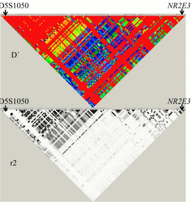
Anin silicoanalysis was performed using the Hapmap/Haploview 4.0 software (http://www. hapmap.org/) to establish the linkage disequilibrium blocks (D´ and r2 parameters) in the genomic region between D15S1050 and theNR2E3gene (Fig 1).
NR2E3: p.Gly56Arg Mutation in adRP Spanish Population
Results
Seven out of 201 adRP studied families were positive for the p.Gly56Arg mutation in the NR2E3gene (five of them identified by direct sequencing of the mutation and two by the adRP
genotyping microarray).
The detection of the dominantNR2E3mutation in seven families, gives us a frequency of 3.5% (7/201) in our adRP studied cohort. All families were of Spanish origin except RP-1650 which was from Venezuela (both grandparents from mother´s side–disease origin–were Venezuelan).
Fig 1. Linkage disequilibrium for region Chr15: 71,980,969–72,110,600 of the Human Genome Assembly GRCh37 (Hapmap/Haploview 4.0
software:http://www.hapmap.org/).Colour image: D´ (red: D´ = 1, the lower the D´ the further away from red). Black & white image: r2(black: r2, the lower
the r2the further away from black).
doi:10.1371/journal.pone.0149473.g001
NR2E3: p.Gly56Arg Mutation in adRP Spanish Population
Phenotypic characteristics of
NR2E3
mutation
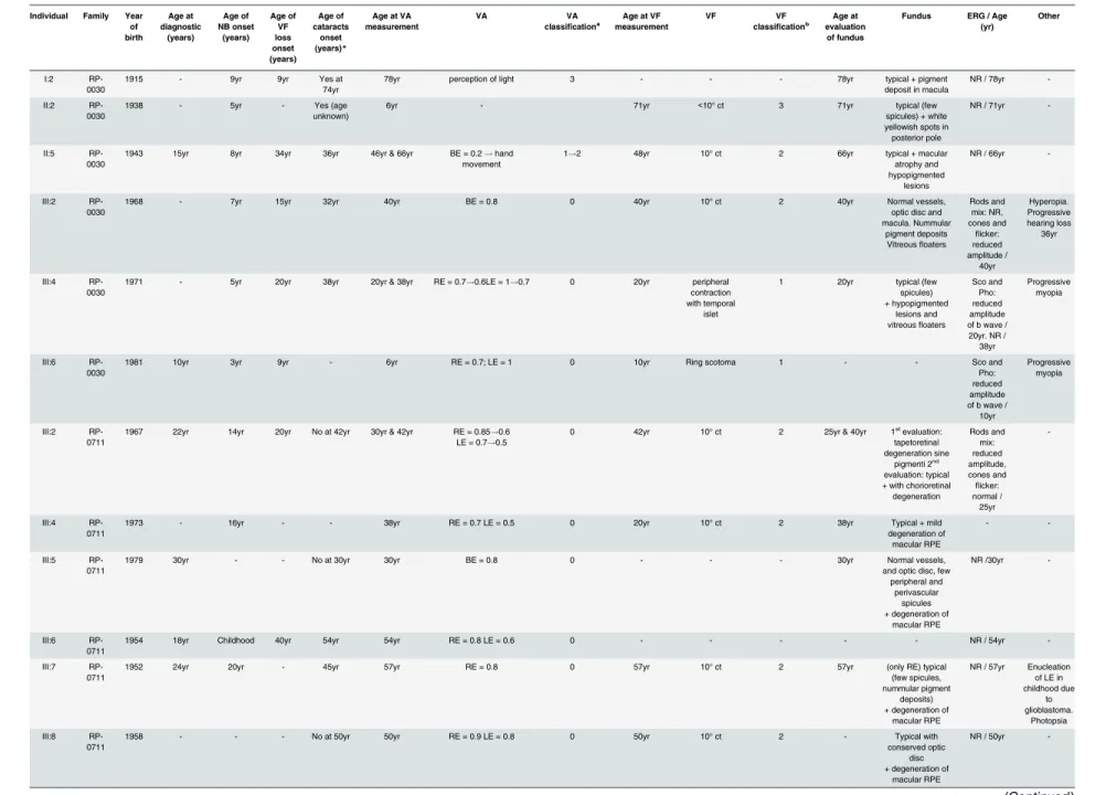
Clinical data were available for 24 subjects from the sevenNR2E3mutated adRP families (Table 1).
In most cases NB was the first noticeable symptom with an onset in pre-adolescence or early twenties (mean age 15.9 ± 10.1 years). Visual field (VF) loss onset was variable, although tubular vision onset tended to occur around the third decade of life (mean age 23.2 ± 11.9 years) and stayed stable until an advanced age. Loss of visual acuity (VA) appeared later in the disease´s evolution, being normal (0.4) until the fifth decade of life (Table 2).
Only two patients presented legal blindness (VA0.1 or VF10°) at 60 and 71 years of age, respectively. Additionally, one patient with RP, who also presented glaucoma, was diag-nosed with complete blindness (neither perception nor projection of light) at the age of 53.
More than half of the patients (58.3%) with available ophthalmological data presented cata-ract (73.7%), most of them (50%) since the third decade of life (Table 2).
All ophthalmoscopic examination data available (19 patients) showed fundus alterations (Fig 2).
These fundus alterations could be detected since the beginning of the vision impairment, although visual acuity was not affected (Table 1).
Although fundus changes showed inter and intrafamiliar variability, most of the patients showed typical RP changes with the progression of the disease. It was also frequent to find mac-ular affectation (47.4% -Table 2-) with preserved visual acuity, even when patients were studied in the first stages of the disease (family RP-0711, IV:2 -Table 1-).
ERG recordings were available for 19 cases. All available ERG recordings (scotopic, phot-opic and flicker) showed alterations from the beginning of the disease, and these changes could be detected around the first decade of life, when studied at that age (family RP-0711 patient IV:2; and family RP-0030 patient III:6—Table 1andS1 Fig). Non recordable ERG occurred around the age of fifty (Table 2). Similarly as for fundus changes, the initial impairment in ERG recordings did not seem to alter visual acuity.
Haplotype analysis
Four of the families (RP-0030, RP-0576, RP-0711 and RP-1182) belonged to the same town in Spain (in the Toledo region, with 2755 inhabitants in 1920 and 3511 inhabitants in 2010, according to the demographic data of the Instituto Nacional de Estadística -INE-http://www. ine.es). In two of these families, RP-0030 and RP-1182, a common ancestor (four generations above the respective family probands) could be identified, but not in RP-0711 and RP-0576 (family trees investigated five generations above the respective probands,Fig 3).
Inferring haplotypes in families RP-1650 and RP-0124 (and even in RP-1996) is very specu-lative. However, the two large families belonging to the same village (RP-0030, RP-0711) shared a common haplotype for the four flanking biomarkers [D15S976 (157bp allele)— D15S1050 (289 bp allele)—D15S204 (127 bp allele)—D15S188 (403 bp allele)] linked to the mutation and RP-1182 shared the same haplotype for the markers
D15S976-D15S1050-D15S204. Besides, RP-0124 and RP-1650 families shared a common hap-lotype defined by the two flanking markers D15S1050 (289bp allele) and D15S204 (127bp allele) whereas the other two families, one from the same city (RP-0576) and RP1996 showed the same proximal marker (D15S1050 289bp) but differed in the distal one (D15S204 119bp allele) indicating that at least two independent mutational events or alternatively a postmuta-tional recombination event, might have occurred (Fig 3).
So, D15S1050 (289bp allele), the closest microsatellite toNR2E3gene, is shared by all patients, leaving a small region of 104Kb comprised between D15S1050 (ensemble genomic
NR2E3: p.Gly56Arg Mutation in adRP Spanish Population
Table 1. Clinical data of 24 affected cases presenting the p.Gly56Arg mutation in theNR2E3gene.
Individual Family Year of birth Age at diagnostic (years) Age of NB onset (years) Age of VF loss onset (years) Age of cataracts onset (years)*
Age at VA measurement
VA VA
classificationa
Age at VF measurement
VF VF classificationb
Age at evaluation of fundus
Fundus ERG / Age (yr)
Other
I:2
RP-0030 1915 - 9yr 9yr Yes at74yr 78yr perception of light 3 - - - 78yr deposit in maculatypical + pigment NR / 78yr
-II:2
RP-0030 1938 - 5yr - unknown)Yes (age 6yr - 71yr
<10° ct 3 71yr typical (few
spicules) + white yellowish spots in posterior pole
NR / 71yr
-II:5
RP-0030
1943 15yr 8yr 34yr 36yr 46yr & 66yr BE = 0.2!hand
movement
1!2 48yr 10° ct 2 66yr typical + macular
atrophy and hypopigmented
lesions
NR / 66yr
-III:2
RP-0030 1968 - 7yr 15yr 32yr 40yr BE = 0.8 0 40yr 10° ct 2 40yr Normal vessels,optic disc and
macula. Nummular pigment deposits Vitreousfloaters
Rods and mix: NR, cones and flicker: reduced amplitude / 40yr Hyperopia. Progressive hearing loss 36yr III:4 RP-0030
1971 - 5yr 20yr 38yr 20yr & 38yr RE = 0.7!0.6LE = 1!0.7 0 20yr peripheral
contraction with temporal
islet
1 20yr typical (few
spicules) + hypopigmented
lesions and vitreousfloaters
Sco and Pho: reduced amplitude of b wave / 20yr. NR / 38yr
Progressive myopia
III:6
RP-0030
1981 10yr 3yr 9yr - 6yr RE = 0.7; LE = 1 0 10yr Ring scotoma 1 - - Sco and
Pho: reduced amplitude of b wave / 10yr
Progressive myopia
III:2
RP-0711
1967 22yr 14yr 20yr No at 42yr 30yr & 42yr RE = 0.85!0.6
LE = 0.7!0.5
0 42yr 10° ct 2 25yr & 40yr 1stevaluation:
tapetoretinal degeneration sine
pigmenti 2nd
evaluation: typical + with chorioretinal degeneration Rods and mix: reduced amplitude, cones and flicker: normal / 25yr -III:4
RP-0711 1973 - 16yr - - 38yr RE = 0.7 LE = 0.5 0 20yr 10° ct 2 38yr degeneration ofTypical + mild
macular RPE
-
-III:5
RP-0711
1979 30yr - - No at 30yr 30yr BE = 0.8 0 - - - 30yr Normal vessels,
and optic disc, few peripheral and
perivascular spicules + degeneration of
macular RPE
NR /30yr
-III:6
RP-0711
1954 18yr Childhood 40yr 54yr 54yr RE = 0.8 LE = 0.6 0 - - - NR / 54yr
-III:7
RP-0711
1952 24yr 20yr - 45yr 57yr RE = 0.8 0 57yr 10° ct 2 57yr (only RE) typical
(few spicules, nummular pigment
deposits) + degeneration of
macular RPE
NR / 57yr Enucleation
of LE in childhood due to glioblastoma. Photopsia III:8 RP-0711
1958 - - - No at 50yr 50yr RE = 0.9 LE = 0.8 0 50yr 10° ct 2 - Typical with
conserved optic disc + degeneration of
macular RPE
NR / 50yr
-(Continued)
Table 1. (Continued)
Individual Family Year of birth Age at diagnostic (years) Age of NB onset (years) Age of VF loss onset (years) Age of cataracts onset (years)*
Age at VA measurement
VA VA
classificationa
Age at VF measurement
VF VF classificationb
Age at evaluation of fundus
Fundus ERG / Age (yr)
Other
IV:2
RP-0711
1991 12yr 11yr 11yr No at 12yr 11yr &18yr RE = 0.8!0.6
LE = 0.7!0.5
0 11yr 30° ct 1 11yr & 18yr 1stevaluation:
tapetoretinal degeneration sine pigmenti Vitreous
floaters 2nd
evaluation: Normal vessels, optic disc, macula and
vitreous. Hypopigmented
lesions in mid periphery.
Rods: NR, mix: reduced amplitude of a and b waves, cones and flicker: normal / 12yr. Rods and mix: NR, cones andflicker very reduced amplitude / 18yr -II:6 RP-1182
1950 51yr 12yr - 51yr 54yr RE = 0.3 LE = 0.6 1 54yr Central &
superior hemifield scotoma
1 58yr Typical
+ degeneration of macular RPE and vitreousfloaters
NR / 58yr
-III:8
RP-1182 1976 26yr 26yr 26yr 31yr 31yr RE = 0.7 LE = 1 0 26 RE = Nasalhemifield
scotoma LE = Normal
1 33yr Normal vessels,
optic disc, macula. Nummular pigment deposits in periphery.
Rods, mix andflicker: NR, cones: very reduced amplitude / 33yr Myopia. Unilateral onset (RE) II:4 RP-0124
1935 57yr 10yr 10yr 57yr** 57yr!61yr RE = 0.1 LE = 0.1 2 57yr RE 10° ct
LE = absolute scotoma
3 57yr Typical NR / 57yr Astigmatism
I:2
RP-0576
1957 38yr 24yr 28yr 52yr 40yr & 52yr RE = 0.7!0.2
LE = 0.9!0.2
0!1 38yr 10° ct 2 38yr Typical Rods, mix
andflicker: NR, cones: very reduced amplitude / 38yr. NR / 40yr
-I:2
RP-1650 - - - 60yr - 2 - - -
-II:2
RP-1650 1958 42yr - - -
-III:1
RP-1650 1978 27yr 29yr 25yr No at 25yr 25yr BE = 0.6 0 32
<20° ct 2 32yr Typical Abnormal /
32yr
-I:2
RP-1996 1924 - - - - 53yr (Neither perception norprojection of light) - - - High IOP
II:4
RP-1996 1953 48yr 38yr 48yr 59yr 57yr!59yr RE = 0.8!0.5; LE = 0.2 1 59
<10° ct 3 59 Degeneration of
macular RPE + macular edema
+ peripheral spicules
Sco, Pho andflicker:
reduced amplitude of b wave / 57yr
High IOP
(Continued)
Table 1. (Continued)
Individual Family Year of birth
Age at diagnostic
(years) Age of NB onset
(years) Age of
VF loss onset (years)
Age of cataracts
onset (years)*
Age at VA measurement
VA VA
classificationa
Age at VF measurement
VF VF classificationb
Age at evaluation of fundus
Fundus ERG / Age (yr)
Other
III:3
RP-1996
1980 30yr 20yr 20yr 32yr 32yr BE = 1 0 30 BE = temporal
hemifield scotoma
1 32 Choroidal
hypopigmentation (no spicules)
- FA: choroidal
silence with granular alteration and
macular pattern / 32yr
Yr: Years. VA: Visual Acuity. VF: Visual Field. Ct: Central. ERG: Electroretinogram. VEP: Visual Evoked Potential. FA: Fluorescein Angiography RPE: retinal pigment epithelium. RE: Right eyes. LE: Left eye. BE = both eyes. Sco = Scotopic. Pho = Photopic.
*All patients with cataracts (except family RP-1996) presented: subcapsular posterior cataracts in BE. **Patients also presented cortical anterior cataract in BE.
***VA after cataract surgery.
#The age at onset of cataracts is not available (mature cataract at diagnosis).
##Legal blindness. VA classi
ficationa: 0 = Normal vision (normal and near normal vision) (0.4), 1 = Moderate low vision (<0.4–>0.1), 2 = Severe low vision (0.1–0.05, legal
blindness), 3 = profound vision loss and blindness (blindness and near blindness,<0.05). VF classificationb0 = normal, 1 = peripheral and ring scotoma, peripheral constriction with
VF20°, 2 =<20°–10° central, 3 =<10° central. Typical fundus: optic disc pallor, attenuation of the retinal vessels and pigmentary deposits resembling bone spicules. IOP:
Intraocular pressure. NR: Non recordable.
doi:10.1371/journal.pone.0149473.t001
NR2E3
:p.Gly56Arg
Mutation
in
adRP
Spanish
Populatio
n
PLOS
ONE
|DOI:10.137
1/journal.p
one.0149473
February
24,
2016
8/1
position Crh15: 71,980,969–71,981,252) and theNR2E3gene (ensemble genomic position Crh15: 72,084,977–72,110,600), which could be common to all of the patients carrying p. Gly56Arg mutation.
Discussion
This paper is an extensive report of frequency of the mutation (p.Gly56Arg) in theNR2E3gene in 24 cases diagnosed of adRP, and the associated phenotype.
The prevalence for this mutation in our present cohort of adRP families is 3.5% which is similar to the rates described in Europe (3.4%) [17], but higher than those described in North America [19].
In our cohort of adRP families, the p.Gly56Arg mutation is the second most common single mutation detected [24] after the p.Pro347Leu mutation in theRHOgene which was found in nine families out of 200, representing a 4.4% [25].
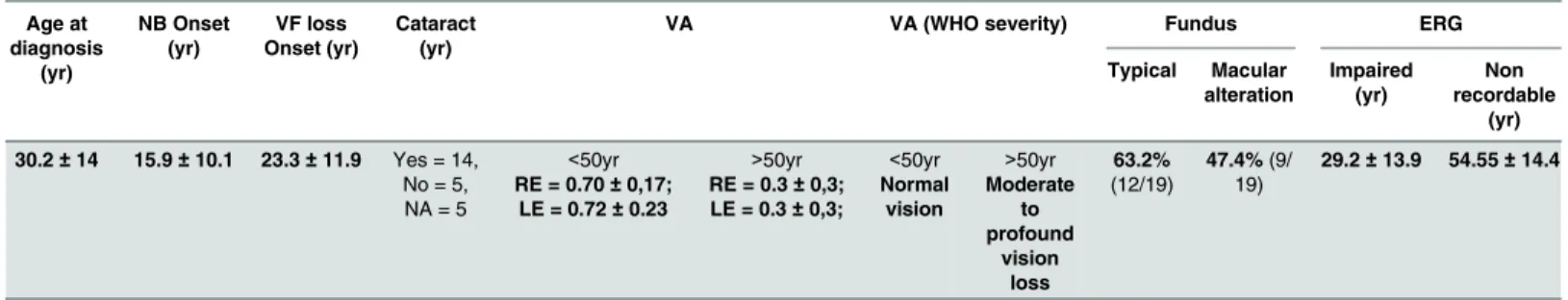
The results of the haplotype analysis are conclusive for the two most informative families belonging to the same small village (Fig 3). We found a common haplotype in those families for the four markers and for three of them in the family RP-1182. These data support an ancient founder effect for the p.Gly56Arg in our families. However one must be cautious because i) one out of four families coming from the same small geographical area did not share the common haplotype. This fact is unexpected and despite there being an association between the 289 allele of D15S1050 and p.Gly56Arg, D15S1050 andNR2E3are in different disequilib-rium blocks, we have to consider that the Spanish population is underrepresented in the Hap-map project and that the allele 289 has a frequency of 0.519 (http://www.genoscope.cns.fr/ externe/gmap/Nature-1995/), so the probability that both the allele 289 and the mutation are located in the chromosome is very high. Furthermore, there are no hot spot recombination sites like the common motif CCNCCNTNNCCNC [26] within theNR2E3gene and their 5’ and 3’UTR regions that could explain the lack of a common ancestral haplotype by a high recombination rate, ii) The frequency of p.Gly56Arg in populations that have evolutionary diverged, such as American, European and Chinese, are high and quite similar (1.2% in Ameri-can [19], 3.4% in European [17] and 1.2% in China [27]) and iii) the mutation c.166G>A is a change GGG>AGG in codon 56 ofNR2E3. This change lies on a CpG dinucleotide and has been reported as highde novomutation site. In fact, the most commonde novomutated codon associated with human disease is a GGG>AGG or CGG mutation in codon 380 ofFGFR3gene Table 2. Phenotypic characteristics (means and standard deviation) of patients with the p.Gly56Arg mutation inNR2E3.
Age at diagnosis
(yr)
NB Onset (yr)
VF loss Onset (yr)
Cataract (yr)
VA VA (WHO severity) Fundus ERG
Typical Macular alteration
Impaired (yr)
Non recordable
(yr)
30.2±14 15.9±10.1 23.3±11.9 Yes = 14, No = 5, NA = 5
<50yr RE = 0.70±0,17;
LE = 0.72±0.23
>50yr RE = 0.3±0,3; LE = 0.3±0,3;
<50yr Normal
vision
>50yr Moderate
to profound
vision loss
63.2% (12/19)
47.4%(9/ 19)
29.2±13.9 54.55±14.4
Yr: Years. VA: Visual Acuity. VF: Visual Field. ERG: Electroretinogram. NA: Not Available. RE: Right Eye. LE: Left Eye. VA classificationa: 0 = Normal
vision (normal and near normal vision) (0.4), 1 = Moderate low vision (<0.4–>0.1), 2 = Severe low vision (0.1–0.05, legal blindness), 3 = profound
vision loss and blindness (blindness and near blindness,<0.05). Typical fundus: optic disc pallor, attenuation of the retinal vessels and pigmentary deposits resembling bone spicules.
doi:10.1371/journal.pone.0149473.t002
NR2E3: p.Gly56Arg Mutation in adRP Spanish Population
[28,29] which results in achondroplasia. In summary, it is tempting to speculate that the p. Gly56Arg arose once in our population as a very ancient hit giving time for a number of recom-bination events that have led to the change in the ancient haplotype.
TheNR2E3gene is associated with both autosomal recessive and dominant retinitis pig-mentosa. TheNR2E3gene recessive mutations present variable phenotypes (ESCS, GFS and Fig 2. Fundus Images: Each patient presents two fundus images per eye.A) Family RP-0030, B) Family RP-0576, C) Family RP-0711.*Fundus image
not available.**Enucleation of the LE due to glioblastoma. RE = Right eye. LE = Left eye.
doi:10.1371/journal.pone.0149473.g002
Fig 3. Pedigrees and Haplotype analysis of adRP families with p.Gly56Arg mutation inNR2E3.Haplotype analysis:!marks theNR2E3gene position.
TheNR2E3extragenic polymorphic markers used are D15S967, D15S1050, D15S204 and D15S188.
doi:10.1371/journal.pone.0149473.g003
NR2E3: p.Gly56Arg Mutation in adRP Spanish Population
CPRD) with variable ophthalmological findings, but all showing night blindness, rudimental or absent rod function, and hyperfunction of the "blue" S-cones [14,20,30]. DominantNR2E3 gene mutation has been associated with RP phenotype [17–20] and to date, the phenotype has only been described in five European [17,18,20] and four North American [19] families char-acterized by presenting RP, but not to other phenotypes associated toNR2E3gene recessive mutations [16,17]. However, some affected members of those families displayed phenotypic similarities to ESCS [17,20].
Although we observe, as it has been previously described [18], inter and intrafamiliar phe-notype variability for the p.Gly56Arg mutation, some gephe-notype-phephe-notype correlation appears to be quite apparent, not only homogeneous progression [19], but occurrence of vision impairment milestones. Patients withNR2E3dominant mutation showed a moderate form of retinal dystrophy. Thus, patients in our cohort present early NB onset around puberty, preser-vation of VF>10° until the 4th-5thdecade of life, normal or moderately low VA (WHO criteria) to an advanced age and preservation of photoreceptor function (recordable ERG) until the 4th decade of life (Table 2).
It is important to notice that this phenotype is milder than the one found in recessive forms [18] and to point that the fundus alterations (additionally to typical RP fundus, atypical find-ings are present as macular changes and choriocapillaris atrophy) and ERG findfind-ings (impaired photopic and flicker records in the first stages of the disease) observed in some of our patients, are not the classical RP changes previously described in the European and North American NR2E3dominant patients [17,19,20].
Moreover these changes did not seem to correlate with visual acuity impairment, at least during the first stages of the disease (impaired ERG and fundus changes with normal VA). However the absence of correlation between ERG and VA is common in RP patients [1].
These fundus changes have recently been described on the ophthalmoscopic findings of NR2E3ESCS (recessive) patients. These changes can lead to a misdiagnosis if family history,
initial symptoms reported by patients and progression of the disease in the early stages are not queried when performing the clinical history. Besides, other ophthalmological signs such as double concentric autofluorescence ring (not performed in our cohort), that has been described as an initial sign of retinal degeneration in patients carrying theNR2E3dominant mutation [20], could help in the molecular diagnostic orientation of adRP patients.
Furthermore, some of these changes such as macular edema, can, at least partially, be treated [31,32], which is important for prognosis, disease outcome and follow up of these patients. Although this event was only observed in one of our patients by ophthalmoscopic examination, we cannot discard that it could be present in other NR2E3 adRP patients, as no OCT data was available for the present cohort.
Conclusion
We believe that the relevance of this study is not only being the single largestNR2E3 genotype-phenotype correlation study performed to date, but also highlighting the importance of p. Gly56Arg in theNR2E3gene associated to adRP.
NR2E3is responsible for two main retinal phenotypes ESCS (including Goldmann-Favre
syndrome) only for recessive forms and RP (including CPRD) related to both dominant and recessive forms. However, in our cohort there is a wide range of phenotypic characteristics that differ from typical RP phenotype and resemble otherNR2E3phenotypes such as ESCS and CPRD, mainly at a fundus level where we found macular disturbance, which is frequently seen in ESCS, in 50% of our evaluated patients and two patients with nummular pigmentation (typi-cal of CPRD phenotype).
NR2E3: p.Gly56Arg Mutation in adRP Spanish Population
In this report, we are providing new clues of a characteristic phenotype for this mutation that allows for making an estimated prognosis of autosomal dominant RP due to p.Gly56Arg mutation in theNR2E3gene. Besides, we believe that this study can improve molecular diagno-sis approach, clinical management, risk assessment, and genetic counselling of adRP patients and their families.
Supporting Information
S1 Fig. Electroretinogram (ERG) recording in IV:2 member of RP-0711 family.A) ERG at 11years of age. B) ERG at 18 years of age.
(DOCX)
Author Contributions
Conceived and designed the experiments: MGH FBK RRA CA. Performed the experiments: MALM PFSJ. Analyzed the data: FBK PFSJ AAF MGH BGS MILM CA. Contributed reagents/ materials/analysis tools: MALM AAF PFSJ. Wrote the paper: FBK PFSJ RRA JMM CA. Oph-thalmologic data evaluation: MILM BGS FBK. Population genetics analysis: JMM FBK MC PFSJ.
References
1. Hamel C. Retinitis pigmentosa. Orphanet J Rare Dis. 2006 Oct; 1:40. PMID:17032466
2. Gawande AA, Donovan WJ, Ginsburg AP, Marmor MF. Photoaversion in retinitis pigmentosa. Br J Ophthalmol 1989 Feb; 73:115–20. PMID:2930757
3. Marmor MF. Visual acuity and field loss in retinitis pigmentosa. Arch Ophthalmol 1991 Jan; 109(1):13– 4. PMID:1987931
4. Marmor MF. Visual loss in retinitis pigmentosa. Am J Ophthalmol 1980 May; 89(5):692–8. PMID:
7377267
5. Marmor MF. The electroretinogram in retinitis pigmentosa. Arch Ophthalmol 1979 Jul; 97(7):1300–4. PMID:454267
6. Haim M. Retinitis pigmentosa: problems associated with genetic classification. Clin Genet 1993 Aug; 44(2):62–70. PMID:8275561
7. Ayuso C, Garcia-Sandoval B, Najera C, Valverde D, Carballo M, Antiñolo G. Retinitis pigmentosa in Spain. The Spanish Multicentric and Multidisciplinary Group for Research into Retinitis Pigmentosa. Clin Genet 1995 Sep; 48(3):120–2. PMID:8556816
8. Ayuso C, editor. Estudio de la Retinosis Pigmentaria en España. Capitulo XI: La Retinosis Pigmentaria en España: estudio clínico y genético. ONCE (Organización Nacional de Ciegos Españoles), 2001.
9. Roduit R, Escher P, Schorderet DF. Mutations in the DNA-binding domain of NR2E3 affect in vivo
dimerization and interaction with CRX. PLoS One 2009 Oct; 4(10):e7379. doi:10.1371/journal.pone. 0007379PMID:19823680
10. Jacobson SG, Marmor MF, Kemp CM, Knighton RW. SWS (blue) cone hypersensitivity in a newly iden-tified retinal degeneration. Invest Ophthalmol Vis Sci. May 1990; 31(5):827–838. PMID:2335450
11. Marmor MF, Jacobson SG, Foerster MH, Kellner U, Weleber RG. Diagnostic clinical findings of a new syndrome with night blindness, maculopathy, and enhanced S cone sensitivity. Am J Ophthalmol. Aug 15 1990; 110(2):124–134. PMID:2378376
12. Nasr YG, Cherfan GM, Michels RG, Wilkinson CP. Goldmann-Favre maculopathy. Retina. 1990; 10 (3):178–180. PMID:2236941
13. Sharon D, Sandberg MA, Caruso RC, Berson EL, Dryja TP. Shared mutations in NR2E3 in enhanced S-cone syndrome, Goldmann-Favre syndrome, and many cases of clumped pigmentary retinal degen-eration. Arch Ophthalmol. 2003 Sep; 121(9):1316–23. PMID:12963616
14. Schorderet DF, Escher P. NR2E3 mutations in enhanced S-cone sensitivity syndrome (ESCS), Gold-mann-Favre syndrome (GFS), clumped pigmentary retinal degeneration (CPRD), and retinitis pigmen-tosa (RP). Hum Mutat 2009 Nov; 30(11):1475–85. doi:10.1002/humu.21096PMID:19718767
NR2E3: p.Gly56Arg Mutation in adRP Spanish Population
15. Haider NB, Jacobson SG, Cideciyan AV, Swiderski R, Streb LM, Searby C, et al. Mutation of a nuclear receptor gene, NR2E3, causes enhanced S cone syndrome, a disorder of retinal cell fate. Nat Genet 2000 Feb; 24(2):127–31. PMID:10655056
16. Yzer S, Barbazetto I, Allikmets R, van Schooneveld MJ, Bergen A, Tsang SH, et al. Expanded clinical spectrum of enhanced S-cone syndrome. JAMA Ophthalmol 2013 Oct; 131(10):1324–30. doi:10.1001/
jamaophthalmol.2013.4349PMID:23989059
17. Coppieters F, Leroy BP, Beysen D, Hellemans J, De Bosscher K, Haegeman G, et al. Recurrent muta-tion in the first zinc finger of the orphan nuclear receptor NR2E3 causes autosomal dominant retinitis pigmentosa. Am J Hum Genet 2007 Jul; 81(1):147–57. PMID:17564971
18. Escher P, Gouras P, Roduit R, Tiab L, Bolay S, Delarive T, et al. Mutations in NR2E3 can cause domi-nant or recessive retinal degenerations in the same family. Hum Mutat 2009 Mar; 30(3):342–51. doi:
10.1002/humu.20858PMID:19006237
19. Gire AI, Sullivan LS, Bowne SJ, Birch DG, Hughbanks-Wheaton D, Heckenlively JR, et al. The Gly56Arg mutation in NR2E3 accounts for 1–2% of autosomal dominant retinitis pigmentosa. Mol Vis 2007 Oct; 13:1970–5. PMID:17982421
20. Escher P, Tran HV, Vaclavik V, Borruat FX, Schorderet DF, Munier FL. Double Concentric Autofluores-cence Ring in NR2E3-p.G56R-Linked Autosomal Dominant Retinitis Pigmentosa. Invest Ophthalmol Vis Sci 2012 Jul; 53(8):4754–64. doi:10.1167/iovs.11-8693PMID:22661467
21. Sullivan LS, Bowne SJ, Reeves MJ, Blain D, Goetz K, Ndifor V, et al. Prevalence of Mutations in eye-GENE Probands With a Diagnosis of Autosomal Dominant Retinitis Pigmentosa. Invest Ophthalmol Vis Sci 2013 Sep; 54(9):6255–61. doi:10.1167/iovs.13-12605PMID:23950152
22. McCulloch DL, Marmor MF, Brigell MG, Hamilton R, Holder GE, Tzekov R, et al. ISCEV Standard for full-field clinical electroretinography (2015 update). Doc Ophthalmol. 2015 Feb: 130(1):1–12 doi:10.
1007/s10633-014-9473-7PMID:25502644
23. Tonisson N, Kurg A, Lohmussaar E, Metspalu A. Arrayed primer extension on the DNA chip-method & applications. In: Schena M, ed. Microarray Biochip Technology. Natick, MA: Eaton Publishing 2000; 247–63.
24. Blanco-Kelly F, García-Hoyos M, Cortón M, Avila-Fernández A, Riveiro-Álvarez R, Giménez A, et al. Genotyping microarray: mutation screening in Spanish families with autosomal dominant retinitis pig-mentosa. Mol Vis 2012; 18:1478–83. PMID:22736939
25. Fernandez-San Jose P, Blanco-Kelly F, Corton M, Trujillo-Tiebas MJ, Gimenez A, Avila-Fernandez A, et al. Prevalence of Rhodopsin mutations in autosomal dominant Retinitis Pigmentosa in Spain: clinical and analytical review in 200 families. Acta Ophthalmol. 2015 Feb; 93(1):e38–44. doi:10.1111/aos.
12486PMID:25408095
26. Myers S, Freeman C, Auton A, Donnelly P, McVean G. A common sequence motif associated with recombination hos spots and genome instability in humans. Nat Genet 2008 Sep; 40(9):1124–29. doi:
10.1038/ng.213PMID:19165926
27. Yang Y, Zhang X, Chen LJ, Chiang SW, Tam PO, Lai TY, et al. Association of NR2E3 but not NRL mutation with retinitis pigmentosa in the Chinese population. Invest Ophthalmol Vis Sci 2010 Apr; 51 (4):2229–35. doi:10.1167/iovs.09-4299PMID:19933183
28. Crow JF. The high spontaneous mutation rate: is it a health risk?. Proc Natl Acad Sci 1997 Aug; 94 (16):8380–86. PMID:9237985
29. Drake JW, Charlesworth B, Charlesworth D, Crow JF. Rates of spontaneous mutation. Genetics 1998 Apr; 148(4):1667–86. PMID:9560386
30. Audo I, Michaelides M, Robson AG, Hawlina M, Vaclavik V, Sandbach JM, et al. Phenotypic variation in enhanced S-cone syndrome. Invest Ophthalmol Vis Sci 2008 May; 49(5):2082–93. doi:10.1167/iovs.
05-1629PMID:18436841
31. Ganesh A, Stroh E, Manayath GJ, Al-Zuhaibi S, Levin AV. Macular cysts in retinal dystrophy. Curr Opin Ophthalmol 2011 Sep; 22(5):332–9. doi:10.1097/ICU.0b013e328349229ePMID:21730849
32. Ikeda Y, Yoshida N, Notomi S, Murakami Y, Hisatomi T, Enaida H, et al. Therapeutic effect of prolonged treatment with topical dorzolamide for cystoid macular oedema in patients with retinitis pigmentosa. Br J Ophthalmol 2013 Sep; 97(9):1187–91. doi:10.1136/bjophthalmol-2012-303005PMID:23782868
NR2E3: p.Gly56Arg Mutation in adRP Spanish Population