Missense Mutations in
Are Liable for
Recessive Congenital Cataracts
Xiaodong Jiaox1☯, Shahid Y. Khan2☯, Bushra Irum2,3, Arif O. Khan4, Qiwei Wang1, Firoz Kabir2, Asma A. Khan3, Tayyab Husnain3, Javed Akram5,6, Sheikh Riazuddin3,5,6, J. Fielding Hejtmancik1☯, S. Amer Riazuddin2☯*
1Ophthalmic Genetics and Visual Function Branch, National Eye Institute, National Institutes of Health, Bethesda, MD, 20892, United States of America,2The Wilmer Eye Institute, Johns Hopkins University School of Medicine, Baltimore, MD, 21287, United States of America,3National Centre of Excellence in Molecular Biology, University of the Punjab, Lahore, 53700, Pakistan,4King Khaled Eye Specialist Hospital, Riyadh, 12329, Saudi Arabia,5Allama Iqbal Medical College, University of Health Sciences, Lahore, 54550, Pakistan,6National Centre for Genetic Diseases, Shaheed Zulfiqar Ali Bhutto Medical University,
Islamabad, Pakistan
☯These authors contributed equally to this work. *[email protected]
Abstract
Purpose
This study was initiated to identify causal mutations responsible for autosomal recessive congenital cataracts in consanguineous familial cases.
Methods
Affected individuals underwent a detailed ophthalmological and clinical examination, and slit-lamp photographs were ascertained for affected individuals who have not yet been oper-ated for the removal of the cataractous lens. Blood samples were obtained, and genomic DNA was extracted from white blood cells. A genome-wide scan was completed with short tandem repeat (STR) markers, and the logarithm of odds (LOD) scores were calculated.
Protein coding exons ofCRYABwere sequenced, bi-directionally. Evolutionary
conserva-tion was investigated by aligningCRYABorthologues, and the expression ofCryabin
embryonic and postnatal mice lens was investigated with TaqMan probe.
Results
The clinical and ophthalmological examinations suggested that all affected individuals had nuclear cataracts. Genome-wide linkage analysis suggested a potential region on
chromo-some 11q23 harboringCRYAB. DNA sequencing identified a missense variation: c.34C>T
(p.R12C) inCRYABthat segregated with the disease phenotype in the family. Subsequent
interrogation of our entire cohort of familial cases identified a second familial case localized
to chromosome 11q23 harboring a c.31C>T (p.R11C) mutation.In silicoanalyses
sug-gested that the mutations identified in familial cases, p.R11C and p.R12C will not be
toler-ated by the three-dimensional structure ofCRYAB. Real-time PCR analysis identified the
a11111
OPEN ACCESS
Citation:Jiaox X, Khan SY, Irum B, Khan AO, Wang Q, Kabir F, et al. (2015) Missense Mutations in
CRYABAre Liable for Recessive Congenital
Cataracts. PLoS ONE 10(9): e0137973. doi:10.1371/ journal.pone.0137973
Editor:Yong-Bin Yan, Tsinghua University, CHINA
Received:April 24, 2015
Accepted:August 25, 2015
Published:September 24, 2015
Copyright:This is an open access article, free of all copyright, and may be freely reproduced, distributed, transmitted, modified, built upon, or otherwise used by anyone for any lawful purpose. The work is made available under theCreative Commons CC0public domain dedication.
Data Availability Statement:The minimal dataset needed for replication can be found and accessed within the paper.
Funding:This study was supported in part by the National Eye Institute, Grant 1R01EY022714 (SAR), Knight Templar Eye Foundation Grant (SAR), King Khaled Eye Specialist Hospital-Johns Hopkins University collaboration grant (SAR), the National Academy of Sciences, Washington DC USA and the Higher Education Commission, Islamabad Pakistan.
expression ofCryabin mouse lens as early as embryonic day 15 (E15) that increased signif-icantly until postnatal day 6 (P6) with steady level of expression thereafter.
Conclusion
Here, we report two novel missense mutations, p.R11C and p.R12C, inCRYABassociated
with autosomal recessive congenital nuclear cataracts.
Introduction
Congenital cataracts are the principal cause of visual impairment in children as they responsi-ble for one-third of cases of blindness in infants worldwide [1,2]. The ocular lens focus the light on the retina and the loss of transparency of the lens comprises this important function, which could lead to permanent blindness, especially during the early developmental periods. Nearly, one-third of the total cases congenital cataract are familial with both autosomal dominant and autosomal recessive inheritance [3]. Congenital cataracts are genetically heterogeneous with genetic loci for both autosomal dominant cataracts (adCC) and autosomal recessive cataracts (arCC) have been localized.
The last decade witnessed localization of multiple loci for congenital cataracts and taken together a total 16 arCC loci have so far been reported [4–19]. Of these genetic loci, causal mutations in eph-receptor type-A2 (EPHA2), connexin50 (GJA8), FYVE and coiled-coil domain containing 1 (FYCO1), glucosaminyl (N-acetyl) transferase 2 (GCNT2), heat-shock transcription factor 4 (HSF4), lens intrinsic membrane protein 2 (LIM2), beaded filament structural protein 1 (BFSP1), crystallin alpha A (CRYAA), crystallin beta B1 (CRYBB1), and crystallin beta B3 (CRYBB3) have been identified [4,6,8,11,13–17,20].
Crystallins constitute nearly 95% of the soluble protein of the vertebrate eye lens as high concentrations of tightly packed crystalline proteins are required for lens transparency and its physiological function [21]. They are sub-divided into three classes, namely alpha, beta and gamma crystallins based upon their elution profile on gel exclusion chromatography.CRYAB
is located on chromosome 11q23 and encodes for a member of the small heat-shock protein family comprising of 175 amino acid protein [22]. The CRYAB protein is expressed in multiple tissues including the ocular lens, heart, skeletal muscle, kidney, lung, and glia in the central ner-vous system [22].
Here, we report two consanguineous familial cases with multiple individuals in both families having congenital cataracts. Ophthalmic examination with a slit lamp confirmed nuclear cata-racts present in the affected individuals. The genome-wide linkage or exclusion analysis local-ized the disease phenotype in two consanguineous familial cases to chromosome 11q23. Bi-directional sequencing identified missense mutations inCRYABthat segregated with the dis-ease phenotype in their respective families and were absent in ethnically matched controls chromosomes. To the best of our knowledge, this is the first report identifying mutations in
CRYABassociated with congenital cataracts in Pakistani families.
Materials and Methods
Recruitment and Clinical Assessment
A total of>200 consanguineous Pakistani families with non-syndromic cataract were recruited
Board (IRB) of National Centre of Excellence in Molecular Biology, Lahore Pakistan, the CNS IRB of the National Eye Institute, Bethesda MD and Johns Hopkins University, Baltimore MD approved for this study. All participating family members provided informed written consent that has been endorsed by the respective IRBs and is consistent with the tenets of the Declara-tion of Helsinki. A detailed clinical and medical history was obtained from the individual fami-lies. The ophthalmic examination was performed with a slit-lamp and phtogrpahs were taken to record the ocular phenotype. A consent to publish ocular phenotype was obtained from the patient and/or the legal guardian. All participating members voluntarily provided blood sample of approximately 10 ml that was stored in 50 ml Sterilin1falcon tubes containing 400μl of 0.5 M EDTA. Blood samples were stored at -20°C for long-term storage.
Genomic DNA Extraction
The genomic DNAs were extracted from white blood cells using a non-organic modified proce-dure as described previously [23]. The concentration of the extracted genomic DNA was esti-mated using a SmartSpec plus Bio-Rad Spectrophotometer (Bio-Rad, Hercules, CA).
Genome-Wide Scan and Exclusion Analysis
The Applied Biosystems MD-10 linkage mapping panels (Applied Biosystems, Foster City, CA) were used to complete a genome-wide scan for family PKCC001. Multiplex polymerase chain reaction (PCR) was completed as described previously [23]. PCR products were mixed with a loading cocktail containing HD-400 size standards (Applied Biosystems) and resolved in an Applied Biosystems 3100 DNA Analyzer. Genotypes were assigned using the Gene Map-per software from the Applied Biosystems. Exclusion analysis was completed for PKCC113 using closely spaced STR markers. The sequences of the primer pairs used for exclusion analy-sis and amplification conditions are available upon request
Linkage Analysis
Linkage analysis was performed with alleles of PKCC001 obtained through the genome-wide scan and alleles of PKCC113 obtained through exclusion analysis using the FASTLINK version of MLINK from the LINKAGE Program Package [24,25]. Maximum LOD scores were calcu-lated using ILINK from the LINKAGE Program Package. arCC was investigated as a
completely penetrant disorder with an affected allele frequency of 0.001.
Mutation Screening
The sequences of primer pairs used to amplify individual exons ofCRYABare available upon request. PCR reactions were completed in 10μl volume containing 20 ng of genomic DNA. The reaction consisted of a denaturation step at 95°C for 5 min followed by a two-step touch-down procedure. The first step of 10 cycles consisted of denaturation at 95°C for 30 seconds, followed by a primer set specific annealing for 30 seconds (annealing temperature decrease by 1°C per cycle) and elongation at 72°C for 45 seconds. The second step of 30 cycles consisted of denaturation at 95°C for 30 seconds followed by annealing (annealing temperature -10°C) for 30 seconds and elongation at 72°C for 45 seconds, followed by a final elongation 72°C for 5 minutes.
Evolutionary Conservation
Evolutionary conservation of the amino acid Arg11 and Arg12 was investigated by aligning the protein sequence of CRYAB orthologues. The evolutionary conservation of amino acid and the possible effect of the amino acid substitution on the structure of the CRYAB protein was exam-ined using SIFT (http://sift.jcvi.org) and PolyPhen2 (http://genetics.bwh.harvard.edu/pph2/
index.shtml) algorithms, respectively.
Biophysical Characteristics
The polarity, optimized matching hydrophobicity, and hydropathicity of the wild type and mutant CRYAB proteins was examined using ProtScale, a bioinformatics tool on ExPASy Server (http://www.expasy.org/tools/protscale.html). Similarly, we used ProtScale to compute the isoelectric point (pI) and the molecular weight (Mw) of wild type and mutant CRYAB pro-teins. The machine-learning algorithm (mCSM) was employed to predict the deleterious effects of missense variants on CRYAB structure. The crystal structure of human CRYAB (PDB code 2YGD) was utilized to predict the impact on protein stability.
Real-Time Expression Analysis
The use of mice in this study was approved by the Johns Hopkins Animal Care and Use com-mittee (ACUC), and all protocols were performed in accordance with a protocol approved by the Johns Hopkins ACUC. Mouse lenses were obtained at different developmental stages including embryonic day 15 (E15), day 18 (E18), at birth, designated as (P0), postnatal day 3 (P3), day 6 (P6), day 9 (P9), day 12 (P12), day 14 (P14), day 21 (P21), day 28 (P28), day 42 (P42), day 56 (P56). Mice were first anesthetized with isoflurane and subsequently euthanized through cervical dislocation. The ocular tissue was extracted, and the lenses were isolated from the retina using forceps under a microscope. The lenses were divided into two pools, each repre-senting a biological replicates for the respective developmental stage. Lenses were dissolved in TRIzol reagent (Invitrogen; Carlsbad, CA) immediately after isolation, and total RNA was extracted from each pool according to manufacturer’s instructions. The quality and quantity of the total RNA was determined on a NanoDrop Lite spectrophotometer (Thermo Scientific, Inc.). First-strand cDNA synthesis was completed using the Superscript III kit (Invitrogen) according to the manufacturer’s instructions. Quantitative real-time PCR analysis was per-formed on a STEP ONE ABI Real-Time PCR System using predesignedCryabTaqMan expres-sion assays (Applied Biosystems).Gapdhwas used as an endogenous internal control. The 2-ΔCT method was used to determine the relative expression normalized toGapdhexpression at each developmental stage.
Results
We were able to enroll a total of four affected individuals along with 19 unaffected members of PKCC001. The large numbers of enrollment augmented the power of the family to generate statistically significant two-point LOD scores during genome-wide linkage. Our theoretical estimates confirmed that PKCC001 can attain a maximum two-point LOD score of 5.30 atθ=
Fig 1. Pedigree drawing with haplotypes of chromosome 11q microsatellite markers.A) Family PKCC001 and B) family PKCC113 with alleles forming the risk haplotype are shown in black, heterozygous alleles part of the risk haplotype are shown in grey and alleles not cosegregating with cataract phenotype are shown in white. Squares: males; circles: females; filled symbols: affected individuals; the double line between individuals: consanguineous mating; and a diagonal line through a symbol: a deceased individual.
0. We completed a genome-wide scan and subsequently calculated two-point LOD scores. Sur-prisingly, we did not identify a single region of statistical significance (LOD>3) or suggestive
linkage (LOD>2) across the entire genome except marker D19S414 on chromosome 19
yield-ing a two-point LOD score of 2.0 atθ= 0. However, markers adjacent to D19S414, D19S226 on the proximal and D19S220 on distal end produced highly negative two-point LOD scores rul-ing out the candidacy of chromosome 19q.
The lack of two-point LOD scores mimicking the theoretical potential of PKCC001 during the genome-wide scan, although surprising, but is certainly not new to us. We have witnessed large familial cases in our cohort yielding huge two-point LOD scores during theoretical simu-lations without presenting any peaks of significant and/or suggestive linkage during genome-wide scans. The most likely explanation being that a high degree of consanguinity and inbreed-ing for many generations may have reduced the critical disease interval below 10 cM resolution of the MD-10 panel.
Thus, we re-evaluated our linkage data and identified a region of chromosome 11q23 with two adjacent panel markers D11S4175 and D11S925 yielding positive two-point LOD scores
Fig 2. Slit lamp photographs of individual 19 of family PKCC001 illustrating nuclear cataracts.
(Table 1A). To establish linkage to chromosome 11q23 region, we chose a marker between D11S4175 and D11S925 from the MD-5 panel, D11S4078 that yielded a two-point LOD score of 4.29 atθ= 0 (Table 1A).
This region harborsCRYAB, a gene previously associated with cardiomyopathy and con-genital cataracts. Bi-directional sequencing ofCRYABidentified a missense variation: c.34C>T; p.R12C that segregated with the disease phenotype in PKCC001 (Figs1Aand3A–
3C). The exome variant server analysis identified the variant in heterozygous form (AA = 0/ AG = 1/GG = 4295) in one individual representing a minor allele frequency (MAF) of 0.0116 in European American population. Likewise, dbSNP analysis revealed MAF score of 0.0010 for rs375933774 (c.34C>T) based on 1000 Genomes database. This variation was not found in
384 control chromosomes of Pakistani and 48 control chromosomes of Saudi decent. A two-point LOD score of 5.53 atθ= 0 was obtained for the causal variant (Table 1A).
To estimate the total genetic load ofCRYABin our cohort of familial cases, we interrogated our cohort of>200 familial cases of congenital cataracts by genotyping closely-spaced STR
markers followed by sequencing all coding exons ofCRYAB. We identified one additional fam-ily, PKCC113 linked to the chromosome 11q23 (Fig 1B) with positive two-point LOD scores
(Table 1B). Bi-directional sequencing ofCRYABidentified a novel missense mutation:
c.31C>T (p.R11C) that segregated with the disease phenotype within the family (Fig 3D–3F).
Likewise, this variation was not found in 384 and 48 control chromosomes of Pakistani and Saudi decent, respectively.
We used SIFT and PolyPhen2 algorithms to evaluate the possible impact of p.R11C and p. R12C mutations on CRYAB. SIFT predictions suggested that both the R11C and R12C substi-tutions will not be tolerated by the native three-dimensional structure of CRYAB. The effect protein function score for R11C and R12C were 0.00 and 0.00, respectively (amino acids with probabilities<.05 are predicted to be deleterious). Likewise, Polyphen2 suggested that both
the R11C and R12C substitutions are probably damaging to the CRYAB structure with a score of 1.00, and 1.00, respectively. We found that both arginines at position 11, and 12 are not only conserved in CRYABmammalian orthologues (Fig 4) but also conserved in CRYAB vertebrate orthologues according to the UCSC genome browser (data not shown).
Subsequently, we examined the impact of these mutations on the physical characteristics of CRYAB. The ProtScale software predicted lower polarity, higher hydrophilicity and hydropho-bicity of the mutant CRYAB compared to the wild type residues in protein secondary structure
(Fig 5). In parallel, we used mCSM, a structure-based algorithm to validate the damaging
nature of both missense variants identified in CRYAB. The analysis predicted the destabilizing
Table 1. Two-point LOD scores of chromosome 11q markers for families A) PKCC001 and B) PKCC113.The asterisk indicates marker included in the genome-wide scan.
Family ID Marker cM Mb 0.00 0.01 0.05 0.10 0.20 0.30 0.40 Zmax θmax
A
PKCC001 D11S4175* 91.47 90.25 1 -2.13 -0.14 0.54 0.85 0.69 0.34 0.85 0.20
PKCC001 c.34C>T 111.78 5.53 5.05 4.54 3.45 2.27 1.04 5.44 5.53 0.00
PKCC001 D11S4078 105.74 112.25 4.29 4.21 3.83 3.35 2.33 1.30 0.39 4.29 0.00
PKCC001 D11S925* 118.47 120.82 1 0.87 1.92 2.08 1.75 1.13 0.47 2.08 0.10
B
PKCC113 D11S4175* 91.47 90.25 0.91 0.88 0.76 0.62 0.34 0.13 0.02 0.91 0.00
PKCC113 c.31C>T 111.78 1.63 1.59 1.41 1.19 0.75 0.37 0.12 1.63 0.00
PKCC113 D11S4078 105.74 112.25 1.21 1.17 1.00 0.80 0.41 0.13 0.02 1.21 0.00
PKCC113 D11S925* 118.47 120.82 1.18 1.15 1.00 0.83 0.51 0.26 0.10 1.18 0.00
nature of both variants (R11C and R12C) that would result in the disruption of the secondary structure of the protein (Table 2). Finally, we estimated the isoelectric point (pI) and computed molecular weight of the mutant CRYAB proteins. We found that both mutant CRYAB proteins had a lower pI (pI: 6.5) compared to the wild type CRYAB (pI: 6.76).
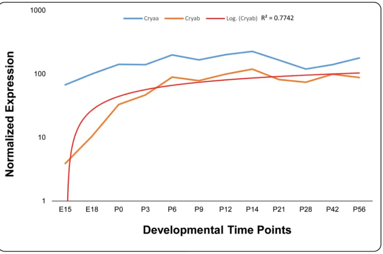
Dubin and colleagues, previously reported the expression ofCRYABin multiple tissues including the ocular lens [22]. We investigated the expression of bothCryaaandCryabin embryonic and postnatal murine lens. As shown inFig 6, we observed expression of bothα
-Fig 3. Sequence chromatograms of causal mutations identified in PKCC001 and PKCC113.Sequence chromatograms of A) Unaffected individual 15 homozygous for wild-type allele; B) unaffected individual 17 heterozygous and C) affected individual 19, homozygous for c.34C>T (p.R12C). Sequence chromatograms of D) unaffected individual 6 homozygous for wild-type allele, E) unaffected individual 8 heterozygous and F) affected individual 10
homozygous for c.31C>T (p.R11C). The arrows point to c.31C and c.34C ofCRYABmutated in PKCC113 and PKCC001, respectively. It is worth to note that mutations identified in PKCC001 and PKCC113 are adjacent amino acids i.e. Arg11, and Arg12.
Fig 4. Sequence alignment ofCRYABin mammalian orthologues illustrating the conservation of Arginine at positions 11 and 12.Primates, Euarchontoglires, Laurasiatheria, and Afrotheria are colored brown, green, purple and orange, respectively.
Crystallins in mouse lens as early as embryonic day 15 (E15); nonetheless, the level ofCryaa
expression was an order of magnitude higher compared with expression ofCryab. In sharp contrast toCryaaof which the expression levels remain nearly steady over the 12 develop-mental stages investigated here, the expression level ofCryabmimics a logarithmic pattern in early stages of increasing significantly up until postnatal day 6 (P6) and from there onwards the expression level remains steady over the remaining time course until two months of age
(Fig 6).
Fig 5. Investigating the physical characteristics of wild-type and mutantCRYABproteins.The polarity (A, D, G), the optimized matching
hydrophobicity (B, E, H), and hydropathicity (C, F, I) plots of the wild-type and mutant CRYAB proteins. Both mutant proteins (R11C and R12C) revealed low polarity (compare A, with D and G), a higher hydrophobicity (compare B, with E and H), and higher hydropathicity (compare C, with F and I), respectively. The x-axis represents the position of amino acids. The y-axis represents the Polarity, hydrophobicity and Hydropathicity values in a default window size of 9. The arrows point to the difference in their respective polarities (1starrow from the left), hydrophobicity (2ndarrow from the left) and hydropathicities (3rdarrow from
the left).
doi:10.1371/journal.pone.0137973.g005
Table 2. The predictive changes ofCRYABmissense variants on the protein stability.RSA: Residue Relative Solvent Accessibility,ΔΔG:Reduction in free energy.
PDB File Chain Wild-type Residue Residue Position Mutant Residue RSA PredicativeΔΔG (Kcal/mol) Outcome
2YGD.pdb A R 11 C 48.9 -1.576 Destabilizing
2YGD.pdb A R 12 C 42.5 -1.448 Destabilizing
Discussion
Here, we report two novel mutations inCRYABassociated with autosomal recessive congenital cataracts identified in consanguineous Pakistani families. Initially, a genome-wide linkage scan localized the critical interval to chromosome 11q23 in PKCC001 while sequencing of the cod-ing exons ofCRYABidentified a novel missense mutation. Subsequently, we identified a second family, PKCC113 harboring a second novel missense mutation inCRYAB. While both muta-tions segregated with the disease phenotype in their respective families, none of these novel variants were present in normal control chromosomes.
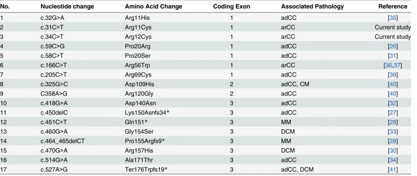
Vicart and colleagues reported a missense mutation (p.R120G) in theCRYABassociated with a desmin-related myopathy with affected individuals exhibiting signs of hypertrophic car-diomyopathy and discrete lens opacities [26]. Subsequently, multiple heterozygous mutations in different ethnic populations were reported with cardiomyopathy and/or cataractogenesis
(Table 3) [26–41]. Among these, Safieh and colleagues reported a missense mutation
c.166C>T (p.R56W) associated with autosomal recessive cataracts in a consanguineous family
of Saudi decent with no clinically significant myopathy [36]. However, one of the older affected individuals presented symptoms of retinal dystrophy and in the later publication the authors showed evidence of clinical rod-cone degeneration only in the adults homozygous for the
Fig 6. Expression profile of alpha-crystallin in developing mouse lens.The expression ofCryaa(blue) andCryab(orange) at different developmental time points was normalized toGapdh. A logarithmic trend line (red) fits theCryabexpression with an R2value of 0.7742. The x-axis and y-axis represent
developmental time points and normalized expression of each mRNA, respectively.
R56W allele who were aphakic since childhood [37]. We did not observe any retinal abnormal-ities in individuals homozygous for R11C and R12C alleles during a follow-up visit; nonethe-less, we cannot rule out the possibility that affected individuals of PKCC001 and/or PKCC113 may develop retinal phenotype in the later years of their lives.
Ghosh and colleagues, recently identified seven interactive sequences for CRYAB chaperone activity through protein pin arrays, which included two sequences in the N-terminal domain, four in the crystallin core domain and one in the C-terminal domain [29]. Interestingly, the first interactive sequence in the N-terminal domain comprising of amino acids 9–20
(WIRRPFFPFHSP) includes Arg11 and Arg12, the two amino acids mutated in PKCC113 and
PKCC001, respectively. Taken together, it is conceivable that these two causal mutations (p. R11C and p.R12C), which substitute a positively charged amino acid for a non-polar amino acid will most likely distort the electrostatic balance of CRYAB. Given the fact that heterozy-gous carriers of these mutations are phenotypically normal, it is safe to assume that both varia-tions render the protein functionless with no gain-of-function or dominant negative effect.
Recently, Chen and colleagues reported a missense mutation involving Arg11, an arginine to histidine substitution (R11H) responsible for autosomal dominant congenital nuclear cata-racts. The mutation responsible for a dominant phenotype results from a positively charged arginine substituting for a positively charged histidine while the recessive phenotype results from polar cysteine substitution. Nevertheless, the biophysical characteristics of the p.R11C mutant CRYAB (Fig 5andTable 2) are not much different compared with p.R11H mutant protein [35]. The precise mechanism of different inheritance patterns resulting from different substitutions for a particular amino acid remains elusive and warrant additional biochemical analyses to decipher the mechanism responsible for different inheritance patterns.
In conclusion, we report two mutations inCRYABassociated with autosomal recessive con-genital cataracts. Identification of causal variants associated with cataractogenesis will help us better understand the biology of the ocular lens including mechanistic details of the mainte-nance of lens transparency.
Table 3. A list of causal mutations reported inCRYABassociated with congenital cataracts and cardiomyopathy.adCC: autosomal dominant con-genital cataracts; arCC: autosomal recessive concon-genital cataracts; DCM: dilated cardiomyopathy; MM: myofibrillar myopathy.
No. Nucleotide change Amino Acid Change Coding Exon Associated Pathology Reference
1 c.32G>A Arg11His 1 adCC [35]
2 c.31C>T Arg11Cys 1 arCC Current study
3 c.34C>T Arg12Cys 1 arCC Current study
4 c.59C>G Pro20Arg 1 adCC [26]
5 c.58C>T Pro20Ser 1 adCC [31]
6 c.166C>T Arg56Trp 1 arCC [36,37]
7 c.205C>T Arg69Cys 1 adCC [39]
8 c.325G>C Asp109His 2 adCC, CM [40]
9 C358A>G Arg120Gly 2 adCC [40]
10 c.418G>A Asp140Asn 3 adCC [32]
11 c.450delC Lys150Asnfs34* 3 adCC [27]
12 c.451C>T Gln151* 3 MM [28]
13 c.460G>A Gly154Ser 3 DCM [33]
14 c.464_465delCT Pro155Argfs9* 3 MM [28]
15 c.470G>A Arg157His 3 DCM [30]
16 c.514G>A Ala171Thr 3 adCC [34]
17 c.527A>G Ter176Trpfs19* 3 adCC, DCM [41]
Acknowledgments
The authors are grateful to the respective families for their participation in this study. This study was supported in part by the National Eye Institute Grant 1R01EY022714 (SAR), the Knight Templar Eye Foundation Grant (SAR), the King Khaled Eye Specialist Hospital-Johns Hopkins University collaboration grant (SAR), the National Academy of Sciences, Washington DC USA and the Higher Education Commission, Islamabad Pakistan.
Author Contributions
Conceived and designed the experiments: XJ SYK QW FK SR JFH SAR. Performed the experi-ments: XJ SYK BI QW FK SAR. Analyzed the data: XJ SYK BI AOK QW FK AAK TH JA SR JFH SAR. Contributed reagents/materials/analysis tools: AOK TH JA SR JFH SAR. Wrote the paper: XJ SYK AOK QW FK TH SR JFH SAR.
References
1. Robinson GC, Jan JE, Kinnis C (1987) Congenital ocular blindness in children, 1945 to 1984 1502. Am J Dis Child 141: 1321–1324. PMID:3687875
2. Hejtmancik JF, Smaoui N (2003) Molecular genetics of cataract. Dev Ophthalmol 37: 67–82. PMID:
12876830
3. Foster A, Johnson GJ (1990) Magnitude and causes of blindness in the developing world. Int Ophthal-mol 14: 135–140. PMID:2188914
4. Kaul H, Riazuddin SA, Shahid M, Kousar S, Butt NH, Zafar AU et al. (2010) Autosomal recessive con-genital cataract linked to EPHA2 in a consanguineous Pakistani family. Mol Vis 16: 511–517. PMID:
20361013
5. Butt T, Yao W, Kaul H, Xiaodong J, Gradstein L, Zhang Y et al. (2007) Localization of autosomal reces-sive congenital cataracts in consanguineous Pakistani families to a new locus on chromosome 1p. Mol Vis 13: 1635–1640. PMID:17893665
6. Ponnam SP, Ramesha K, Tejwani S, Ramamurthy B, Kannabiran C (2007) Mutation of the gap junction protein alpha 8 (GJA8) gene causes autosomal recessive cataract. J Med Genet 44: e85. PMID: 17601931
7. Pras E, Pras E, Bakhan T, Levy-Nissenbaum E, Lahat H, Assia EI et al. (2001) A gene causing autoso-mal recessive cataract maps to the short arm of chromosome 3. Isr Med Assoc J 3: 559–562. PMID:
11519376
8. Pras E, Raz J, Yahalom V, Frydman M, Garzozi HJ, Pras E et al. (2004) A nonsense mutation in the glu-cosaminyl (N-acetyl) transferase 2 gene (GCNT2): association with autosomal recessive congenital cataracts. Invest Ophthalmol Vis Sci 45: 1940–1945. PMID:15161861
9. Kaul H, Riazuddin SA, Yasmeen A, Mohsin S, Khan M, Nasir IA et al. (2010) A new locus for autosomal recessive congenital cataract identified in a Pakistani family. Mol Vis 16: 240–245. PMID:20161816
10. Heon E, Paterson AD, Fraser M, Billingsley G, Priston M, Balmer A et al. (2001) A progressive autoso-mal recessive cataract locus maps to chromosome 9q13-q22. Am J Hum Genet 68: 772–777. PMID:
11179024
11. Smaoui N, Beltaief O, BenHamed S, M'Rad R, Maazoul F, Ouertani A et al. (2004) A homozygous splice mutation in the HSF4 gene is associated with an autosomal recessive congenital cataract. Invest Ophthalmol Vis Sci 45: 2716–2721. PMID:15277496
12. Riazuddin SA, Yasmeen A, Zhang Q, Yao W, Sabar MF, Ahmed Z et al. (2005) A new locus for autoso-mal recessive nuclear cataract mapped to chromosome 19q13 in a Pakistani family. Invest Ophthalmol Vis Sci 46: 623–626. PMID:15671291
13. Pras E, Levy-Nissenbaum E, Bakhan T, Lahat H, Assia E, Geffen-Carmi N et al. (2002) A missense mutation in the LIM2 gene is associated with autosomal recessive presenile cataract in an inbred Iraqi Jewish family. Am J Hum Genet 70: 1363–1367. PMID:11917274
14. Pras E, Frydman M, Levy-Nissenbaum E, Bakhan T, Raz J, Assia EI et al. (2000) A nonsense mutation (W9X) in CRYAA causes autosomal recessive cataract in an inbred Jewish Persian family. Invest Ophthalmol Vis Sci 41: 3511–3515. PMID:11006246
16. Riazuddin SA, Yasmeen A, Yao W, Sergeev YV, Zhang Q, Zulfiqar F et al. (2005) Mutations in betaB3-crystallin associated with autosomal recessive cataract in two Pakistani families. Invest Ophthalmol Vis Sci 46: 2100–2106. PMID:15914629
17. Cohen D, Bar-Yosef U, Levy J, Gradstein L, Belfair N, Ofir R et al. (2007) Homozygous CRYBB1 dele-tion mutadele-tion underlies autosomal recessive congenital cataract. Invest Ophthalmol Vis Sci 48: 2208–
2213. PMID:17460281
18. Sabir N, Riazuddin SA, Butt T, Iqbal F, Nasir IA, Zafar AU et al. (2010) Mapping of a new locus associ-ated with autosomal recessive congenital cataract to chromosome 3q. Mol Vis 16: 2634–2638. PMID:
21179239
19. Sabir N, Riazuddin SA, Kaul H, Iqbal F, Nasir IA, Zafar AU et al. (2010) Mapping of a novel locus associ-ated with autosomal recessive congenital cataract to chromosome 8p. Mol Vis 16: 2911–2915. PMID:
21203409
20. Chen J, Ma Z, Jiao X, Fariss R, Kantorow WL, Kantorow M et al. (2011) Mutations in FYCO1 cause autosomal-recessive congenital cataracts. Am J Hum Genet 88: 827–838. S0002-9297(11)00201-1
[pii]; doi:10.1016/j.ajhg.2011.05.008PMID:21636066
21. Delaye M, Tardieu A (1983) Short-range order of crystallin proteins accounts for eye lens transparency. Nature 302: 415–417. PMID:6835373
22. Dubin RA, Ally AH, Chung S, Piatigorsky J (1990) Human alpha B-crystallin gene and preferential pro-moter function in lens. Genomics 7: 594–601. PMID:2387586
23. Khan SY, Ali S, Naeem MA, Khan SN, Husnain T, Butt NH et al. (2015) Splice-site mutations identified in PDE6A responsible for retinitis pigmentosa in consanguineous Pakistani families. Mol Vis 21: 871–
882. PMID:26321862
24. Lathrop GM, Lalouel JM (1984) Easy calculations of lod scores and genetic risks on small computers. Am J Hum Genet 36: 460–465. PMID:6585139
25. Schaffer AA, Gupta SK, Shriram K, Cottingham RW Jr. (1994) Avoiding recomputation in linkage analy-sis. Hum Hered 44: 225–237. PMID:8056435
26. Vicart P, Caron A, Guicheney P, Li Z, Prevost MC, Faure A et al. (1998) A missense mutation in the alphaB-crystallin chaperone gene causes a desmin-related myopathy. Nat Genet 20: 92–95. PMID:
9731540
27. Berry V, Francis P, Reddy MA, Collyer D, Vithana E, MacKay I et al. (2001) Alpha-B crystallin gene (CRYAB) mutation causes dominant congenital posterior polar cataract in humans. Am J Hum Genet 69: 1141–1145. PMID:11577372
28. Selcen D, Engel AG (2003) Myofibrillar myopathy caused by novel dominant negative alpha B-crystallin mutations. Ann Neurol 54: 804–810. doi:10.1002/ana.10767PMID:14681890
29. Ghosh JG, Estrada MR, Clark JI (2005) Interactive domains for chaperone activity in the small heat shock protein, human alphaB crystallin. Biochemistry 44: 14854–14869. doi:10.1021/bi0503910
PMID:16274233
30. Inagaki N, Hayashi T, Arimura T, Koga Y, Takahashi M, Shibata H et al. (2006) Alpha B-crystallin muta-tion in dilated cardiomyopathy. Biochem Biophys Res Commun 342: 379–386. S0006-291X(06)
00238-5 [pii]; doi:10.1016/j.bbrc.2006.01.154PMID:16483541
31. Liu M, Ke T, Wang Z, Yang Q, Chang W, Jiang F et al. (2006) Identification of a CRYAB mutation asso-ciated with autosomal dominant posterior polar cataract in a Chinese family. Invest Ophthalmol Vis Sci 47: 3461–3466. 47/8/3461 [pii]; doi:10.1167/iovs.05-1438PMID:16877416
32. Liu Y, Zhang X, Luo L, Wu M, Zeng R, Cheng G et al. (2006) A novel alphaB-crystallin mutation associ-ated with autosomal dominant congenital lamellar cataract. Invest Ophthalmol Vis Sci 47: 1069–1075.
47/3/1069 [pii]; doi:10.1167/iovs.05-1004PMID:16505043
33. Pilotto A, Marziliano N, Pasotti M, Grasso M, Costante AM, Arbustini E (2006) alphaB-crystallin muta-tion in dilated cardiomyopathies: low prevalence in a consecutive series of 200 unrelated probands. Biochem Biophys Res Commun 346: 1115–1117. S0006-291X(06)01197-1 [pii]; doi:10.1016/j.bbrc.
2006.05.203PMID:16793013
34. Devi RR, Yao W, Vijayalakshmi P, Sergeev YV, Sundaresan P, Hejtmancik JF (2008) Crystallin gene mutations in Indian families with inherited pediatric cataract. Mol Vis 14: 1157–1170. PMID:18587492
35. Chen Q, Ma J, Yan M, Mothobi ME, Liu Y, Zheng F (2009) A novel mutation in CRYAB associated with autosomal dominant congenital nuclear cataract in a Chinese family. Mol Vis 15: 1359–1365. 143 [pii].
PMID:19597569
36. Safieh LA, Khan AO, Alkuraya FS (2009) Identification of a novel CRYAB mutation associated with autosomal recessive juvenile cataract in a Saudi family. Mol Vis 15: 980–984. 103 [pii]. PMID:
37. Khan AO, Abu SL, Alkuraya FS (2010) Later retinal degeneration following childhood surgical aphakia in a family with recessive CRYAB mutation (p.R56W). Ophthalmic Genet 31: 30–36. doi:10.3109/
13816810903452047PMID:20141356
38. Del Bigio MR, Chudley AE, Sarnat HB, Campbell C, Goobie S, Chodirker BN et al. (2011) Infantile mus-cular dystrophy in Canadian aboriginals is an alphaB-crystallinopathy. Ann Neurol 69: 866–871. doi:
10.1002/ana.22331PMID:21337604
39. Sun W, Xiao X, Li S, Guo X, Zhang Q (2011) Mutation analysis of 12 genes in Chinese families with congenital cataracts. Mol Vis 17: 2197–2206. 238 [pii]. PMID:21866213
40. Sacconi S, Feasson L, Antoine JC, Pecheux C, Bernard R, Cobo AM et al. (2012) A novel CRYAB mutation resulting in multisystemic disease. Neuromuscul Disord 22: 66–72. S0960-8966(11)01308-3
[pii]; doi:10.1016/j.nmd.2011.07.004PMID:21920752