The Effects of Dietary Iron and Capsaicin on
Hemoglobin, Blood Glucose, Insulin
Tolerance, Cholesterol, and Triglycerides, in
Healthy and Diabetic Wistar Rats
Adriana Márquez-Ibarra1☯, Miguel Huerta1☯, Salvador Villalpando-Hernández2,
Mónica Ríos-Silva1, María I. Díaz-Reval1, Humberto Cruzblanca1, Evelyn Mancilla1, Xóchitl Trujillo1
*
1Unidad de Investigación Dr. Enrico Stefani, Centro Universitario de Investigaciones Biomédicas, Universidad de Colima, Col. Villas San Sebastián, Colima, Colima, México,2Centro de Investigación en Nutrición y Salud, Instituto Nacional de Salud Pública, Universidad No. 655 Colonia Santa María Ahuacatitlán, Cerrada Los Pinos y Caminera C.P., Cuernavaca, Morelos, México
☯These authors contributed equally to this work. *[email protected]
Abstract
Objective
Our aim was to assess the effects of dietary iron, and the compound capsaicin, on hemoglo-bin as well as metabolic indicators including blood glucose, cholesterol, triglycerides, insu-lin, and glucose tolerance.
Materials and Methods
Our animal model was the Wistar rat, fed a chow diet, with or without experimentally induced diabetes. Diabetic males were fed control, low, or high-iron diets, the latter, with or without capsaicin. Healthy rats were fed identical diets, but without the capsaicin supplement. We then measured the parameters listed above, using the Student t-test and ANOVA, to com-pare groups.
Results
Healthy rats fed a low-iron diet exhibited significantly reduced total cholesterol and triglyceride levels, compared with rats fed a control diet. Significantly reduced blood lipid was also pro-voked by low dietary iron in diabetic rats, compared with those fed a control diet. Insulin, and glucose tolerance was only improved in healthy rats fed the low-iron diet. Significant increases in total cholesterol were found in diabetic rats fed a high-iron diet, compared with healthy rats fed the same diet, although no statistical differences were found for triglycerides. Hemoglobin levels, which were not statistically different in diabetic versus healthy rats fed the high-iron diet, fell when capsaicin was added. Capsaicin also provoked a fall in the level of cholesterol and triglycerides in diabetic animals, versus diabetics fed with the high iron a11111
OPEN ACCESS
Citation:Márquez-Ibarra A, Huerta M, Villalpando-Hernández S, Ríos-Silva M, Díaz-Reval MI, Cruzblanca H, et al. (2016) The Effects of Dietary Iron and Capsaicin on Hemoglobin, Blood Glucose, Insulin Tolerance, Cholesterol, and Triglycerides, in Healthy and Diabetic Wistar Rats. PLoS ONE 11(4): e0152625. doi:10.1371/journal.pone.0152625
Editor:Zane Andrews, Monash University, AUSTRALIA
Received:April 22, 2015 Accepted:March 16, 2016 Published:April 11, 2016
Copyright:© 2016 Márquez-Ibarra et al. This is an open access article distributed under the terms of the Creative Commons Attribution License, which permits unrestricted use, distribution, and reproduction in any medium, provided the original author and source are credited.
Data Availability Statement:Data are from the PRESENT study whose authors may be contacted at UNIVERSIDAD DE COLIMA, Centro Universitario de Investigaicones Biomédicas, av. 25 de julio No. 965, Col. Villas San Sebastian, 28040-Colima, Colima, Mexico, e-mail:[email protected].
diet alone. In conclusion, low levels of dietary iron reduced levels of serum triglycerides, hemoglobin, and cholesterol, and significantly improved insulin, and glucose tolerance in healthy rats. In contrast, a high-iron diet increased cholesterol significantly, with no significant changes to triglyceride concentrations. The addition of capsaicin to the high-iron diet (for dia-betic rats) further reduced levels of hemoglobin, cholesterol, and triglycerides. These results suggest that capsaicin, may be suitable for the treatment of elevated hemoglobin, in patients.
Introduction
The possibility that iron plays a role in the development of diabetes has been suggested by sev-eral authors [1]. Multiple recent studies have shown that iron accumulation increases the risk of developing type-2 diabetes mellitus, with its depletion shown to be protective [1–3]. Mecha-nistically, the major iron-regulating hormone, hepcidin, is known to regulate intestinal iron absorption, and modulates erythropoiesis [4]. Also of relevance is the renal dysfunction that accompanies diabetes, which can also modify hemoglobin regulation [5,6].
The increased consumption of dietary iron leads to elevated iron storage in the body, bound by the protein, ferritin. Increased iron storage correlates with the development of diabetes [7,8], with dietary iron overload shown to interfere with both plasma lipid transport (which increases triglyceride and cholesterol levels), and blood lipids (in Sprague Dawley rats [9,10]). In mice, increased dietary iron induces insulin resistance, and elevated levels of hepcidin [11]; iron depletion, achieved using phlebotomy, iron chelators, or a low-iron diet, increases insulin sensitivity, and secretion [12]. In addition, reduced iron intake, or the depletion of iron stores, improves the lipid profile; an effect that can be reversed by high levels of dietary iron, which also adversely influences total lipid level [13–15]. Interestingly, the vanilloid receptor 1 (also termed the capsaicin receptor, or the transient receptor potential cation channel subfamily V member 1 (TRPV1)), participates in the regulation of pancreatic beta cell function [16], with capsaicin itself acting to enhance intestinal iron uptake in experiments in vitro[17], which, of itself, promotes a potent antioxidant effect [18].
This study aimed to determine whether low- or high-iron diets, the latter with or without capsaicin, could affect insulin tolerance, levels of glucose, cholesterol, triglycerides, and hemo-globin. For this investigation, we used healthy and diabetic Wistar rats.
Materials and Methods
Animals
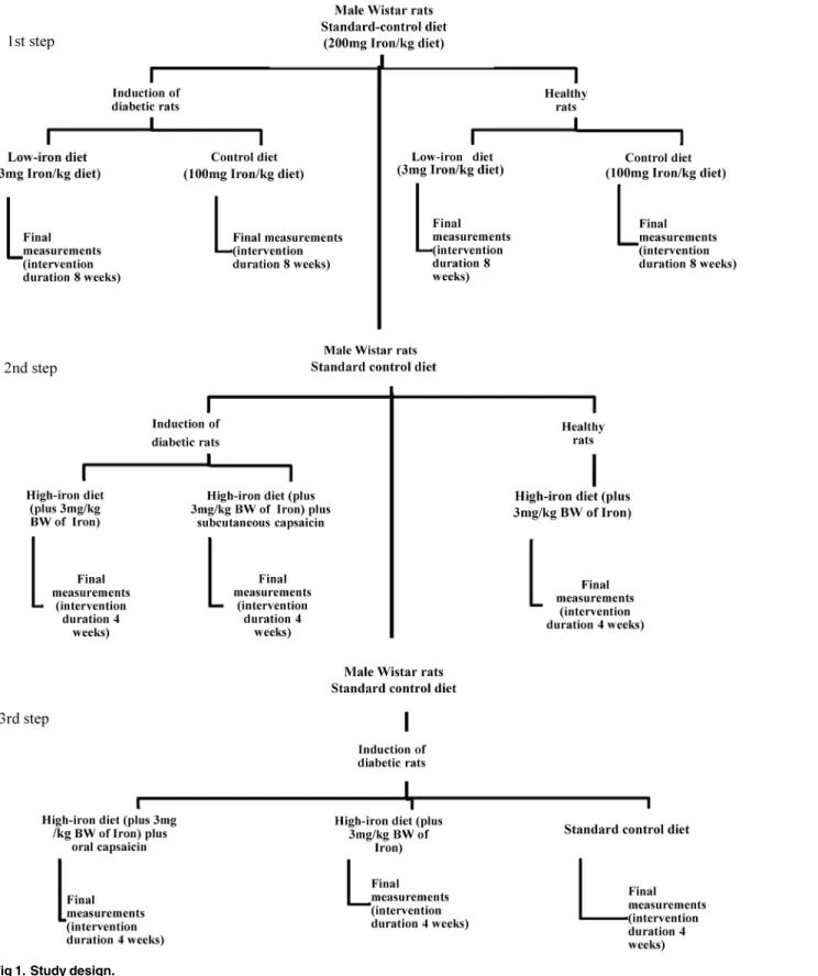
Male Wistar rats (Harlan Laboratories, Inc. Stoughton Rd, USA), weighing 250 to 350 g each, and aged approximately two months, were split into several randomized groups. All animals were maintained at the Laboratory Animals of the Centro Universitario de Investigaciones Bio-médicas, Universidad de Colima, México. Animals were housed in standard photoperiod con-ditions (12 h light /12 h dark), a 22 ± 2°C environmental temperature, and fedad libitumwith water and rodent food (200 mg iron/kg diet) (Harlan Laboratories, Inc. Stoughton Rd, USA; standard control diet) (Table 1). The experimental design of the present study comprised three components. The first involved experiments using low-iron and control diets, for healthy and diabetic rats. The second step was to conduct experiments in diabetic rats, using a high-iron diet with- or without subcutaneous capsaicin and healthy rats with high iron-diet. Finally, stud-ies were performed using diabetic rats alone fed with a high-iron diet plus oral capsaicin, a
Colima, México. AMI received a fellowship for PhD studies from CONACyT-Mexico.
standard control diet (Harlan Laboratories, Inc. Stoughton Rd, USA, 200 mg iron/kg diet), or a high-iron diet without capsaicin (Fig 1).
When diabetes was successfully induced in rats (fasting glucose200 mg/dL), interven-tional diets were applied. Low-iron (low-iron diet 3 mg iron/kg diet) and control diets (100 mg iron/kg diet) for healthy and diabetic rats were commenced simultaneously. All experimental protocols and animal management were in accordance with the ethical standards of the Mexi-can Official Norm technical specifications for the production, care, and use of laboratory ani-mals (NOM-062-ZOO-1999); additional recommendations in the care and use of laboratory animals were from the National Institutes of Health. The condition of the animals was moni-tored daily. The reporting of this animal research follows the ARRIVE guidelines (S1 File) [19].
Method of Euthanasia
Rats were euthanized without pain or distress by increasing anesthesia doses (intraperitoneal lethal doses with Pentobarbital Sodium, Pets pharm Mexico). The Ethics Committee from the Universidad de Colima approved all protocols (2011–09).
The experimental induction of diabetes
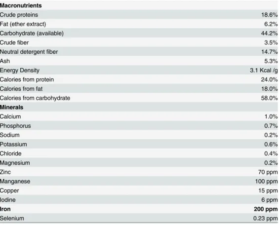
To induce diabetes, rats received a single intraperitoneal administration dose of 45 mg/kg body weight of streptozotocin (STZ, Sigma-Aldrich CO. St. Louis MO, USA) [20]. STZ damages Table 1. Standard control diet¥. The high-iron diet consists of 200 mg iron/kg diet (standard control diet)
plus 3 mg polymalthosed iron, administered orally.
Macronutrients
Crude proteins 18.6%
Fat (ether extract) 6.2%
Carbohydrate (available) 44.2%
Crudefiber 3.5%
Neutral detergentfiber 14.7%
Ash 5.3%
Energy Density 3.1 Kcal /g
Calories from protein 24.0%
Calories from fat 18.0%
Calories from carbohydrate 58.0%
Minerals
Calcium 1.0%
Phosphorus 0.7%
Sodium 0.2%
Potassium 0.6%
Chloride 0.4%
Magnesium 0.2%
Zinc 70 ppm
Manganese 100 ppm
Copper 15 ppm
Iodine 6 ppm
Iron 200 ppm
Selenium 0.23 ppm
¥Harlan Rodent Diet Catalog #2018.
Fig 1. Study design.
pancreatic beta cells, which alters serum glucose. A fasting blood glucose measurement of higher or equal to 200 mg/dL was used to confirm a diabetic state [20,21]. Diseased rats, or those aged less than two months, were excluded from our study.
Experimental protocol
Low-iron diet. The low-iron diet (D03072501) contained 3 mg/kg diet (Research Diets, Inc. New Brunswick, NJ, USA); the interventional control diet (D12050203) contained iron at 100 mg/kg diet (Research Diets, Inc.) (Table 2). These diets were similar to those used by Mina-miyama et al. [14]; their low-iron diet contained 3.2 mg/kg diet, and the control diet, 100.9 mg/ kg diet. Each diet was used for eight weeks [22].
High-iron diet, with or without capsaicin. The standard control diet contained iron at 200 mg/kg diet (Harlan Laboratories, Inc.). Rats were divided into three groups: two (groups 1 and 2) with STZ-induced diabetes, with one healthy group (group 3). All groups were fed with a standard control diet (Harlan Laboratories, Inc.), with iron-regulated diets as described below. Only one group of STZ-induced diabetic rats received capsaicin via a subcutaneous route (1 mg/kg body weight/day); this treatment was of four weeks duration, to enable compar-ison with previous studies [23,24]. No adverse effects were observed. To explore the role of dia-betes on hemoglobin, in the context of a standard control diet and a high-iron diet with or without capsaicin, we used three groups of rats: 1) one group of diabetic rats fed a standard diet (control); 2) a second group of diabetic rats fed with a high-iron diet, and 3), a third group of diabetic rats fed a high-iron diet plus capsaicin, administered by oral gavage at a dose of 1 mg/ kg body weight/day for four weeks.
Iron administration. Polymaltose iron (Takeda, Mexico, SA de CV, under license of Vifor International, Inc. Switzerland) was administered by oral gavage (Industrial Medical Plastica Silice SA de CV, Mexico) at a dose of 3 mg iron/kg body weight/day for four weeks; a dose proven to cause a significant accumulation of iron [25]. This treatment was used for rats on the high-iron diet (both healthy and diabetic groups).
Capsaicin administration. Capsaicin (Sigma-Aldrich, St. Louis, MO, USA) was dissolved in 10% Tween 80 (Sigma-Aldrich, St. Louis, MO, USA) and 10% ethanol. We added a 0.9% saline solution [26] to this mix, which was administered immediately, subcutaneously, at a dose of 1 mg/kg body weight/day [18], over four weeks. For oral use, capsaicin was dissolved in saline solution and immediately administered using an orogastric catheter (Industrial Medical Plastica Silice SA. de CV., Mexico).
Biochemical measurements
determined in blood samples after 12 h fasting [28–30] with the Accutrend Plus auto-analyzer (Roche, Mannheim, Germany). According to the manufacturer, this instrument had an intra-assay precision of 3.7% for total cholesterol, and 3.4% for triglycerides. Using controls, we cali-brated the intra-assay precision as 5% for total cholesterol, and 2.4% for triglycerides.
Statistical analyses
We used descriptive statistical analyses. The expressed variables were reported as medians with standard error; p-values<0.05 were considered statistically significant. The Stata software
(version 11, StataCorp LP, USA) was used to perform our analyses. We calculated the area under the curve (AUC) for ITT and OGTT tests using the mathematical TAI model [31]. Paired Student’s t-test were used to assess differences in mean values at the beginning and end of each intervention. The Student t-test for independent samples was used to assess differences in the mean values recorded for dietary intervention and controls diets for rats with experimen-tally induced diabetes. The same analyses were applied to compare mean values at the begin-ning and end of the intervention, and to compare the mean values between healthy groups of animals. ANOVA was used to assess differences in mean values for high-iron dietary groups.
Results
Effects of low dietary iron on diabetic rats
After 8 weeks of either the control diet, or dietary intervention, both groups of rats with STZ-induced diabetes exhibited increased fasting glucose compared with initial values. Rats on the Table 2. Diets used in the low–iron diet experiments.These diets were similar to those used by Minamiyama [14]. SeeDiscussion.
Diet Catalog D030725011 D12050203€
g% kcal%
Protein 20.3 20.3 20.3 20.3
Carbohydrate 63.9 63.9 63.9 63.9
Fat 7 15.8 7 15.8
Kcal/gm 4 4
Ingredient g Kcal ppm Iron g Kcal ppm Iron
Casein 200 800 1.7 200 800 1.7
L-Cystine 3 12 0 3 12 0
Corn Starch 397.486 1589.944 0.4 397.486 1589.944 0.3974
Maltodextrin 10 132 528 0.11 132 528 0.1056
Sucrose 100 400 0,08 100 400 0,08
Avicel, PH101 50 0 0.015 50 0 0.015
Soybean Oil 70 630 0 70 630 0
t-Butylhydroquinone 0.014 0 0.00014 0.014 0 0.00014
Mineral Mix S18706 (no added iron) 35 0 0.56 35 0 0.56
Ferric Citrate (17% Iron) 0 0 0 0.5715 0 97.155
Vitamin Mix V10037 10 40 0.009 10 40 0.009
Choline Bitartrate 2.5 0 0.0005 2.5 0 0.0005
Red Dye, FD&C#40 0.05 0 0.0065 0 0 0
Blue Dye, FD&C#1 0 0 0 0.05 0 0.0033
Total 1000.05 4000 2.9 1000.622 4000 100
1
Low–Iron diet 3mg/kg diet, €
Control diet 100mg /kg diet Research Diets.
low-iron diet expressed an initial mean fasting glucose concentration of 344.3 ± 15.4 mg/dL, which increased to 495.5 ± 15.3 mg/dL after 8 weeks on the diet (p<0.001). Rats on the
con-trol diet had an initial mean fasting glucose concentration of 322.0 ± 30.2 mg /dL, which increased to 542.8 ± 28.1 mg /dL after 8 weeks (p<0.001). Fasting glucose levels at the end of
the 8-week diet period were lower in the low-iron cohort compared to animals on the control diet (seeTable 3), but these differences were not found to be significant (p = 0.17).
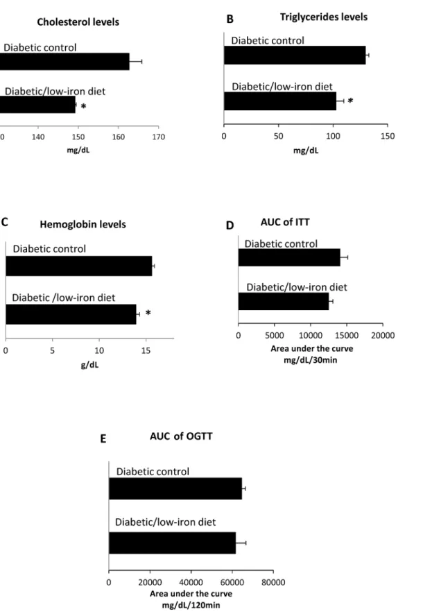
The AUC of OGTT in diabetic rats was not statistically different when comparing the low-ironvs. control diet groups (p = 0.2) (Table 3,Fig 2E). Similar results were obtained for diabetic rats using the AUC of ITT, when comparing data for the eight-week low-ironvs. control diets (Table 3,Fig 2D). Significant reductions in cholesterol and triglycerides were found after 8-weeks of the low-iron diet, compared with rats on a control diet (Fig 2A and 2B). As expected, hemoglobin levels fell significantly in the group that received the low-iron diet, com-pared with the control group. After 8 weeks, mean hemoglobin levels differed significantly between the low-diet and control diet groups (Table 3; p<0.05) (Fig 2C). These effects could
not be explained by changes in body weight, as this was not substantially altered at the end of the study (232.8 ± 11.9vs. 227.2 ± 6.4 g for diabetic rats on either the low-iron or control diet, respectively (p = 0.6);Table 3).
Effects of low dietary iron on healthy rats
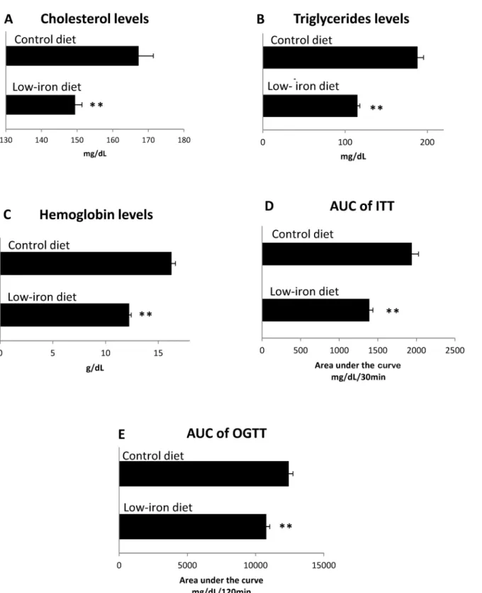
Cholesterol and triglyceride concentrations were significantly lower (Fig 3A and 3B,Table 3) in the dietary intervention group than in the control diet group, following 8 weeks (p<0.001)
(cholesterol) and p<0.001 (triglycerides). As expected, after 8 weeks of the low-iron diet,
hemoglobin levels in healthy rats decreased from 16.1 ± 0.8 to 12.2 ± 0.2 (p<0.001). Final
hemoglobin concentrations were significantly lower in the dietary intervention group than in the control group (p<0.001;Fig 3C,Table 3). The areas under the ITT curves were
1390 ± 46.03 mg/dL/min for the intervention group, and 1941 ± 86.1 mg/dL/min for the con-trol group (p<0.05;Fig 3D,Table 3). At the end of the eight-week diet, the areas under the
OGTT curves of the dietary intervention group exhibited a higher glucose tolerance than the control diet group (10786.3 ± 251.6 mg/dL/minvs. 12432.2 ± 305.3 mg/dL/min, respectively; p<0.001;Table 3,Fig 3E).
Effects of a high-iron diet, with or without capsaicin, on cholesterol,
triglyceride, and hemoglobin concentrations
Mice with a form of hereditary iron overload (hemochromatosis) are more likely to develop diabetes [3]. With this in mind, we investigated whether a high iron diet, with or without cap-saicin, could alter glucose, cholesterol, triglyceride, or hemoglobin levels. After four weeks of a high-iron diet, cholesterol was significantly increased in diabetic rats versus healthy rats fed the high-iron diet (p<0.05) (Fig 4A Table 4); triglycerides did not increase significantly (Fig 4B,
Table 4). However, cholesterol levels in diabetic rats fed the high-iron diet could be reduced (p<0.05) by the administration of subcutaneous capsaicin (at a dose of 1 mg/kg body
weight/day) to levels comparable with those seen in healthy rats fed the high-iron diet (Fig 4A,
Table 4). Capsaicin also significantly reduced triglyceride (p<0.001) and hemoglobin
(p<0.001) concentration, compared to either the diabetic or healthy test groups fed a
high-iron diet (Table 4,Fig 4B and 4C), although glucose levels were not significantly decreased (Table 4).
In terms of hemoglobin concentration, tend to be lower, but not statistically significant differ-ences were found for values acquired initially, versus those after four weeks of treatment (17.6 ± 0.5 vs. 17.3 ± 0.5 g/dL respectively; p = 0.8; n = 7). This outcome was also seen for dia-betic rats fed with a high-iron diet (18.3 ± 0.4 vs. 17.7 ± 0.2 g/dL for initial and 4 weeks of hemoglobin levels, respectively, p = 0.08;Table 4). However, for diabetic rats fed with a high iron diet plus oral capsaicin, hemoglobin levels were significantly reduced when comparing the initial values with those after four weeks of treatment (17.4 ± 0.4 vs. 15.8 ± 0.4 g/dL, respec-tively; p = 0.02; n = 7). These results show that oral capsaicin significantly reduced the levels of hemoglobin, replicating the effects seen with subcutaneous administration (Table 4).
Discussion
The results of this study using a rat chow diet showed that a low-iron diet could reduce total cholesterol and triglyceride levels to the same degree in both healthy and diabetic rats. In addi-tion, the low-iron diet was associated with a significantly improved insulin and glucose toler-ance in healthy rats.
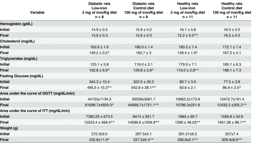
One mechanistic insight for the role of dietary iron has been previously published by Meh-dad et al. 2015; who showed that reduced dietary iron enhanced insulin receptor expression in Table 3. Diabetic and healthy rats fed with low and control iron diet.p value from paired Student t-test at the beginning and end of intervention. Values are mean±standard error of the mean. Two rats died during the study. Initial refers to values before intervention. Final refers to values after 8 weeks of dietary intervention. The paired Student t-test was calculated with the lower n indicated at the top of the Table.
Diabetic rats Diabetic rats Healthy rats Healthy rats Low-iron Control diet Low-iron Control diet Variable 3 mg of iron/Kg diet 100 mg of iron/Kg diet 3 mg of iron/Kg diet 100 mg of iron/Kg diet
n = 8 n = 8 n = 11 n = 11
Hemoglobin (g/dL)
Initial 14.9±0.5 15.9±0.2 16.1±0.8 16.0±0.5
Final 13.9±0.3 15.6±0.3 12.2±0.2** 16.2±0.3
Cholesterol (mg/dL)
Initial 162.6±1.6 166.0±1.4 180.0±7.4 172.1±1.4
Final 149.2±0.2* 162.7±3 149.4±1.9* 167.2±4.1
Triglycerides (mg/dL)
Initial 123.1±5.8 119.0±3.1 179.9±7.1 185.1±6.3
Final 102.8±6.9* 129.8±2.8* 115.0±2.8** 188.1±7.3
Fasting Glucose (mg/dL)
Initial 344.3±15.4 322.0±30.2 80.7±3.0 77.5±3.8
Final 495.5±15.3** 542.8±28.1** 83.6±2.1 86.9±2.5*
Area under the curve of OGTT (mg/dL/min)
Initial 44733±1134.3 30039±3061.7 10662.2±173.9 10472.7±191.4 Final 61698.7±4929.3* 64668.7±1721.1** 10786.3±251.6 12432.2±305.3** Area under the curve of ITT (mg/dL/min)
Initial 7360.25±673.5 8414±931.7 1883±60.7 1556.8±52.6
Final 12523.4±569.4** 14099.6±1034.9** 1390±46.03** 1941.36±86.1** Weight (g)
Initial 272.3±9.5 267.5±5.1 261.21±6.5 257±7.4
Final 232.8±11.9* 227.2±6.4** 336.8±6.1** 328.9±8.6**
*p<0.05,
**p<0.001, paired Student t-test. Look for cross group comparison in Figs2and3.
Fig 2. Effects of a low-iron diet on rats with STZ-induced diabetes.Diabetic rats were analyzed after 8 weeks on a low-iron diet (n = 8), or a control diet (n = 8). (A) Cholesterol levels. (B) Triglyceride levels. (C) Hemoglobin levels. (D) Area under insulin curve. (E) Area under glucose tolerance curve. Bars represent the mean and standard error; p-values assessed with Student’s t-test;*p<0.05.
Fig 3. Effects of a low-iron diet on healthy rats.Healthy rats were analyzed after 8 weeks on a low-iron diet (n = 10), or control diet (n = 10). (A) Cholesterol levels. (B) Triglyceride levels. (C) Hemoglobin levels. (D) Area under the insulin tolerance test curve. E) Area under glucose tolerance curve. Bars represent the mean and standard error; p-values assessed with Student’s t-test;*p<0.05,**p<0.001.
Fig 4. Effects of a high-iron diet, with or without capsaicin, on rats with STZ-induced diabetes.Diabetic and healthy rats were analyzed after 4 weeks on a high-iron diet (n = 10), or a high-iron diet plus capsaicin (3mg/kg body weight/day) (n = 10) (A) Cholesterol levels. (B)Triglyceride levels. (C) Hemoglobin levels. Bars represent the mean and standard error; p- values assessed with ANOVA. For (A) p<0.05 when comparing group 1vs. groups 2, and 3; for (B)
p<0.001 when comparing group 2vs. group 1, and 3 (C) p<0.001 when comparing group 2vs. groups 1, and 3.**p<0.001,*p<0.05.
skeletal muscle [32]. On the other hand, our results agree with those of Minamiyama et al. [14], in which the effects of iron depletion on lipid profiles and glycosylated hemoglobin (HbA1c) levels were examined. Otsuka Long-Evans Tokushima Fatty (OLETF) rats were used in that study [12], age-paired with Long-Evans Tokushima Otsuka (LETO) rats. Low dietary iron (3.2 mg iron/kg diet), or phlebotomy, were used to reduce iron levels in the blood, with compari-sons made with rats fed a control iron diet (100.9 mg iron/kg diet). The results of the present study also agree with an investigation using obese mice (ob/ob,lep-/-), conducted by Cooksey et al. [12]. At the end of that study, mice that received a low-iron diet exhibited a diminution in triglyceride and cholesterol levels, and increased insulin sensitivity, relative to controls. One possible mechanism for these effects was suggested by a study by Kamei et al. [33], in which the transcriptional response to the experimental induction of anemia was studied in Sprague Dawley rats. In that study, changes in the transcription of genes involved in metabolism were identified. With respect to our data collected with low dietary iron, we could speculate that altered gene expression, possibly acting directly on cholesterol and triglyceride synthesis, could be relevant. However, we should emphasize that in our study, the low-iron diet was moderate, such that anemia was avoided (<10 g hemoglobin/dL). Additional studies would be needed to
establish the timeframe over which a low-iron diet would be needed to induce anemia. On the other hand, at the end of eight-weeks, for the diabetic rats there exists a significant loss in weight. Previous studies in type 2 diabetic patients showed weight loss when these patients failed to receive treatment for diabetes control [34]. More experiments are now needed to determine if the model we use corresponds more accurately to type 2 or type 1 of diabetes.
The experimental designs of previous studies have differed from ours mainly in terms of the animal models used. For example, both ob/ob mice and OLETF rats are natural models of obe-sity and develop hyperglycemia later in life; both models are genetically predisposed to obeobe-sity. A possible explanation for the discrepancies between our data and previous studies is that they Table 4. Diabetic and healthy rats fed with high iron diet plus capsaicin.p value from paired Student t-test at the beginning and at the end of interven-tion. The dose of capsaicin used was 1 mg/kg body weight/day administered subcutaneously. Initial refers to values before interveninterven-tion. Final refers to values after 4 weeks of intervention. To confirm the effects of diabetes on hemoglobin in the high-iron group, an additional group of seven rats were tested; those data have been added£n = 17. The paired Student t-test was calculated with the lower n indicated at the top of the Table. In diabetic rats plus capsaicin the
differences between initial and final values corresponds to death of one rat. In healthy rats with high iron the missing values at the beginning were due to tech-nical difficulties in the measurements.
Variable Diabetic rats High-iron diet plus Capsaicin Diabetic rats High-iron diet Healthy rats High-iron diet
n = 9 n = 10 n = 8
Hemoglobin (g/dL)
Initial 19.2±0.3 18.3±0.4£ 18.3±0.4
Final 16.3±0.4** 17.7±0.2£ 18.55±0.1
Cholesterol (mg/dL)
Initial 152.5±1.7 154.4±2.8 150.2±0.9
Final 157.3±4.2 169.4±1.2** 154±5.1
Triglycerides(mg/dL)
Initial 103.3±9.1 109±12.5 135.8±19.7
Final 116.5±3.8 155.6±6.1* 140.1±5.3
Fasting Glucose (mg/dL)
Initial 409.3±30.5 397.5±30.6 90.6±2.6
Final 393.6±43.7 412.5±17.3 92.9±2.1
*p<0.05,
**p<0.001, paired Student t-test. Look for cross group comparison inFig 4.
were performed using animal models of type 2 diabetes with hyperinsulinemia, while the pres-ent study was performed in an animal model of hypoinsulinemia. Our findings of a lack of any dietary effect on either fasting glucose concentration may be related to exposure time. In con-trast, rats fed with a high-iron diet exhibited increased in total cholesterol level. To provide mechanistic context, some authors have linked chronic dietary iron overload and lipid peroxi-dation, with impaired plasma lipid transport, and therefore increased cholesterol [9]. Another credible possibility is that excess stored iron might cause oxidative stress, with reactive oxygen species subsequently damaging cell membranes, lipids, and proteins. The resultant lesions could damage tissue, and organs, including the liver and pancreas [35,36]. Graham et al. [37] also suggested that hepatic iron overload could increase hepatic cholesterol biosynthesis. Clearly, the mechanistic interactions between iron and cholesterol remain unclear.
To summarize, we now show that a low-iron diet reduced triglyceride and cholesterol levels in healthy rats, and in rats with experimentally induced diabetes. The low-iron diet also improved insulin and glucose tolerance in healthy rats. In contrast, these same biochemical parameters were worsened in the context of anemia (<10 g hemoglobin/dL) [38], which is
clin-ically relevant given that both anemia, and hemochromatosis, have been shown to increase morbidity and mortality through heart failure and thrombosis [39,40].
Our results suggest that a low-iron diet reduce the dyslipidemia, and improve insulin and glucose tolerance in healthy rats. Showing that context is important, Ikeda et al. [22] have also demonstrated that a low-iron diet prevented diabetic nephropathy in diabetic rats, which was partly attributable to reduced oxidative stress. With respect to capsaicin, Prakash and Sriniva-san [17] reported that acute administration (60 min) of capsaicin intake enhanced the intesti-nal uptake of iron in segments of isolated intestine. While previous studies failed to find any substantial effects on cholesterol metabolism that could be attributed to acute capsaicin intake (in conjunction with a standard diet), [41] the oxidation of low-density lipoprotein (in rats) has been reported [42]. We now demonstrate that capsaicin reduces hemoglobin when admin-istered as part of a high-iron diet [5]. As previously mentioned, diabetes per se may modify hemoglobin concentration, mainly via renal dysfunction which alters erythropoietin synthesis, and subsequently hemoglobin level [5,6]. Our data show that the reduction in hemoglobin level achieved using both oral and subcutaneous capsaicin, were greater than that achieved by the diabetic state alone. Additionally, the levels of hemoglobin in diabetic rats, on both the high-iron and standard control diets (Harlan), were not significantly different. Further research is now required to investigate the mechanisms for capsaicin’s action on hemoglobin, lipid metabolism, and iron regulation, as well as possible therapeutic uses.
Conclusions
Our results suggest that a low-iron diet could improve lipid homeostasis in diabetes, and that capsaicin reduces hemoglobin levels in rats fed a high-iron diet.
Supporting Information
S1 File. NC3Rs ARRIVE Guidelines Checklist-FILLED FINAL.pdf.
(PDF)
Acknowledgments
to MScs. Sandra Oseguera-Bernal and Germán Pérez from University Center for Multivariant Statistic (CIEMA-UdeC) for helping in the statistics analysis of the data.
Author Contributions
Conceived and designed the experiments: XT MH SVH AMI. Performed the experiments: AMI EM MRS HC MIDR. Analyzed the data: AMI EM MRS XT MH SVH HC MIDR. Con-tributed reagents/materials/analysis tools: HC MIDR SVH XT MH. Wrote the paper: XT MH HC MIDR SVH.
References
1. Fernández-Real JM, López-Bermejo A, Ricart W. Cross-talk between iron metabolism and diabetes. Diabetes. 2002; 51(8): 2348–2354. PMID:12145144
2. Zhao Z, Li S, Liu G, Yan F, Ma X, Huang Z, et al. Body iron stores and heme-iron intake in relation to risk of type 2 diabetes: a systematic review and meta-analysis. PLOS ONE. 2012; 7(7): e41641. doi: 10.1371/journal.pone.0041641PMID:22848554
3. Huang J, Jones D, Luo B, Sanderson M, Soto J, Abel ED, et al. Iron overload and diabetes risk: a shift from glucose to Fatty Acid oxidation and increased hepatic glucose production in a mouse model of hereditary hemochromatosis. Diabetes. 2011; 60(1): 80–87. doi:10.2337/db10-0593PMID:20876715
4. Ganz T, Nemeth E. Hepcidin and iron homeostasis. Biochim Biophys Acta. 2012; 1823(9): 1434–1443.
doi:10.1016/j.bbamcr.2012.01.014PMID:22306005
5. Thomas MC, MacIsaac RJ, Tsalamandris C, Power D, Jerums G. Unrecognized anemia in patients with diabetes: a cross-sectional survey. Diabetes Care. 2003; 26:1164–1169. PMID:12663591
6. Panjeta M, Tahirovic, Karamehic J, Sofic E, Ridic O, Coric J. The relation of erythropoietin towards hemoglobin and hematocrit in varying degrees of renal insufficiency. Mater Sociomed. 2015; 27:144–
148. doi:10.5455/msm.2015.27.144-148PMID:26236158
7. Salonen JT, Tuomainen TP, Nyyssönen K, Lakka HM, Punnonen K. Relation between iron stores and non-insulin dependent diabetes in men: case-control study. BMJ 1998; 317(7160): 727. PMID: 9732340
8. Smotra S, Kudyar RP. Relationship between serum ferritin and type-2 diabetes mellitus. JK Science. 2008; 10(4):170–174.
9. Brunet S, Thibault L, Delvin E, Yotov W, Bendayan M, Levy E. Dietary iron overload and induced lipid peroxidation are associated with impaired plasma lipid transport and hepatic sterol metabolism in rats. Hepatology. 1999; 29:1809–1817 PMID:10347124
10. Whittaker P, Chanderbhan RF. Effect of increasing iron supplementation on blood lipids in rats. Br J Nutr. 2001; 86:587–592. PMID:11737956
11. Dongiovanni P, Ruscica M, Rametta R, Recalcati S, Steffani L, Gatti S, et al. Dietary iron overload induces visceral adipose tissue insulin resistance. Am J Pathol. 2013; 182:2254–2263. doi:10.1016/j. ajpath.2013.02.019PMID:23578384
12. Cooksey RC, Jones D, Gabrielsen S, Huang J, Simcox JA, Luo B, et al. Dietary iron restriction or iron chelation protects from diabetes and loss of beta-cell function in the obese (ob/ob Lep-/-) mouse. Am J Physiol Endocrinol Metab. 2010; 298(6): 1236–1243.
13. Jiang R, Manson JE, Meigs JB, Ma J, Rifai N, Hu FB. Body iron stores in relation to risk of type 2 diabe-tes in apparently healthy women. JAMA. 2004; 291(6): 711–717. PMID:14871914
14. Minamiyama Y, Takemura S, Kodai S, Shinkawa H, Tsukioka T, Ichikawa H, et al. Iron restriction improves type 2 diabetes mellitus in Otsuka Long-Evans Tokushima fatty rats. Am J Physiol Endocrinol Metabol. 2010; 298(6): 1140–1149.
15. Borel MJ, Beard JL, Farrell PA. Hepatic glucose production and insulin sensitivity and responsiveness in iron-deficient anemic rats. Am J Physiol. 1993; 264(3): 380–390.
16. Uchida K, Tominaga M. The role of thermosensitive TRP (transient receptor potential) channels in insu-lin secretion. Endocr J. 2011; 58:1021–1028. PMID:21785227
17. Prakash UN, Srinivasan K. Enhanced intestinal uptake of iron, zinc and calcium in rats fed pungent spice principles-piperine, capsaicin and ginger (Zingiber officinale). J Trace Elem Med Biol. 2013; 27 (3): 184–190. doi:10.1016/j.jtemb.2012.11.003PMID:23332714
19. Kilkenny C, Browne WJ, Cuthill IC, Emerson M, Altman DG (2010) Improving Bioscience Research Reporting: The ARRIVE Guidelines for Reporting Animal Research. PLoS Biol 8(6): e1000412. doi: 10.1371/journal.pbio.1000412PMID:20613859
20. Zolghadri Y, Fazeli M, Kooshki M, Shomali T, Karimaghayee N, Dehghani M. Achillea Millefolium L. Hydro- Alcoholic Extract Protects Pancreatic Cells by Down Regulating IL- 1βand iNOS Gene Expres-sion in Diabetic Rats. Int J Mol Cell Med. 2014; 3(4):255–262. PMID:25635252
21. Ríos-Silva M, Trujillo X, Trujillo-Hernández B, Sánchez-Pastor E, Urzúa Z, Mancilla E, et al. Effect of chronic administration of forskolin on glycemia and oxidative stress in rats with and without experimen-tal diabetes. Int J Med Sci. 2014; 11(5):448–452. doi:10.7150/ijms.8034PMID:24688307
22. Ikeda Y, Enomoto H, Tajima S, Izawa-Ishizawa Y, Kihira Y, Izhizawa K, et al. Dietary iron restriction inhibits progression of diabetic nephropathy in db/db mice. Am J Physiol Renal Physiol. 2013; 304(7): 1028–1036.
23. Thompson K, Molina R, Donaghey T, Brain JD, Wessling-Resnick M. The influence of high-iron diet on rat lung manganese absorption. Toxicol Appl Pharmacol. 2006; 210(1–2):17–23. PMID:15993455
24. Abd Allah ES, Ahmed MA, Abdel Mola AF. Comparative study of the effect of verapamil and vitamin D on iron overload-induced oxidative stress and cardiac structural changes in adult male rats. Pathophys-iology. 2014; 21(4):293–300. doi:10.1016/j.pathophys.2014.06.002PMID:25092628
25. Maaroufi K, Had-Aissouni L, Melon C, Sakly M, Abdelmelek H, Poucet B, et al. Effects of prolonged iron overload and low frequency electromagnetic exposure on spatial learning and memory in the young rat. Neurobiol Learn Mem. 2009; 92(3): 345–355. doi:10.1016/j.nlm.2009.04.002PMID:19394433
26. Zhou XF, Livett BG. Effect of capsaicin-sensitive sensory nerves on plasma glucose and catechol-amine levels during 2-deoxyglucose-induced stress in conscious rats. Br J Pharmacol. 1990; 100(3): 523–529. PMID:2390676
27. Miron VR, Bauermann L, Morsch AL, Zanin RF, Correa M, da Silva AC, et al. Enhanced NTPDase and 5´nucleotidase activities in diabetes mellitus and iron-overload model. Mol Cell Biochem. 2007; 298(1–
2):101–107. PMID:17119848
28. Cherng JY, Shih MF. Preventing dyslipidemia by Chlorella pyrenoidosa in rats and hamsters after chronic high fat diet treatment. Life Sci. 2005; 76(26): 3001–3013. PMID:15850594
29. Zhang SY, Sun XJ, Zheng JB, Wang W, Liu D, Chen NZ, et al. Preserve common limb in duodenal-jeju-nal bypass surgery benefits rats with type 2-like diabetes. Obes Surg. 2014; 24(3): 405–411. doi:10. 1007/s11695-013-1103-zPMID:24190437
30. de Almeida MM, Luquetti SC, Sabarense CM, do Amaral Corrêa JO, dos Reis LG, Santos da Concei-ção EP, et al. Butter naturally enriched in cis-9, trans-11 CLA prevents hyperinsulinemia and increases both serum HDL cholesterol and triacylglycerol levels in rats. Lipids Health Dis. 2014; 13: 200. doi:10. 1186/1476-511X-13-200PMID:25534067
31. Tai MM A. Mathematical model for the determination of total area under glucose tolerance and other metabolic curves. Diabetes Care. 1994; 17(2): 152–154. PMID:8137688
32. Mehdad A, Campos NA, Arruda SF, Siqueira EM. Iron Deprivation May Enhance Insulin Receptor and Glut4 Transcription in Skeletal Muscle of Adult Rats. J Nutr Health Aging. 2015; 19(8): 846–854. doi: 10.1007/s12603-015-0541-9PMID:26412289
33. Kamei A, Watanabe Y, Ishijima T, Uehara M, Arai S, Kato H, et al. Dietary iron-deficient anemia induces a variety of metabolic changes and even apoptosis in rat liver: a DNA microarray study. Physiol Geno-mics. 2010; 42(2):149–156. doi:10.1152/physiolgenomics.00150.2009PMID:20388835
34. Lee CG, Boyko EJ, Barrett-Connor E, Miljkovic I, Hoffman AR, Everson-Rose SA, et al. Osteoporotic Fractures in Men (MrOS) Study Research Group. Insulin sensitizers may attenuate lean mass loss in older men with diabetes. Diabetes Care. 2011; 34(11): 2381–2386. doi:10.2337/dc11-1032PMID: 21926282
35. Kundu D, Roy A, Mandal T, Bandyopadhyay U, Ghosh E, Ray D. Relation of iron stores to oxidative stress in type 2 diabetes. Niger J Clin Pract. 2013; 16(1): 100–103. doi:10.4103/1119-3077.106776
PMID:23377481
36. Rajpathak SN, Crandall JP, Wylie-Rosett J, Kabat GC, Rohan TE, Hu FB. The role of iron in type 2 dia-betes in humans. Biochem Biophys Acta. 2009; 1790(7): 671–681. doi:10.1016/j.bbagen.2008.04.005
PMID:18501198
37. Graham RM, Chua AC, Carter KW, Delima RD, Johnstone D, Herbison CE, et al. Hepatic iron loading in mice increases cholesterol biosynthesis. Hepatology. 2010; 52(2): 462–471. doi:10.1002/hep.23712
PMID:20683946
39. Alexandrakis MG, Tsirakis G. Anemia in heart failure patients. ISRN Hematol. 2012;246915 doi:10. 5402/2012/246915PMID:22536520
40. Meroño T, Sorroche P, Brites FD. [Increased iron store and its relationship with cardiovascular disease] Medicina (B Aires) 2011; 71(6): 566–572.
41. Srinivasan MR, Chandrasekhara N. Comparative influence of vanillin & capsaicin on liver & blood lipids in the rat. Indian J Med Res. 1992; 96: 133–135. PMID:1428053
![Table 2. Diets used in the low–iron diet experiments. These diets were similar to those used by Minamiyama [14]](https://thumb-eu.123doks.com/thumbv2/123dok_br/18288168.346311/6.918.57.867.129.552/table-diets-used-iron-experiments-diets-similar-minamiyama.webp)