RNA Interference-Guided Targeting of
Hepatitis C Virus Replication with Antisense
Locked Nucleic Acid-Based Oligonucleotides
Containing 8-oxo-dG Modifications
Margit Mutso1,2‡, Andrei Nikonov1,2‡, Arno Pihlak2‡, EvaŽusinaite1,2, Liane Viru1,2, Anastasia Selyutina1, Tõnu Reintamm2,3, Merike Kelve2,3, Mart Saarma4, Mati Karelson2,5,
Andres Merits1*
1Institute of Technology, University of Tartu, Tartu, Estonia,2GeneCode, Ltd., Tallinn, Estonia, 3Department of Gene Technology, Tallinn University of Technology, Tallinn, Estonia,4Institute of Biotechnology, University of Helsinki, Helsinki, Finland,5Institute of Chemistry, University of Tartu, Tartu, Estonia
‡These authors are joint first authors on this work.
Abstract
The inhibitory potency of an antisense oligonucleotide depends critically on its design and the accessibility of its target site. Here, we used an RNA interference-guided approach to select antisense oligonucleotide target sites in the coding region of the highly structured hepatitis C virus (HCV) RNA genome. We modified the conventional design of an antisense oligonucleotide containing locked nucleic acid (LNA) residues at its termini (LNA/DNA gap-mer) by inserting 8-oxo-2’-deoxyguanosine (8-oxo-dG) residues into the central DNA re-gion. Obtained compounds, designed with the aim to analyze the effects of 8-oxo-dG modifications on the antisense oligonucleotides, displayed a unique set of properties. Com-pared to conventional LNA/DNA gapmers, the melting temperatures of the duplexes formed by modified LNA/DNA gapmers and DNA or RNA targets were reduced by approximately 1.6-3.3°C per modification. Comparative transfection studies showed that small interfering RNA was the most potent HCV RNA replication inhibitor (effective concentration 50 (EC50):
0.13 nM), whereas isosequential standard and modified LNA/DNA gapmers were approxi-mately 50-fold less efficient (EC50: 5.5 and 7.1 nM, respectively). However, the presence of
8-oxo-dG residues led to a more complete suppression of HCV replication in transfected cells. These modifications did not affect the efficiency of RNase H cleavage of antisense oli-gonucleotide:RNA duplexes but did alter specificity, triggering the appearance of multiple cleavage products. Moreover, the incorporation of 8-oxo-dG residues increased the stability of antisense oligonucleotides of different configurations in human serum.
OPEN ACCESS
Citation:Mutso M, Nikonov A, Pihlak A,Žusinaite E, Viru L, Selyutina A, et al. (2015) RNA Interference-Guided Targeting of Hepatitis C Virus Replication with Antisense Locked Nucleic Acid-Based
Oligonucleotides Containing 8-oxo-dG Modifications. PLoS ONE 10(6): e0128686. doi:10.1371/journal. pone.0128686
Academic Editor:Emanuele Buratti, International Centre for Genetic Engineering and Biotechnology, ITALY
Received:January 13, 2015
Accepted:April 29, 2015
Published:June 3, 2015
Copyright:© 2015 Mutso et al. This is an open access article distributed under the terms of the
Creative Commons Attribution License, which permits unrestricted use, distribution, and reproduction in any medium, provided the original author and source are credited.
Data Availability Statement:All relevant data are within the paper.
Introduction
The application of complementary DNA or RNA molecules or their derivatives for the modu-lation biological functions of specific RNA(s) is referred to as antisense technology. Antisense oligonucleotides (ASOs) are the major class of antisense agents used for sequence-specific RNA knockdown [1], and they can also be used to modulate RNA synthesis, maturation and transport. Two different mechanisms account for the inhibitory properties of ASOs. The first mechanism is typically mediated by the steric inhibition of translation machinery operating on the targeted RNA. In general, this mechanism is not associated with the destruction of targeted molecules, and, accordingly, it is most effective for coding RNAs if the ASO target site overlaps with or is located upstream of the initiation codon [2]. The second mechanism relies on the ability of ribonuclease H (RNase H), a ubiquitous group of cellular enzymes, to cleave the RNA part of the heteroduplexes formed between DNA ASOs and targeted RNA [3,4]. This mecha-nism results in the degradation of the targeted RNA and is therefore effective regardless of the position of the ASO binding site [2].
The activity of ASOs depends on many factors, including the efficiency of cell entry, the sta-bility of the complex formed with the targeted RNA and the resistance of the ASO to enzymatic degradation. The low potency of standard RNA and DNA ASOs results from their poor uptake and extremely short intracellular and serum half-lives. Sugar moiety and phosphate backbone modifications have been used to increase the resistance of ASOs to degradation. Some of these modifications also increase the binding efficiency of ASOs to their target sequences [5] and/or may be beneficial for cell entry. However, only phosphorothioate-s [6], boranophosphate- [7], oxepane- [8], cyclohexene- [9], and fluoro-arabino (FANA)-modified ASOs [10] have been re-ported to activate RNase H upon binding to targeted mRNA. In contrast, fully modified N3’, P5’-phosphoramidates [11], morpholinos [12], peptide nucleic acids (PNA) [13], tricyclo-DNA [14], 2’-O-methyl locked nucleic acids (LNA) and 2’-O-methoxyethyl RNAs [15] lack this property. To overcome this issue, co-polymers of 2’-O-methyl RNA [16], FANA [17], PNA or LNA [18–20] with DNA have been developed. ASOs containing LNA residues at their termini (hereafter, ASOs with several terminal LNA monomers and internal DNA residues are termed LNA/DNA gapmers) are more effective activators of RNase H-mediated cleavage than 2’-O-methyl RNA/DNA gapmers or all-DNA ASOs [19].
The nucleobase moiety represents an alternative option for ASO modification. Several het-erocyclic base modifications in ASOs have been described (reviewed in [21]). However, only a few of those modifications have been analyzed for their ability to activate RNase H. Thus far, ASOs with modified nucleobases (such as 5-(N-aminohexyl)carbamoyl-2’-dU [22] and G-clamps [23]) have been found to be worse RNase H activators than non-modified DNA oligo-nucleotides. The majority of sugar moiety, phosphate backbone, and nucleobase modifications increase the melting temperature (Tm) of ASO duplexes with DNA and RNA [24,25]. Further-more, ASOs containing both LNA bases and phosphorothioate modifications possess excellent serum stability and longin vivohalf-lives, enabling their successful use in clinical trials [26].
The 8-oxo-2’-deoxyguanosine (8-oxo-dG) residue contains a minimally modified nucleo-base, which is naturally occurring and can result from oxidative DNA damage. In the context of ASO, 8-oxo-guanine forms 3- to 4-fold weaker bonds with complementary cytosine (com-pared to non-modified guanine) [27], which results in a decrease in the Tm of the ASO:DNA duplexes [28–30]. However, both 8-oxo-dG [31] and 5-hydroxy-2’-deoxycytidine (5-OH-dC) [32], another product of DNA oxidization, have not only major but also minor zwitterionic and ionic tautomeric isomers, respectively (Fig 1A). Interestingly, theoretical quantum chemi-cal chemi-calculations performed on the minor tautomeric form of 8-oxo-dG and anion of 5-OH-dC suggest that they exhibit abnormally strong bonding with their respective normal nucleobases
collection and analysis, decision to publish, or preparation of the manuscript. Co-authors AP, TR, MM, AN, LV, EZ, M. Kelve and M. Karelson were employed by GeneCode, Ltd at some period of time when the work results of which have been included into this manuscript was in progress. GeneCode, Ltd. provided support in the form of salaries for authors AP, TR, MM, AN, LV, EŽand M. Kelve, but did not have any additional role in the study design, data collection and analysis, decision to publish, or preparation of the manuscript. The specific roles of these authors are articulated in the‘author contributions’section.
Competing Interests:Co-authors AP, TR, MM, AN, LV, EZ, M. Kelve and M. Karelson were employed by GeneCode, Ltd. during the initiation of the study (2008-2012). Currently no author of this manuscript is employed by Genecode Ltd. M. Karelson, MS and AM are inventors in the following patent/patent applications related to the modified nucleotides. 1) US Patent US7786292 B2 (“Antisense agents combining strongly bound base-modified oligonucleotide and artificial nuclease”; inventors: M. Karelson, MS and MP; owner: Baltic Technology Development, Ltd. (now GeneCode AS);http://www. google.com/patents/US7786292). 2) Patent application (“Use of oligonucleotides with modified bases as antiviral agents”; inventors: MS, AM and M. Karelson; owner: Baltic Technology Development, Ltd. (now GeneCode AS,http://www.
in an aqueous environment [33]. It is not known how these effects might contribute to the effi-ciency of ASOs containing such modified residues.
Thein vivodelivery of therapeutic nucleic acids to target tissues and organs represents an-other important problem that has been only partly solved (reviewed [34]). In the absence of specific delivery vehicles, the main target of ASOs in both rodent [35] and non-human primate [36,37] models is liver. Targeting other organs, including kidney [35,37], heart, diaphragm, lung, fat, gastrocnemius and quadriceps, is less efficient [35]. Clinical trials have shown that miravirsen, an experimental ASO drug that targets the microRNA miR122, spontaneously en-ters the liver [26].
As ASOs have a clear potential for the treatment of liver-associated pathologies, we chose hepatitis C virus (HCV) RNA as a target for modified ASOs. HCV is a major human pathogen that affects the lives of over one hundred million people. Its positive polarity RNA genome is
>9000 nucleotides (nt) long and contains strong RNA secondary structures [38]. Thus far,
only ASOs targeting the 350-nt region at the 5’-terminus of HCV RNA, which contains the in-ternal ribosome entry site (IRES), are efficient inhibitors of viral replication [15,39–46]. Hence, HCV RNA represents an important but difficult target for ASOs, and it may enable the deter-mination of the positive and negative impacts of ASO modifications.
Here, we demonstrate that the incorporation of 8-oxo-dG residues destabilizes not only ASO:DNA but also ASO:RNA duplexes. These modifications also slow down the formation of all-DNA ASO:RNA duplexes, but they have little effect on the formation of duplexes between
Fig 1. Incorporation of 8-oxo-dG, but not 5-OH-dC, reduces the Tm of ASO:DNA and ASO:RNA duplexes. (A)Structures of 8-oxo-dG, its zwitterionic (minor) form and 5-OH-dC (common and minor forms). (B, C)The effects of modified nucleobases on the Tm of ASO:DNA(B)and ASO:RNA(C)duplexes as measured by FRET. Target DNA or RNA oligonucleotides (Table 2) were labeled with Cy3 at the 3’-end, and modified oligonucleotide probes and controls were labeled with FITC at the 5’-end. Probe:target hybridization was quantified by measuring the decrease in FITC fluorescence due to the energy transfer to the Cy3 fluorochrome attached to the hybridized target. Increasing target concentrations (25, 50, 100, 200, and 400 nM) were used (x-axis), whereas the probe concentration remained constant at 50 nM in all experiments. The experimentally measured Tm is presented on they-axis. Y (X) and YY (XX) probes contained one or two 8-oxo-dG (5-OH-dC) modifications, respectively. C, unmodified control probe. The error bars represent the standard deviation of three independent experiments.
an LNA/DNA gapmer ASO and its target RNA. Furthermore, the presence of 8-oxo-dG resi-dues does not have a negative impact on the efficiency of RNase H-mediated cleavage of the RNA target. Simultaneously, this modification alters the specificity of RNase H cleavage and increases the stability of ASOs in human serum. RNA interference (RNAi)-guided target site selection was used to identify novel sites in the coding region of the HCV genome that could be efficiently targeted with small interfering RNAs (siRNAs) and ASOs. The incorporation of 8-oxo-dG residues into the DNA region of an LNA/DNA gapmer oligonucleotide that targets such sites led to the development of ASOs that exhibit this novel mechanism of antisense action.
Materials and Methods
Oligonucleotides and modified oligonucleotides
siRNAs targeting the coding region of the HCV genome were designed using an algorithm that was developed in-house and were obtained fromSigma-Aldrich(USA). Validated controls, in-cluding non-targeting siRNAs (AM4611 or AM4635) and a combination of siRNAs against firefly luciferase (Luc) [47] (AM4629), were obtained fromLife Technologies(USA). If not stat-ed otherwise, the non-modifistat-ed all-DNA oligonucleotides (hereafter designatstat-ed as“D”), modi-fied all-DNA oligonucleotides (designated as“DM”), LNA/DNA gapmers (designated as“LD”) and LNA/DNA gapmers with modified nucleobases (designated as“LDM”) were synthesized byExiqon A/S(Denmark). All the obtained oligonucleotides were dissolved in sterile water, ali-quoted and stored at -85°C.
The batches of modified oligonucleotides obtained from different commercial providers were of variable quality. Accordingly, they failed to produce consistent and reproducible results in biologi-cal assays. For these reasons, multi-step quality control and purification protocols were developed. The quality of each oligonucleotide batch was independently verified in-house by RP-HPLC analy-sis using an HPLC LC20-A Prominence system (Shimadzu, Japan) equipped with an SPD-M20A absorbance detector. A Phenomenex Luna 5μm C18 100A column (250x4.6 mm, SecurityGuard
C18 4x3 mm precolumn) was temperature-regulated at 45°C and operated at a flow rate of 1 ml/ min. The oligonucleotides were eluted using a methanol gradient in 0.02 M ammonium phosphate buffer, pH 7.0 [48]. For chromatogram analysis, Shimadzu LCsolution software was used. For pre-parative purification, inorganic phosphate was removed from the RP-HPLC fractions by the IE-HPLC method using LiClO4as the eluent. The fractions were precipitated and washed with
ace-tone. Depending on the downstream application, the oligonucleotide preparations were re-precipi-tated using ethanol/ammonium acetate (pH 7.0) or ethanol/sodium acetate. The oligonucleotides used in the subsequent analyses contained only trace quantities of impurities.
Mass spectrometric analysis of oligonucleotides was performed as described previously [48] using a Bruker Daltonics Autoflex instrument. Briefly, the matrix solution consisted of 7.8 mg/ml 2,4,6-trihydroxyacetophenone and 12 mM diammonium citrate in acetonitrile/water (1:1). An al-iquot of the concentrated chromatographic fraction (or original preparation of oligonucleotide) was mixed with the same volume of matrix solution. One microliter of the resultant mixture was deposited on the target and dried in the air. The samples were analyzed in negative ion mode with the linear configuration.
Melting curve analysis using Förster Resonance Energy Transfer
(FRET)
(hereafter designated as“target”) were purchased fromDNA Technology(Denmark) or Sigma-Aldrich(USA). Complementary oligonucleotides (hereafter designated as“probes”) were la-beled in-house with FITC at the 5’-end or purchased with a 5’FAM label fromExiqon A/S
(Denmark). Hybridization was quantified by FRET between the FITC and Cy3 labels or the FAM and TYE563 labels (detected as a decrease in FITC or FAM fluorescence) when they were brought in close proximity due to the formation of a duplex between the probe and its target. The target oligonucleotides were used at concentrations of 25, 50, 100, 200 and 400 nM, and the probe was always used at a concentration of 50 nM. The probe-target interactions were measured in a 384-well optical plate in a volume of 20μl in buffer containing 150 mM NaCl
and 50 mM Tris-HCl (pH 8.0) using an ABI7900HT Real-Time PCR instrument (Life Technol-ogies, USA). The temperature was rapidly increased to 95°C, and the complexes were allowed to melt for 10 min. Then, the temperature was decreased to 5°C over 45 min, stabilized for 5 min and increased slowly (over 45 min) to 95°C. The Tm values were calculated from the ob-tained melting curves.
Preparation of ASO:RNA duplexes
An ssRNA with the sequence 5’-GGC UUU ACC GGC GAU UUC GAC UCA GUG AUC GAC UGC A-3’(Exiqon A/S, Denmark) was labeled at the 5´ terminus usingγ-33P ATP (
Per-kin Elmer, USA) and phage T4 polynucleotide kinase (Thermo Scientific, USA) according to the manufacturers’protocols, purified, and dissolved in buffer containing 10 mM HEPES, pH 7.2, and 20 mM KCl. The labeled RNA was mixed with partially complementary D4676, DM4676, LD4676, LDM4676 or MixLD4676 oligonucleotides (Table 1). The obtained mix-tures were heated to 95°C and allowed to slowly cool to 35°C. The appropriate volume of 5x non-denaturing loading buffer containing 50% glycerol, 0.1% bromophenol blue and 0.1% xy-lene cyanol was added to the samples, after which ASO:RNA duplexes were purified and quan-tified as described previously [50].
To analyze the spontaneous formation of duplexes between target ssRNA and ASOs, 5 fmol of33P-labeled ssRNA was mixed with 50 fmol of D4676, DM4676, LD4676 or LDM4676 (Table 1) in buffer containing 10 mM HEPES, pH 7.2, and 20 mM KCl [50]. Samples were col-lected immediately (0 time point) or after incubation for 0.5, 1, 2, 4 or 8 h at 37°C. The appro-priate volume of 5x non-denaturing loading buffer was added, and the samples were analyzed
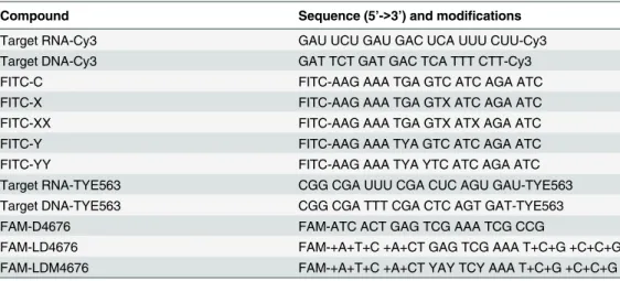
Table 1. Sequences of DNA and LNA/DNA gapmer oligonucleotides.
Compound Sequence (5’->3’) and modifications
D4676 NH2-ATC ACT GAG TCG AAA TCG CCG
D4676inv NH2-GCC GCT AAA GCT GAG TCA CTA
LD4676 NH2-+A+T+C +A+CT GAG TCG AAA T+C+G +C+C+G
LD4676inv NH2-+G+C+C +G+CT AAA GCT GAG T+C+A +C+T+A
DM4676 NH2-ATC ACT YAY TCY AAA TCG CCG
LDM3570 NH2-+A+T+G +A+TA GAX AGT XXA A+C+A +C+A+C
LDM3570inv NH2-+C+A+C +A+CA AXX TGA XAG AT+A +G+T+A
LDM4676 NH2-+A+T+C +A+CT YAY TCY AAA T+C+G +C+C+G
LDM4676inv NH2-+G+C+C +G+CT AAA YCT YAY T+C+A +C+T+A
MixLD4676 NH2-+AT+C AC+T GAG +TCG +AAA +TC+G C+C+G
NH2 = 5’amino modifier C6. This group was present only in the oligonucleotides used for melting curve determination and for the analysis of delivery into cells. + = prefix for LNA; X = 5-OH-dC; Y = 8-oxo-dG.
by PAGE in 15% native gels. The gels were dried, exposed to a storage phosphor screen and vi-sualized using a Typhoon Trio scanner (GE Healthcare, UK).
In vitro
RNase H assay
Target RNA (designated as FR3131), consisting of 269 nt from the 5’end of the HCV genome and the region spanning positions 3081 to 5943, was synthesizedin vitrousing an mMESSAGE mMACHINE T7 Transcription Kit (Life Technologies, USA) according to the manufacturer’s instructions. RNase H assays were performed as described by Kurreck and co-authors [19]. Briefly, the reaction mixture contained 1x RNase H buffer, 0.5 U of bacterial RNase H (Thermo Scientific, USA), 5 pmol of ASO and 500 ng of FR3131 RNA. The reaction mixture was incu-bated at 37°C for 0, 1, 5, 10, 20 or 60 min. At each time point, a 10μl aliquot was collected. The
reaction was stopped by adding 10μl of 2x stop buffer (50 mM EDTA and 1% SDS) and
subse-quent heating to 95°C for 2 min. The reaction products were separated on a 0.8% TAE agarose gel and detected with ethidium bromide staining.
The kinetics of ASO:RNA duplex cleavage were analyzed using pre-formed ASO:RNA du-plexes. Briefly, 1 fmol of the labeled duplexes was mixed with 0.05 U of RNase H in 1x RNase H buffer, and the reaction was performed at 37°C. Aliquots were collected immediately after mixing the substrate and enzyme (0 time point) or after incubation for 10 s or 0.5, 1, 5, 10 or 20 min. The reactions were stopped by adding an equal volume of 2x stop buffer. The samples were then heated to 95°C for 2 min, cooled rapidly on ice and separated by PAGE in native 15% gels. The gels were dried, exposed to a storage phosphor screen and visualized using a Ty-phoon Trio scanner (GE Healthcare, UK). Quantitative analyses were performed using Image-Quant TL Software (GE Healthcare, UK).
Analysis of the stability of ASOs in human serum
To analyze the stability of D4676, DM4676, LD4676, and LDM4676 (Table 1) in human serum, these compounds were labeled with33P as described above for RNA oligonucleotides. Five fmol of each33P-labeled ASO was incubated in human serum (Human Serum, Off the clot, Type AB;PAA, Germany) at 37°C. Aliquots were collected immediately after preparation of the mixtures (0 time point) or after incubation for 0.25, 0.5, 1, 2, 4 or 6 h. Next, 2x stop solu-tion was added to each aliquot. The full-length oligonucleotides and their degradasolu-tion products were separated and analyzed as described above. The average half-lives of each type of ASO were obtained from three independent experiments by fitting the obtained values to an expo-nential decay function.
HCV replicon cells
Huh-luc/neo-ET cells, which harbor the I389/NS3-3’/LucUbiNeo-ET replicon of HCV geno-type 1b (Con1 isolate) (Fig 2A), and replicon-free Huh7-cure cells were obtained from ReBli-kon GmbH(Germany). The cells were maintained in Dulbecco’s modified Eagle’s medium supplemented with penicillin, streptomycin, 0.5 mg/ml G418, 10% fetal calf serum and 2 mM L-glutamine (GE Healthcare, UK).
using a First-Strand cDNA Synthesis kit (Thermo Scientific). HCV-specific cDNA fragment containing the mutation site was PCR-amplified using of primers flanking the mutated region. Obtained PCR products were purified and sequenced using Sanger sequencing.
Transfection of cells with siRNAs or ASOs
For RNAi-guided screening, Huh-luc/neo-ET cells were reverse-transfected with siRNAs (100 nM each) using Lipofectamine RNAiMAX reagent (Life Technologies, USA). Luc activity was measured 48 h post-transfection (p.t.) using reagents and protocols fromPromega(USA). The total protein content in the cell lysates was measured by Bradford micro-assay (Bio-Rad, USA).
Lipofectamine 2000, Lipofectamine RNAiMAX, Lipofectamine LTX (Life Technologies, USA), DOTAP, FuGENE HD (Roche, Switzerland) and TurboFect (Thermo Scientific, USA) re-agents were used to optimize the transfection of Huh-luc/neo-ET cells. Various amounts of these reagents and forward- or reverse-transfection protocols were used to deliver ASOs (Table 1) conjugated to Alexa Fluor 568 into the cells. The transfection efficiencies were
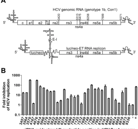
Fig 2. RNAi-guided oligonucleotide target-site selection in the coding region of HCV RNA. (A) Schematic of the HCV genome and the luc/neo-ET (I389/NS3-3’/LucUbiNeo-ET) replicon. The numbers above the HCV genomic RNA indicate the positions of the start codons for the non-structural proteins NS3-NS5B. Luc/neo, firefly luciferase/neomycin phosphotransferase cassette; E-I, encephalomyocarditis virus IRES element.(B)Inhibitory effects of thirty-two different siRNAs targeting the NS3-NS5B region of the luc/neo-ET replicon. The siRNAs (Table 3, guide strands are indicated on thex-axis;“+”, combination of control siRNAs targeting the Luc reporter gene) were transfected into Huh-luc/neo-ET cells at a concentration of 100 nM. At 48 h p.t., the total protein content and Luc activities in cell lysates were determined. The Luc activities were first normalized to total protein content; next, the obtained values were normalized to the value obtained for control cells transfected with non-targeting negative control siRNA (designated as“-”), which was set to 1. They-axis indicates the fold inhibition of HCV replication achieved using the corresponding siRNAs (note the logarithmic scale). The error bars represent the standard deviation of three independent
experiments.
analyzed using an LSRII flow cytometer (BD Biosciences, USA). Cytotoxic effects (or lack there-of) were observed using a Nikon Eclipse confocal microscope (Nikon, Japan).
Quantitation of the inhibitory effects of ASOs
Cells were collected and lysed at selected time points. The total protein content in the lysate and Luc activity were measured. For the normalization of HCV replication, which is propor-tional to Luc activity, the following calculations were performed. First, to enable the compari-son of the average Luc activity per living cell, the total protein content of the cells was used to normalize the Luc activity as previously reported [51]. Thus, the HCV replication signal was expressed as relative light units per microgram of protein (RLU/μg protein). Second, the
ob-tained normalized Luc values were divided by those obob-tained for the negative controls:“-”
siRNA- or mock-transfected cells. The averages and standard deviations of seven independent experiments were obtained. Subsequently, the dimensionless average values were fitted with a four-parameter dose-response equation (variable slope model) using Prism 5 (GraphPad Soft-ware,Inc., San Diego, CA, USA) to estimate the effective concentration 50 (EC50) values.
Results
Thermal stability of the all-DNA ASO:RNA duplex is reduced upon
incorporation of 8-oxo-dG residues
8-oxo-dG residues have been reported to destabilize ASO:DNA duplexes [27,29,30]. However, ASOs are generally used to target RNA rather than DNA molecules. To analyze the effects of 8-oxo-dG residues on the binding of ASOs to DNA and RNA molecules, a set of all-DNA oli-gonucleotides was prepared in which none, one, or two of the centrally located dG residues were substituted with 8-oxo-dG residues. For comparison, a set of ASOs containing 5-OH-dC residues was prepared because of the similarity to 8-oxo-dG; the 5-OH-dC minor tautomeric form (Fig 1A) was predicted to have abnormally strong bonding to dG residues [33]. Neverthe-less, the introduction of 5-OH-dC residues simultaneously into ASO and target DNA also re-sults in decreased ASO:DNA duplex stability [28]. All the oligonucleotides were 21 nt long and had identical sequences (Table 2). Duplex formation between these oligonucleotides and their
Table 2. Sequences of oligonucleotides used to determine the melting temperatures by FRET.
Compound Sequence (5’->3’) and modifications
Target RNA-Cy3 GAU UCU GAU GAC UCA UUU CUU-Cy3
Target DNA-Cy3 GAT TCT GAT GAC TCA TTT CTT-Cy3
FITC-C FITC-AAG AAA TGA GTC ATC AGA ATC
FITC-X FITC-AAG AAA TGA GTX ATC AGA ATC
FITC-XX FITC-AAG AAA TGA GTX ATX AGA ATC
FITC-Y FITC-AAG AAA TYA GTC ATC AGA ATC
FITC-YY FITC-AAG AAA TYA YTC ATC AGA ATC
Target RNA-TYE563 CGG CGA UUU CGA CUC AGU GAU-TYE563
Target DNA-TYE563 CGG CGA TTT CGA CTC AGT GAT-TYE563
FAM-D4676 FAM-ATC ACT GAG TCG AAA TCG CCG
FAM-LD4676 FAM-+A+T+C +A+CT GAG TCG AAA T+C+G +C+C+G
FAM-LDM4676 FAM-+A+T+C +A+CT YAY TCY AAA T+C+G +C+C+G
NH2 = 5’amino modifier C6; + = prefix for LNA; X = 5-OH-dC; Y = 8-oxo-dG.
targets was monitored by FRET. In this setup, any difference in the Tm of the formed duplexes is attributable to the presence of the 8-oxo-dG or 5-OH-dC modifications.
When the 8-oxo-dG modification (FITC-Y) was introduced, the Tm values of both the ASO:DNA and ASO:RNA duplexes were reduced by ~1.6–1.8°C compared to those of the du-plexes formed by control oligonucleotides. Increasing the number of 8-oxo-dG modifications (FITC-YY) resulted in further reduction of the Tm by ~1.6°C for ASO:DNA and by ~2.5°C for ASO:RNA duplexes. Thus, destabilization resulting from the insertion of 8-oxo-dG modifica-tions occurs not only for ASO:DNA duplexes (Fig 1B) but also for ASO:RNA duplexes (Fig 1C). In contrast, duplexes formed by oligonucleotides with one or two 5-OH-dC residues had nearly the same Tm as duplexes formed by unmodified control oligonucleotides (Fig 1B and 1C). Thus, the introduction of the 5-OH-dC modification into an ASO alone had virtually no effect on the stability of duplexes formed with DNA or RNA.
RNAi-guided selection reveals potential ASO target sites in the coding
region of HCV RNA
Known targets for ASOs are located in a 350-nt region at the 5’-terminus of HCV RNA. Clear-ly, targeting a region that comprises less than four percent of the virus genome is a bottleneck that hinders the development of the most efficient ASOs. RNAi technology was used to search for highly accessible target sites in the HCV coding region. Twenty-eight different siRNAs tar-geting the HCV genome were designed (Table 3). Each siRNA had a 19-nt duplex region with 2-nt 3’-overhangs [52]. In addition, four siRNAs (3564, 7749, 7805, and 7983) that were previ-ously reported to efficiently inhibit HCV replication [53,54] were used for comparison. Non-targeting siRNAs (AM4611 or AM4635) and a combination of siRNAs against a sequence en-coding Luc marker (AM4629) [47] were used as negative and positive controls, respectively.
The level of Luc activity in Huh-luc/neo-ET cells is directly proportional to the copy num-ber and replication efficiency of the HCV subgenomic replicon, making it an efficient tool for analyzing the anti-HCV efficiencies of the obtained siRNAs [55,56]. At a concentration of 100 nM, the majority of the designed HCV-specific siRNAs induced less of an effect that the posi-tive controls (Fig 2B). Moreover, similarly to the negative control siRNA, several siRNAs (4167, 7512, 8685, and 9336) did not have any effect on HCV replication. The effects of three siRNAs (4814, 7790 and 8161) were comparable to those of the positive controls (70- to 100-fold inhibition), and two siRNAs (3570 and 4676) were more potent (approximately 300-fold inhibition).
As the high inhibitory potential of an siRNA indicates the accessibility of the corresponding target sites, it was concluded that RNAi-guided screening enabled the selection of several po-tential ASO target sites in the HCV coding region. However, an all-DNA ASO based on the se-quence of the guide strand of siRNA 4676 was essentially unable to suppress HCV replication. Therefore, HCV-specific 21-mer LNA/DNA gapmer oligonucleotides that contained five LNA monomers at each end and three modified residues in the DNA region were designed (Tables1
and2). As the target site of siRNA 4676 contained three C-residues in its central region, it was targeted by ASOs containing three 8-oxo-dG nucleotides. In contrast, the target site for siRNA 3570 contained three G-residues, and hence, a control ASO was generated that contained three 5-OH-dC residues (Table 1).
Inhibitory efficiency of modified ASOs is reduced by point mutation in the
target site
the target region) have no such effect. The only mutation in the selected target sites, for which the viability of the mutant replicon has been previously demonstrated, is located in the target site of siRNA 3570 and results in a Thr54!Ala change in NS3 [57]. Therefore, this mutation was introduced into the HCV replicon that was used to generate a Huh-luc/neo-ET-3570mut cell line (Fig 3A). As the central region of the target site of siRNA 3570 contains only two C-residues, an ASO similar to LDM4676 (which contains three 8-oxo-dG residues;Table 1) could not be designed against this site. Therefore, a control LNA/DNA gapmer containing three 5-OH-dC residues (LDM3570;Fig 3A) was used instead. Oligonucleotides with inverted se-quences (LDM4676inv and LDM3570inv;Table 1) were used as controls.
Huh-luc/neo-ET and Huh-luc/neo-ET-3570mut cells were transfected with different concen-trations of siRNA 3570, siRNA 4676, LDM4676, LDM4676inv, LDM3570 and LDM3570inv. A
Table 3. Sequences of oligonucleotides in siRNA duplexes.
Positiona Antisense strand (5'->3') Sense siRNA strand (5'->3')
3457 UAG UGA UGA UGC AGC CAA GUA C UUG GCU GCA UCA UCA CUA GC 3564 GAC AGU CCA ACA CAC GCC AUU U GGC GUG UGU UGG ACU GUC UA 3570 AUG AUA GAC AGU CCA ACA CAC G UGU UGG ACU GUC UAU CAU GG 4167 UAC CCC GGU UCU GAU GUU AGG U AAC AUC AGA ACC GGG GUA AG 4676 AUC ACU GAG UCG AAA UCG CCG G CGA UUU CGA CUC AGU GAU CG 4814 AUG CCC AUC CUG CCC CUA CCA G UAG GGG CAG GAU GGG CAU UU 5066 UUG UCU CCU GCC UGC UUA GUC C UAA GCA GGC AGG AGA CAA CU 5378 AUC CUG CCC ACA AUG ACC ACG U GGU CAU UGU GGG CAG GAU CA 5518 UUG CCU UCU GUU UGA AUU GUU C AAU UCA AAC AGA AGG CAA UC 5622 AUU CCA CAU AUG CUU CGC CCA G GCG AAG CAU AUG UGG AAU UU 5939 AUC UCG CCG CUC AUG ACC UUA A GGU CAU GAG CGG CGA GAU GC 5978 AUA GCA GGG AGU AGG UUA ACC U UAA CCU ACU CCC UGC UAU CC 6274 AUA UCC AAU CCC AAA CAU CUC G AUG UUU GGG AUU GGA UAU GC 6365 UAC CCA CGU UGA CAU GAG AAG U CUC AUG UCA ACG UGG GUA CA 6590 UAC UCC UCA GCA GCC ACC CGC G GGU GGC UGC UGA GGA GUA CG 7043 AUG UUC CCG CCC AUC UCC UGC A GGA GAU GGG CGG GAA CAU CA 7125 UAC UUC CCU CUC AUC CUC CUC G GAG GAU GAG AGG GAA GUA UC 7512 AUC CCC CGG CUC CCC CUC AAG U GAG GGG GAG CCG GGG GAU CC 7699 UUG UAG CAU AGA CCA AGU UGU A ACU UGG UCU AUG CUA CAA CA 7749 CAG UCU GUC AAA GGU GAC CUU G GUC ACC UUU GAC AGA CUG CA 7790 AUC UCC UUG AGC ACG UCC CGG G GGA CGU GCU CAA GGA GAU GA 7805 GAC GCC UUC GCC UUC AUC UCC A GAU GAA GGC GAA GGC GUC CA 7983 GUC AAU UGG UGU CUC AGU GUC C ACU GAG ACA CCA AUU GAC AC 8155 AUC CGU AUG AAG AGC CCA UCA A UGG GCU CUU CAU ACG GAU UC 8161 AUU GGA AUC CGU AUG AAG AGC U CUU CAU ACG GAU UCC AAU AC 8468 AAG UAA CAU GUG AGG GUA UUA A UAC CCU CAC AUG UUA CUU GA 8657 AAG UCG UAU UCU GGU UUG GGC C CAA ACC AGA AUA CGA CUU GG 8674 AUG AUG UUA UCA ACU CCA AGU U UGG AGU UGA UAA CAU CAU GC 8685 AUU GGA GGA GCA UGA UGU UAU A ACA UCA UGC UCC UCC AAU GU 8819 AUG AUG AUG UUG CCU AGC CAG G GCU AGG CAA CAU CAU CAU GU 8873 AUG GAG AAG AAA UGA GUC AUC U GAC UCA UUU CUU CUC CAU CC 9336 AUA GAU GCC UAC CCC UAC AGA U GUA GGG GUA GGC AUC UAU CU
a
Position refers to the terminal 3’-end nucleotide position of the siRNA antisense strand in the HCV Con1 genome (GenBank accession number: AJ238799).
Huh-point mutation in HCV RNA that changes the classical A:U base pair in the siRNA guide-strand: target-RNA duplex to the G:U wobble base pair resulted in a marked (10-fold) decrease in the in-hibitory efficiency of siRNA 3570 (Fig 3B). As expected, this mutation did not alter the inhibitory efficiency of siRNA 4676 or LDM4676. Transfecting cells with different amounts of LDM4676inv resulted in increased HCV replication regardless of the presence or absence of the mutation in the siRNA 3570 target site (Fig 3C). In contrast to LDM4676, the inhibitory activity of LDM3570 was clearly reduced by the mutation (Fig 3B). At concentrations up to 62.5 nM, the control oligo-nucleotide LDM3570inv was unable to suppress HCV replication, and its effects (if any) were not diminished by the mutation in the siRNA 3570 target site (Fig 3B). Therefore it can be concluded that at these concentrations the potency of LDM3570 was indeed specifically reduced by muta-tion of its target site. These findings are consistent with the antisense mechanism of acmuta-tion of modified oligonucleotides; furthermore, the data indicate that the observed suppression of HCV replication was not caused by side effects of the ASOs. However, at concentrations 125 or 250 nM, LDM3570inv exhibited toxicity and inhibited HCV replication (Fig 3B).
Incorporation of 8-oxo-dG residues into LNA/DNA gapmer
oligonucleotides has no negative impact on their antisense potency
At the highest concentrations, LDM4676 and LDM3570 visibly damaged the transfected cells. Although these ASOs lack the nucleotide sequence descriptors (TCC and TGC motifs) charac-teristic of hepatotoxic LNA/DNA gapmer oligonucleotides [58], cytotoxicity may result from additional factors and their combinations. These factors include the transfection protocol, the presence of modified nucleobases and the cytotoxicity of the transfection reagent. As cytotoxic-ity represents an obstacle for more detailed studies, more suitable transfection methods were sought. Huh7 cells and their derivatives were difficult to transfect with DNA or LNA/DNA gapmer oligonucleotides without causing cell damage. Therefore, the results obtained using dif-ferent amounts of six transfection reagents (seeMaterials and Methods) and reverse- or direct-transfection protocols were compared. Based on the results of this comparison, an optimized Lipofectamine 2000-mediated reverse-transfection protocol was selected that enabled the deliv-ery of siRNA and ASOs into 70–75% of the cells. Further increases in transfection efficiency re-quired higher amounts of transfection reagent, which resulted in decreased viability of
transfected cells. Thus, in subsequent experiments, the reduction of HCV replication to 25–
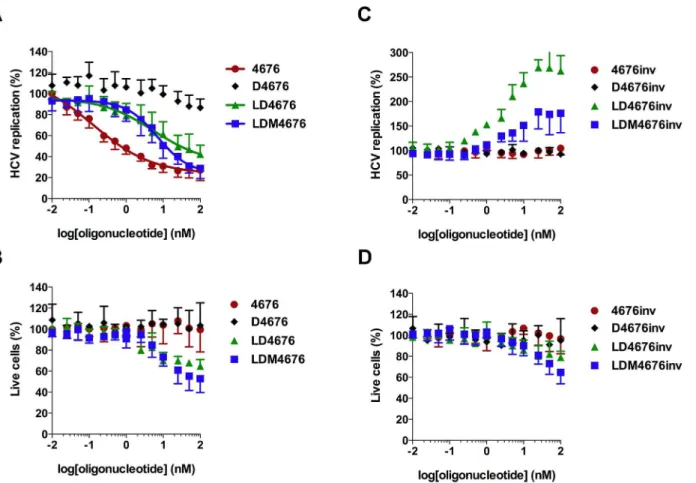
30% of its original level roughly corresponded to complete suppression of HCV replication in every siRNA- or ASO-transfected cell. Even using the selected protocol, higher concentrations of LD4676, LDM4676 and LDM4676inv exhibited some cytotoxicity, as determined by mea-suring the total protein content in the lysates of transfected cells (Fig 4B–4D). Cell viability measurements using the xCELLigence system (ACEA Biosciences, USA) revealed the same trend. Therefore, as in all previous experiments (Figs2B,3B and 3C), the replication signal (Luc activity) was normalized to the total protein content of the cell lysate (essentially, to the number of living cells). The quantitative evaluation by four-parameter dose-response curve fit-ting (variable slope model) of such values obtained in seven independent experiments is shown inFig 4A, and the obtained EC50values for different ASOs are summarized inTable 4.
luc/neo-ET-3570mut (white bars) cells were transfected with increasing concentrations (x-axis) of LDM3570, LDM3570inv and siRNA 3570(B)or LDM4676, LDM4676inv and siRNA 4676(C). The HCV replication values (y-axis) were calculated as described forFig 2B. The obtained values were subsequently normalized to those from mock-transfected control cells, which was set to 100%. Each panel represents data from one of two reproducible independent experiments.
The obtained data revealed that D4676 did not considerably reduce HCV RNA replication, confirming a previous observation that the all-DNA ASOs are not efficient inhibitors. siRNA 4676 had an EC50of 0.13 nM, whereas the EC50values for LD4676 and LDM4676 were
approx-imately 50-fold higher (Table 4). Interestingly, despite the roughly equal mean EC50values of
Fig 4. Effects of 8-oxo-dG residues on antisense potency and off-target effects of LNA/DNA gapmer oligonucleotides.Huh-luc/neo-ET cells were transfected with increasing concentrations (x-axis) of various oligonucleotides (Table 1) targeting the 4676 site(A, B)or with inverted non-targeting control oligonucleotides(C, D). The error bars represent the standard deviation of seven independent experiments.(A, C)The effects of the oligonucleotides on HCV replication are shown on they-axis. Transfection and normalization of Luc activity were performed as described forFig 2B. The obtained values were subsequently normalized to those from mock-transfected control cells, which were set to 100%. The values for siRNA 4676, LD4676, and LDM4676 were fitted with a four-parameter dose-response equation (variable slope model); estimated EC50values are shown inTable 4.(B, D)Percentage of living cells in transfected cell cultures. The total protein content in the lysates of transfected Huh-luc/neo-ET cells was normalized to that of mock-transfected cells (set to 100%) to obtain the percentage of living cells (y-axis).
doi:10.1371/journal.pone.0128686.g004
Table 4. EC50, CI and R2values for LD4676, LDM4676, and siRNA 4676 estimated from seven indepen-dent experiments.
ON/siRNA EC50(nM) CI (nM) R2
D4676 ND ND ND
LD4676 5.5 0.9–32.9 0.77
LDM4676 7.1 4.0–12.5 0.87
siRNA 4676 0.13 0.03–0.5 0.95
CI = 95% confidence interval; R2= goodness of a four-parametric nonlinear regression curve
fit; ND = not determined.
LD4676 and LDM4676, the shape of the LD4676 inhibitory curve at higher concentrations was linear rather than non-linear, which was also evident by the goodness of fit with the variable slope non-linear regression curve (Table 4). Consequently, at the highest concentrations, LDM4676 inhibited the HCV replication signal to a approximately 27% residual level. As ap-proximately 25% inhibition was achieved with siRNA 4676 (Fig 4A), which is capable of nearly completely inhibiting HCV replication in positively transfected cells (Fig 2B), it was calculated that 100 nM LDM4676 suppressed HCV replication by at least 95% in ASO-transfected cells. In contrast, the residual HCV replication level in the presence of the highest concentration of LD4676 was 42% (Fig 4A), indicating that HCV replication in ASO-transfected cells was inhib-ited by no more than 80%. To confirm that the observed inhibitory effects (Fig 4A) were se-quence-specific, control oligonucleotides with inverted sequences (Table 1) were transfected into Huh-luc/neo-ET cells. Importantly, despite an observation of mild cytotoxicity at the highest concentrations, none of the control oligonucleotides inhibited HCV replication (Fig 4C and 4D); these data indicated that the observed effects of the ASOs targeting the siRNA 4676 site were sequence-specific. These results also demonstrated that under certain conditions, LDM4676 might be a more efficient inhibitor of HCV RNA replication than LD4676. However, in general, the incorporation of 8-oxo-dG residues did not result in significant gains in anti-sense potency in a cell-based HCV replication assay.
8-oxo-dG residues reduce the Tm of the LNA/DNA gapmer ASO:RNA
duplex but have little effect on duplex formation
The Tm values of the LDM4676:DNA and LDM4676:RNA duplexes were determined and compared to the Tm values of duplexes composed of non-modified all-DNA ASO (D4676) or LNA/DNA gapmer ASO (LD4676) (Table 1).
Numerous studies have shown that the incorporation of LNA residues strongly increases the binding of ASOs to their targets [20,59]. Consistent with this, the melting temperature of the LD4676:DNA duplex was 20°C higher than the Tm of the D4676:DNA duplex (Fig 5A). The effect of LNA residues on the Tm of the ASO:RNA duplex was even more prominent: the Tm increase was greater than 30°C at all analyzed target RNA concentrations (Fig 5B). Consis-tent with the results obtained for all-DNA ASOs (Fig 1B, 1C), the incorporation of 8-oxo-dG residues reduced the Tm of duplexes of LNA:DNA gapmer ASOs with both DNA (Fig 5A) and RNA (Fig 5B) targets. For both targets, the decrease in Tm (LDM4676versusLD4676) was be-tween 5 and 10°C (Fig 5A and 5B).
To investigate how the reduced Tm affects the efficiency of ASO:RNA duplex formation, a 37-nt ssRNA that contains the target site of siRNA 4676 (Fig 5C) was labeled with33P and in-cubated with D4676, DM4676, LD4676 or LDM4676 (Table 1) at physiological temperature (37°C). ASO:RNA duplexes were detected immediately after the mixing of the ssRNA target and ASO (Fig 5C). The 8-oxo-dG residues in all-DNA ASOs clearly reduced the efficiency of duplex formation. This effect was likely due to the reduced Tm of DM4676. The LNA/DNA gapmer ASO formed duplexes at least as efficiently as non-modified all-DNA ASO. Interest-ingly, 8-oxo-dG residues did not inhibit LNA/DNA gapmer ASO:RNA duplex formation (Fig 5C), most likely because the Tm of the LDM4676:RNA duplex remained sufficiently high to ensure its effective formation.
8-oxo-dG residues have no adverse effects on RNase H-mediated
cleavage of ASO:RNA duplexes
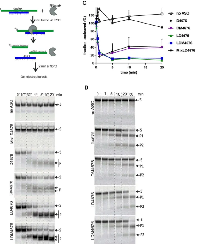
assay, LDM4676 was a somewhat more efficient inhibitor of HCV replication (Fig 4A). There-fore, we asked whether there were any differences in the ability of these duplexes to undergo RNase H-mediated target RNA cleavage. As human RNase H enzymes are not commercially available, we used bacterial RNase H, which has very similar fold and active center organization [60] and shares many enzymatic properties with human RNase H enzymes [61,62].
Pre-formed ASO:RNA duplexes, consisting of D4676, DM4676, LD4676, LDM4676 or an LNA/DNA mixomer oligonucleotide designated MixLD4676 (Table 1), and33P-labeled 37-nt target RNA molecules were used to estimate the kinetics of RNase H-mediated cleavage (Fig 6A). As expected, RNase H had no effect on ssRNA (Fig 6B and 6C). Similarly, due to the ab-sence of the obligatory 6-bp DNA:RNA duplex stretch required for RNase H activation [19,62], RNase H could not cleave the RNA strand in the MixLD4676:RNA duplex (Fig 6B and 6C). In contrast, D4676:RNA and DM4676:RNA duplexes were rapidly cleaved. However, after 0.5 min, the reaction plateaued, leaving 20–30% of the substrate uncleaved. The cleavage of
Fig 5. 8-oxo-dG residues reduce the Tm of duplexes between LNA/DNA gapmers and their targets. The effects of 8-oxo-dG residues on the Tm of LNA/DNA gapmer ASO:DNA(A)and LNA/DNA gapmer ASO: RNA (B) duplexes were measured by FRET. Target DNA or RNA oligonucleotides (Table 2) were labeled with TYE563 at the 3’-end; the D4676, LD4676 and LDM4676 probes had FAM at the 5’-end. The
measurements were performed, and the data are presented as described forFig 1(C). The effect of 8-oxo-dG residues on ASO:RNA duplex formation. Upper: schematic of the experimental setup. Applicable for some ASOs: Y, 8-oxo-dG residue; +, LNA sugar base. Lower: the33P-labeled 37-nt ssRNA target was mixed with the indicated ASOs. The samples were collected immediately (“0”) or after incubation at 37°C for the indicated times. The obtained probes were resolved by native PAGE in 15% gels and imaged using a Typhoon Trio instrument. The positions of the ASO:RNA duplexes (“duplex”) and ssRNA are shown at right. (A-C) Each panel represents data from one of three reproducible independent experiments.
duplexes containing LD4676 or LDM4676 initially followed similar kinetics, but cleaved prod-ucts continued to accumulate for 5 more min, reducing the levels of intact substrates to 10% of the initial amount (Fig 6B and 6C). As no significant differences in the cleavage of duplexes formed by D4676 and LD4676 and their respective modified ASOs were observed (Fig 6C), it was concluded that the incorporation of 8-oxo-dG into the ASO had no effect on the overall ef-ficiency of RNase H-mediated cleavage of pre-formed ASO:RNA duplexes.
8-oxo-dG modifications alter the specificity of ASO-mediated RNase H
cleavage of target RNA
Upon closer examination of the RNase H cleavage assay results, we noticed that the pattern of cleavage products generated by ASOs with and without 8-oxo-dG modification dramatically differed. In particular, a single major cleavage product was clearly dominant for the LD4676: RNA duplex, whereas for the LDM4676:RNA duplex, at least two major cleavage products of roughly the same abundance were observed (Fig 6B). A similar effect, although less pro-nounced, was observed for duplexes containing the all-DNA oligonucleotides D4676 and DM4676. Thus, the presence of 8-oxo-dG residues in ASOs triggered multiple cleavages by RNase H in the targeted DNA:RNA duplex region.
Efficiency of RNase H-mediated cleavage of target RNA molecules
correlates with the efficiency of ASO:RNA duplex formation
Next, we studied the effect of 8-oxo-dG residues on ASO:target RNA duplex formation and subsequent cleavage by RNase H in a single reaction. To account for the possible influence of RNA secondary structure, full-length HCV replicon RNA was used as a target. However, this RNA underwent slow degradation in the absence of ASOs. Therefore, FR3131, anin vitro -syn-thesized 3131-nt fragment of HCV replicon RNA, was used as a target. This target RNA was pre-incubated with D4676, DM4676, LD4676 or LDM4676 for 10 min at 37°C; next, RNase H was added to the reaction mixture. In the absence of ASO, FR3131 RNA remained stable (Fig 6D). In the presence of D4676, the targeted RNA was cleaved into two fragments of the ex-pected sizes (Fig 6D). 8-oxo-dG residues clearly reduced the cleavage efficiency in the presence of all-DNA ASOs (Fig 6D). As the modification had a similar effect on ASO:RNA duplex for-mation (Fig 5C) but did not affect the degradation of pre-formed ASO:RNA duplexes (Fig 6B and 6C), it was concluded that the rate-limiting step for RNase H-mediated target RNA cleav-age was ASO:RNA duplex formation. In the presence of LNA/DNA gapmer ASOs, nearly com-plete cleavage of FR3131 RNA occurred (Fig 6D). This result is consistent with the more complete degradation of pre-formed LNA/DNA gapmer ASO:RNA duplexes (Fig 6C). In the context of LNA/DNA gapmers, 8-oxo-dG residues had little, if any, effect on RNase H-mediat-ed cleavage of target RNA (Fig 6D). Due to the large sizes of the FR3131 cleavage products (ap-proximately 1250- and 1850-nt), the effect of 8-oxo-dG residues on the precise cleavage positions within the ASO:RNA duplex could not be observed in this experiment.
average of three independent experiments. Error bars indicate the standard deviation.(D)Cleavage of FR3131 RNA by RNase H in the presence of different ASOs. The RNA and ASOs were mixed and incubated at 37°C for 10 min; then, RNase H was added to the reaction mixture. RNA samples were collected at the indicated time points and analyzed by electrophoresis in native 0.8% TAE agarose gels. The results from one of three independent reproducible experiments are shown. S: substrate; P1 and P2: cleavage products.
Incorporation of 8-oxo-dG residues increases the stability of
oligonucleotides in human serum
Another factor that affects the potency of ASOs is their stability in biological environments. To determine whether 8-oxo-dG residues affected the stability of all-DNA and LNA/RNA gapmer ASOs,33P-labeled D4676, DM4676, LD4676 and LDM4676 were incubated in human serum for 0, 0.25, 0.5, 1, 2, 4 and 6 h. Consistent with previous data [19], the LNA/DNA gapmer oligo-nucleotides were much more stable than their all-DNA counterparts (Fig 7A and 7B). The half-life of D4676 was less than 8 min, whereas the half-life of LD4676 was more than 10 times longer. Importantly, the incorporation of 8-oxo-dG residues significantly increased the stability of both types of oligonucleotides (Fig 7B). The half-life of DM4676 (15 min) was nearly twice of that of D4676. Similarly, 8-oxo-dG modification increased the half-life of the LNA/DNA gapmer oligonucleotide from 90 min to 130 min. The mechanism(s) responsible for this stabili-zation are outside of the scope of current study and remain unknown.
Discussion
The difficulties hampering the clinical use of ASO drugs include their low efficiency, low bio-availability, rapid degradation and unwanted side effects (typically off-target effects). Thus, synthetic ASOs often contain one or more modifications aimed at improving the properties of the compound. The common theme in ASO design is to achieve increased ASO:target duplex
Fig 7. 8-oxo-dG residues increase the stability of ASOs in human serum. (A)33P-labeled D4676, DM4676, LD4676 and LDM4676 oligonucleotides (Table 1) were incubated in human serum at 37°C. Aliquots were collected at the indicated time points and analyzed by PAGE in native 15% gels. The results from one of three independent reproducible experiments are shown.(B)Quantitative representation of the stability of the oligonucleotides. The amounts of radioactivity remaining in the uncleaved ASOs were quantified using a Typhoon Trio instrument. Quantifications were performed for each gel. The obtained values were normalized to the radioactivity present in the substrate before incubation in human serum (set to 100%). The fraction of remaining ASO is shown as a single exponential decay function. Each point corresponds to the average from three independent experiments. Error bars indicate the standard deviation;*p<0.05 and**p<0.01 (Student’s t-test).
stability [5], which is typically associated with higher ASO potency [63]. However, ASOs with a very high Tm also tend to bind to secondary targets, and their ability to trigger RNase H-medi-ated target RNA degradation may be reduced or even completely abolished [25]. Consequently, such ASOs are generally highly efficient only if they target short microRNAs or if their binding site overlaps with the region directly involved in translation initiation. This limitation drastical-ly reduces the number of suitable ASO binding sites in mRNA molecules. Here, we provide a novel platform for ASO target site selection and show that the destabilization of the ASO:tar-get-RNA duplex by the introduction of 8-oxo-dG modifications has several advantages over conventional LNA/DNA gapmer ASO design.
The initial idea of using 8-oxo-dG residues was based on theoretical quantum calculations. It was hypothesized that the antisense activity of oligonucleotides containing 8-oxo-dG may be increased due to the ability of corresponding nucleobases to acquire tautomeric forms, which enables enhanced binding to complementary normal nucleobases [33]. However, experimental data revealed that the incorporation of 8-oxo-dG residues reduced the Tm of ASO:DNA and ASO:RNA duplexes (Figs1,5A and 5B), probably because the major tautomer of 8-oxo-dG in aqueous solution is the 6,8-diketo form [64], which binds to complementary dC residues more weakly than does the standard dG residue [29]. Thus, the contribution of the strongly binding minor zwitterionic tautomer of 8-oxo-dG (Fig 1A) may be too small to be detected. The overall oligonucleotide Tm analysis, however, did not exclude the possibility that different tautomeric forms of 8-oxo-dG might play an important role in base pairing.
The efficiency of ASOs critically depends on the physical accessibility of its target site. Very limited information is available about the actual topological structures of mRNA in cells, where they form RNA-protein complexes. In this study, the attempts to target sequences with specific nucleotide compositions or regions lacking predicted secondary structures (calculated using the Minimal Free-Energy method) were unsuccessful. High-throughput selective 20-hydroxyl
acylation analyzed by primer extension (SHAPE) has been used to resolve the secondary struc-ture of the HIV-1 RNA genome [65]. However, for HCV, only the structures of short non-cod-ing terminal fragments of the genome have been analyzed usnon-cod-ing SHAPE [66–68]. Furthermore, some viral RNAs exist in different conformations, and corresponding changes are integral to the regulation of the viral infection cycle [69,70]. Hence, there is the need for empirical screen-ing of targeted RNA.
The low efficiency of all-DNA ASOs (Fig 4A) hinders their use as screening tools. Therefore, we reasoned that siRNAs, which can be highly efficient and are relatively inexpensive, might represent suitable tools for performing such screening. This approach led to the identification of two potent siRNAs that were 3-fold more efficient than positive control siRNAs targeting the non-structured reporter part of the replicon RNA construct (Fig 2). Therefore, we hypothe-sized that the ability of these siRNAs to suppress HCV replication would serve as a good indi-cation that the corresponding regions of the HCV RNA genome are accessible not only to the cellular RNA silencing machinery but also to ASOs. An additional benefit of this approach is that it allows the direct comparison of the effects of ASOs and siRNAs targeting the same se-quences. It would be interesting to compare the siRNA mapping data with the SHAPE-based structures of corresponding RNAs. If there is a correlation between the observed efficiencies of siRNAs and the determined high-order structures of mRNAs (or a viral RNA genomes), siR-NAs might become useful tools for probing the high-order structure of RsiR-NAs inside the cell.
Experiments with anin vitro-synthesized fragment of HCV RNA (Fig 6D) and with the HCV replicon cell line (Fig 3) confirmed that modified LNA/DNA gapmer oligonucleotides use an antisense mode of action. Although the insertion of three 8-oxo-dG residues into the LNA/DNA gapmer ASO did not reduce the EC50, it somewhat increased the inhibition of virus
to the increased stability of modified compounds in a biologically relevant environment (Fig 7).
Modified ASOs, where LNA residues are dispersed over the length of the compound, were found to lack an antiviral effect. Consistent with previous studies [71], MixLD4676 and other similarly designed ASOs were unable to trigger RNase H-mediated degradation of the RNA strand in ASO:RNA duplexes (Fig 6B and 6C). These data, similar to those published by Laxton and co-workers [72], highlighted the importance of RNase H-mediated cleavage for the anti-HCV activity of ASOs. RNase H-mediated RNA degradation also depends on the ability of an ASO to form a duplex with its target. Using a short target RNA molecule, duplex formation was shown to be fast, and, as expected, its efficiency correlated with the ASO Tm (Fig 5). Some-what unexpectedly, the initial speed of degradation of pre-formed ASO:RNA duplexes de-pended little, if at all, on the modifications introduced into the ASO (Fig 6C). Instead, there was a clear correlation between the efficiency of ASO:RNA duplex formation (Fig 5C) and the efficiency of RNase H-mediated cleavage of FR3131 RNA (Fig 6D), suggesting that in thein vitroRNA cleavage experiment, the efficiency and speed of ASO:RNA duplex formation was the rate-limiting step. However, due to different conditions, including differences in the speci-ficity and abundance of RNase H enzymes in living human cells, the possibility cannot be ex-cluded that thein vivoactivity of ASOs does not necessarily correlate with its binding to small model substrates. Indeed, human cells have two different RNase H enzymes. Although the human RNase H1 shares many enzymatic properties with the bacterial enzyme, there are dif-ferences. Human RNase H1 binds to A-type RNA:DNA duplexes with much greater activity than bacterial RNase H and displays a strong positional preference for cleavage, i.e., it cleaves between 8 and 12 nucleotides from the 50-RNA-30-DNA terminus of the duplex [62].
There-fore, it would be interesting to study whether the presence of 8-oxo-dG residues affects the cleavage specificity of human RNase H enzymes. If this indeed is the case, then such modifica-tions might be particularly useful for constructing ASOs that target viruses that rapidly develop resistance to siRNAs [73,74] or ASOs against RNAs with pre-existing variations in the target site.
Although LDM4676 displayed high activity in differentin vitroassays, its value as a poten-tial HCV inhibitor critically depends on itsin vivoperformance. However, such studies are hampered by the lack of low-cost small-animal models and by the high costs of ASOs contain-ing LNA bases, 8-oxo-dG residues and phosphorothioate modifications (needed for increased
in vivostability) in its backbone. However, should such types of compounds be highly activein vivo, they could contribute to the development of ASO-based HCV treatments. Miravirsen, the first experimental drug of this type, has already been successfully used in clinical trials
[26,75,76]. However, miravirsen targets an important cofactor of HCV genome expression and replication, whereas the LDM4676-type ASO targets the HCV genome itself. Combinations of drugs with different mechanisms of action have been key for the successful treatment of chron-ic infections caused by viruses capable of rapidly developing drug resistance. It is also likely that despite recent progress in the development of orally deliverable oligonucleotide drugs [77,78], a subcutaneous injection will remain the main delivery method for ASOs or their com-binations. Therefore, the long half-life of ASOs, allowing once-a-week administration [26,79], represents an important property of such compounds. Correspondingly, increases in the serum half-life, resulting from the insertion of 8-oxo-dG residues (Fig 7), may represent another im-portant benefit of modified ASO compounds.
reliable general method for ASO target site selection. This study also provides an important set of findings concerning the use of naturally occurring, minimally modified nucleobases in ASOs. In contrast to nucleobases that contain bulky modifications, the 8-oxo-dG residues re-duced the Tm of ASO:RNA duplexes and had no negative effects on RNase H-mediated degra-dation of RNA strands in ASO:RNA duplexes. Instead, 8-oxo-dG residues facilitated cleavage by RNase H at multiple positions within the target region. Furthermore, the incorporation of 8-oxo-dG residues increased the stability of ASOs in human serum. These effects, possibly combined with other properties of modified nucleobases (such as strong base-pairing of their minor tautomeric forms), outweigh the negative effects on the overall Tm of the ASO. This en-abled us to obtain modified LNA/DNA gapmer oligonucleotides with EC50values similar to
their non-modified counterparts but capable of almost completely inhibiting HCV replication in replicon cell lines at higher concentrations.
Author Contributions
Conceived and designed the experiments: MM AN AP EŽLV AS TR M. Kelve MS M. Karelson AM. Performed the experiments: MM AN AP TR M. Kelve. Analyzed the data: MM AN AP TR M. Kelve M. Karelson AM. Contributed reagents/materials/analysis tools: MM AN AP TR M. Kelve MS M. Karelson AM. Wrote the paper: MM AN AP TR M. Kelve MS M. Karelson AM.
References
1. Kling J. Safety signal dampens reception for mipomersen antisense. Nat Biotechnol. 2010; 28: 295– 297. doi:10.1038/nbt0410-295PMID:20379153
2. Toulmé JJ. New candidates for true antisense. Nat Biotechnol. 2001; 19: 17–18. doi:10.1038/83451
PMID:11135542
3. Hausen P, Stein H. Ribonuclease H. An enzyme degrading the RNA moiety of DNA-RNA hybrids. Eur J Biochem FEBS. 1970; 14: 278–283. PMID:5506170
4. Stein H, Hausen P. Enzyme from calf thymus degrading the RNA moiety of DNA-RNA Hybrids: effect on DNA-dependent RNA polymerase. Science. 1969; 166: 393–395. PMID:5812039
5. Deleavey GF, Damha MJ. Designing chemically modified oligonucleotides for targeted gene silencing. Chem Biol. 2012; 19: 937–954. doi:10.1016/j.chembiol.2012.07.011PMID:22921062
6. Furdon PJ, Dominski Z, Kole R. RNase H cleavage of RNA hybridized to oligonucleotides containing methylphosphonate, phosphorothioate and phosphodiester bonds. Nucleic Acids Res. 1989; 17: 9193–9204. PMID:2555787
7. Rait VK, Shaw BR. Boranophosphates support the RNase H cleavage of polyribonucleotides. Anti-sense Nucleic Acid Drug Dev. 1999; 9: 53–60. PMID:10192289
8. Sabatino D, Damha MJ. Oxepane nucleic acids: synthesis, characterization, and properties of oligonu-cleotides bearing a seven-membered carbohydrate ring. J Am Chem Soc. 2007; 129: 8259–8270. doi:
10.1021/ja071336cPMID:17559214
9. Verbeure B, Lescrinier E, Wang J, Herdewijn P. RNase H mediated cleavage of RNA by cyclohexene nucleic acid (CeNA). Nucleic Acids Res. 2001; 29: 4941–4947. PMID:11812823
10. Damha MJ, Wilds CJ, Noronha A, Brukner I, Borkow G, Arion D, et al. Hybrids of RNA and Arabinonu-cleic Acids (ANA and 2‘F-ANA) Are Substrates of Ribonuclease H. J Am Chem Soc. 1998; 120: 12976–12977. doi:10.1021/ja982325+
11. Heidenreich O, Gryaznov S, Nerenberg M. RNase H-independent antisense activity of oligonucleotide N3‘–>P5 â™phosphoramidates. Nucleic Acids Res. 1997;25: 776–780.
12. Summerton J. Morpholino antisense oligomers: the case for an RNase H-independent structural type. Biochim Biophys Acta. 1999; 1489: 141–158. PMID:10807004
13. Hanvey JC, Peffer NJ, Bisi JE, Thomson SA, Cadilla R, Josey JA, et al. Antisense and antigene proper-ties of peptide nucleic acids. Science. 1992; 258: 1481–1485. PMID:1279811
15. Brown-Driver V, Eto T, Lesnik E, Anderson KP, Hanecak RC. Inhibition of translation of hepatitis C virus RNA by 2-modified antisense oligonucleotides. Antisense Nucleic Acid Drug Dev. 1999; 9: 145– 154. PMID:10355821
16. Monia BP, Lesnik EA, Gonzalez C, Lima WF, McGee D, Guinosso CJ, et al. Evaluation of 2’-modified oligonucleotides containing 2’-deoxy gaps as antisense inhibitors of gene expression. J Biol Chem. 1993; 268: 14514–14522. PMID:8390996
17. Lok C-N, Viazovkina E, Min K-L, Nagy E, Wilds CJ, Damha MJ, et al. Potent gene-specific inhibitory properties of mixed-backbone antisense oligonucleotides comprised of 2’-deoxy-2’-fluoro-D-arabinose and 2’-deoxyribose nucleotides. Biochemistry (Mosc). 2002; 41: 3457–3467. PMID:11876654
18. Grünweller A, Wyszko E, Bieber B, Jahnel R, Erdmann VA, Kurreck J. Comparison of different anti-sense strategies in mammalian cells using locked nucleic acids, 2’-O-methyl RNA, phosphorothioates and small interfering RNA. Nucleic Acids Res. 2003; 31: 3185–3193. PMID:12799446
19. Kurreck J, Wyszko E, Gillen C, Erdmann VA. Design of antisense oligonucleotides stabilized by locked nucleic acids. Nucleic Acids Res. 2002; 30: 1911–1918. PMID:11972327
20. Wahlestedt C, Salmi P, Good L, Kela J, Johnsson T, Hökfelt T, et al. Potent and nontoxic antisense oli-gonucleotides containing locked nucleic acids. Proc Natl Acad Sci U S A. 2000; 97: 5633–5638. PMID:
10805816
21. Herdewijn P. Heterocyclic modifications of oligonucleotides and antisense technology. Antisense Nu-cleic Acid Drug Dev. 2000; 10: 297–310. doi:10.1089/108729000421475PMID:10984123
22. Ueno Y, Kumagai I, Haginoya N, Matsuda A. Effects of 5-(N-aminohexyl)carbamoyl-2’-deoxyuridine on endonuclease stability and the ability of oligodeoxynucleotide to activate RNase H. Nucleic Acids Res. 1997; 25: 3777–3782. PMID:9380497
23. Flanagan WM, Wolf JJ, Olson P, Grant D, Lin KY, Wagner RW, et al. A cytosine analog that confers en-hanced potency to antisense oligonucleotides. Proc Natl Acad Sci U S A. 1999; 96: 3513–3518. PMID:
10097067
24. Kurreck J. Antisense technologies. Improvement through novel chemical modifications. Eur J Biochem FEBS. 2003; 270: 1628–1644. PMID:12694176
25. Zamaratski E, Pradeepkumar PI, Chattopadhyaya J. A critical survey of the structure-function of the an-tisense oligo/RNA heteroduplex as substrate for RNase H. J Biochem Biophys Methods. 2001; 48: 189–208. PMID:11384757
26. Janssen HLA, Reesink HW, Lawitz EJ, Zeuzem S, Rodriguez-Torres M, Patel K, et al. Treatment of HCV infection by targeting microRNA. N Engl J Med. 2013; 368: 1685–1694. doi:10.1056/ NEJMoa1209026PMID:23534542
27. Jin Q, Fleming AM, Ding Y, Burrows CJ, White HS. Structural Destabilization of DNA Duplexes Con-taining Single-Base Lesions Investigated by Nanopore Measurements. Biochemistry (Mosc). 2013; 52: 7870–7877. doi:10.1021/bi4009825PMID:24128275
28. Ganguly M, Szulik MW, Donahue PS, Clancy K, Stone MP, Gold B. Thermodynamic signature of DNA damage: characterization of DNA with a 5-hydroxy-2’-deoxycytidine2’-deoxyguanosine base pair. Bio-chemistry (Mosc). 2012; 51: 2018–2027. doi:10.1021/bi3000269
29. Hamm ML, Parker AJ, Steele TWE, Carman JL, Parish CA. Oligonucleotide incorporation and base pair stability of 9-deaza-2’-deoxyguanosine, an analogue of 8-oxo-2’-deoxyguanosine. J Org Chem. 2010; 75: 5661–5669. doi:10.1021/jo1010763PMID:20669985
30. Singh SK, Szulik MW, Ganguly M, Khutsishvili I, Stone MP, Marky LA, et al. Characterization of DNA with an 8-oxoguanine modification. Nucleic Acids Res. 2011; 39: 6789–6801. doi:10.1093/nar/gkr275
PMID:21572101
31. Shibutani S, Takeshita M, Grollman AP. Insertion of specific bases during DNA synthesis past the oxi-dation-damaged base 8-oxodG. Nature. 1991; 349: 431–434. doi:10.1038/349431a0PMID:1992344
32. Suen W, Spiro TG, Sowers LC, Fresco JR. Identification by UV resonance Raman spectroscopy of an imino tautomer of 5-hydroxy-2’-deoxycytidine, a powerful base analog transition mutagen with a much higher unfavored tautomer frequency than that of the natural residue 2’-deoxycytidine. Proc Natl Acad Sci U S A. 1999; 96: 4500–4505. PMID:10200291
33. Karelson M, Lomaka A. Quantum-chemical modeling of the tautomeric equilibria of modified anionic nu-cleic acid bases. Ark 2001 Iii 51–62. 2002; 2: 51–62.
34. Yin H, Kanasty RL, Eltoukhy AA, Vegas AJ, Dorkin JR, Anderson DG. Non-viral vectors for gene-based therapy. Nat Rev Genet. 2014; 15: 541–555. doi:10.1038/nrg3763PMID:25022906
36. Hildebrandt-Eriksen ES, Aarup V, Persson R, Hansen HF, Munk ME,Ørum H. A locked nucleic acid
oli-gonucleotide targeting microRNA 122 is well-tolerated in cynomolgus monkeys. Nucleic Acid Ther. 2012; 22: 152–161. doi:10.1089/nat.2011.0332PMID:22545703
37. Lanford RE, Hildebrandt-Eriksen ES, Petri A, Persson R, Lindow M, Munk ME, et al. Therapeutic silenc-ing of microRNA-122 in primates with chronic hepatitis C virus infection. Science. 2010; 327: 198–201. doi:10.1126/science.1178178PMID:19965718
38. Simmonds P, Tuplin A, Evans DJ. Detection of genome-scale ordered RNA structure (GORS) in ge-nomes of positive-stranded RNA viruses: Implications for virus evolution and host persistence. RNA N Y N. 2004; 10: 1337–1351. doi:10.1261/rna.7640104PMID:15273323
39. Alt M, Renz R, Hofschneider PH, Paumgartner G, Caselmann WH. Specific inhibition of hepatitis C viral gene expression by antisense phosphorothioate oligodeoxynucleotides. Hepatol Baltim Md. 1995; 22: 707–717.
40. Caldarelli SA, Mehiri M, Di Giorgio A, Martin A, Hantz O, Zoulim F, et al. A cyclic PNA-based compound targeting domain IV of HCV IRES RNA inhibits in vitro IRES-dependent translation. Bioorg Med Chem. 2005; 13: 5700–5709. doi:10.1016/j.bmc.2005.06.008PMID:16061387
41. Hanecak R, Brown-Driver V, Fox MC, Azad RF, Furusako S, Nozaki C, et al. Antisense oligonucleotide inhibition of hepatitis C virus gene expression in transformed hepatocytes. J Virol. 1996; 70: 5203– 5212. PMID:8764029
42. Lima WF, Brown-Driver V, Fox M, Hanecak R, Bruice TW. Combinatorial screening and rational optimi-zation for hybridioptimi-zation to folded hepatitis C virus RNA of oligonucleotides with biological antisense ac-tivity. J Biol Chem. 1997; 272: 626–638. PMID:8995306
43. McCaffrey AP, Meuse L, Karimi M, Contag CH, Kay MA. A potent and specific morpholino antisense in-hibitor of hepatitis C translation in mice. Hepatol Baltim Md. 2003; 38: 503–508. doi:10.1053/jhep. 2003.50330
44. Mizutani T, Kato N, Hirota M, Sugiyama K, Murakami A, Shimotohno K. Inhibition of hepatitis C virus replication by antisense oligonucleotide in culture cells. Biochem Biophys Res Commun. 1995; 212: 906–911. doi:10.1006/bbrc.1995.2055PMID:7626129
45. Nulf CJ, Corey D. Intracellular inhibition of hepatitis C virus (HCV) internal ribosomal entry site (IRES)-dependent translation by peptide nucleic acids (PNAs) and locked nucleic acids (LNAs). Nucleic Acids Res. 2004; 32: 3792–3798. doi:10.1093/nar/gkh706PMID:15263060
46. Wakita T, Wands JR. Specific inhibition of hepatitis C virus expression by antisense oligodeoxynucleo-tides. In vitro model for selection of target sequence. J Biol Chem. 1994; 269: 14205–14210. PMID:
8188703
47. Elbashir SM, Harborth J, Lendeckel W, Yalcin A, Weber K, Tuschl T. Duplexes of 21-nucleotide RNAs mediate RNA interference in cultured mammalian cells. Nature. 2001; 411: 494–498. doi:10.1038/ 35078107PMID:11373684
48. Lopp A, Reintamm T, Kuusksalu A, de Rosa S, Kelve M. A novel endoribonuclease from the marine sponge Tethya aurantium specific to 2’,5’-phosphodiester bonds. Biochimie. 2012; 94: 1635–1646. doi:
10.1016/j.biochi.2012.04.002PMID:22542997
49. You Y, Tataurov AV, Owczarzy R. Measuring thermodynamic details of DNA hybridization using fluo-rescence. Biopolymers. 2011; 95: 472–486. doi:10.1002/bip.21615PMID:21384337
50. Das PK, Merits A, Lulla A. Functional cross-talk between distant domains of chikungunya virus non-structural protein 2 is decisive for its RNA-modulating activity. J Biol Chem. 2014; 289: 5635–5653. doi:
10.1074/jbc.M113.503433PMID:24407286
51. Nikonov A, Juronen E, Ustav M. Functional characterization of fingers subdomain-specific monoclonal antibodies inhibiting the hepatitis C virus RNA-dependent RNA polymerase. J Biol Chem. 2008; 283: 24089–24102. doi:10.1074/jbc.M803422200PMID:18574240
52. Elbashir SM, Martinez J, Patkaniowska A, Lendeckel W, Tuschl T. Functional anatomy of siRNAs for mediating efficient RNAi in Drosophila melanogaster embryo lysate. EMBO J. 2001; 20: 6877–6888. doi:10.1093/emboj/20.23.6877PMID:11726523
53. Kapadia SB, Brideau-Andersen A, Chisari FV. Interference of hepatitis C virus RNA replication by short interfering RNAs. Proc Natl Acad Sci U S A. 2003; 100: 2014–2018. doi:10.1073/pnas.252783999
PMID:12566571
54. Wilson JA, Jayasena S, Khvorova A, Sabatinos S, Rodrigue-Gervais IG, Arya S, et al. RNA interference blocks gene expression and RNA synthesis from hepatitis C replicons propagated in human liver cells. Proc Natl Acad Sci U S A. 2003; 100: 2783–2788. doi:10.1073/pnas.252758799PMID:12594341