and Apoptotic Cell Death Mediated by Activation of JNK
and Reactive Oxygen Species in HT1080 Human
Fibrosarcoma Cells
Aeyung Kim, Minju Im, Nam-Hui Yim, Taesoo Kim, Jin Yeul Ma*
Korean Medicine (KM)-Based Herbal Drug Development Group, Korea Institute of Oriental Medicine (KIOM), Daejeon, Republic of Korea
Abstract
KIOM-C was recently demonstrated to have anti-metastatic activity in highly malignant cancer cells via suppression of NF-kB-mediated MMP-9 activity. In addition, it was reported to be effective for clearance of the influenza virus by increasing production of anti-viral cytokines, such as TNF-aand IFN-c, and efficacious in the treatment of pigs suffering from porcine circovirus-associated disease (PCVAD). In this study, we investigated whether KIOM-C induces cancer cell death and elucidated the underlying anti-cancer mechanisms. In addition, we examined whether KIOM-C oral administration suppressesin vivotumor growth of HT1080 cells in athymic nude mice. We initially found that KIOM-C at concentrations of 500 and 1000mg/ml caused dose- and time-dependent cell death in cancer cells, but not normal hepatocytes, to
approximately 50% of control levels. At the early stage of KIOM-C treatment (12 h), cells were arrested in G1phase, which was accompanied by up-regulation of p21 and p27, down-regulation of cyclin D1, and subsequent increases in apoptotic and autophagic cells. Following KIOM-C treatment, the extent of caspase-3 activation, PARP cleavage, Beclin-1 expression, and LC3-II conversion was remarkably up-regulated, but p62 expression was down-regulated. Phosphorylation of AMPK, ULK, JNK, c-jun, and p53 was increased significantly in response to KIOM-C treatment. The levels of intracellular ROS and CHOP expression were also increased. In particular, the JNK-specific inhibitor SP600125 blocked KIOM-C-induced ROS generation and CHOP expression almost completely, which consequently almost completely rescued cell death, indicating that JNK activation plays a critical role in KIOM-C-induced cell death. Furthermore, daily oral administration of 85 and 170 mg/kg KIOM-C efficiently suppressed the tumorigenic growth of HT1080 cells, without systemic toxicity. These results collectively suggest that KIOM-C efficiently induces cancer cell death by both autophagy and apoptosis via activation of JNK signaling pathways, and KIOM-C represents a safe and potent herbal therapy for treating malignancies.
Citation:Kim A, Im M, Yim N-H, Kim T, Ma JY (2014) A Novel Herbal Medicine, KIOM-C, Induces Autophagic and Apoptotic Cell Death Mediated by Activation of JNK and Reactive Oxygen Species in HT1080 Human Fibrosarcoma Cells. PLoS ONE 9(5): e98703. doi:10.1371/journal.pone.0098703
Editor:Guillermo Velasco, Complutense University, Spain
ReceivedNovember 5, 2013;AcceptedMay 6, 2014;PublishedMay 30, 2014
Copyright:ß2014 Kim et al. This is an open-access article distributed under the terms of the Creative Commons Attribution License, which permits unrestricted use, distribution, and reproduction in any medium, provided the original author and source are credited.
Funding:This work has been supported by a Grant K13050 awarded to Korea Institute of Oriental Medicine (KIOM) from Ministry of Education, Science and Technology (MEST), Republic of Korea. The funders had no role in study design, data collection and analysis, decision to publish, or preparation of the manuscript.
Competing Interests:The authors have declared that no competing interests exist. * E-mail: jyma@kiom.re.kr
Introduction
During tumor development, controlled cell proliferation and cell death are frequently disrupted by mutations in oncogenes or tumor suppressor genes [1]. These acquired mutations and consequent alterations in the associated signaling pathways lead to resistance to chemotherapy or radiotherapy. In general, current chemotherapy regimens are associated with significant side effects and dose-limiting toxicities [2,3]. Therefore, identification of agents targeting the programmed cell death (PCD) pathway without causing adverse effects to normal cells is critical for improving cancer treatment.
PCD is classified based on morphological changes, and can be defined as apoptosis (type I), autophagy (type II), or programmed necrosis (type III). PCD plays a pivotal role in regulating organism development, tissue homeostasis, stress responses, and elimination of damaged cells [4]. Under conditions such as nutrient deprivation, hypoxia, and metabolic, oxidative, and genotoxic stresses, autophagy provides the energy required for cellular
Previous studies have shown that reactive oxygen species (ROS) participate in both apoptosis and autophagy triggered by anti-cancer agents [12]. Interestingly, ROS act as a strong signal for the activation of the mitogen-activated protein kinase (MAPK) family of signaling proteins, including c-jun-N-terminal kinase (JNK), p38, and ERK [13]. Sustained p38, ERK, and/or JNK activation, along with an increase in intracellular ROS production, induce autophagy and apoptosis [14,15]. Under stress conditions such as oxidative stress, glucose starvation, and inhibition of protein glycosylation, the endoplasmic reticulum (ER) initiates the unfolded protein response (UPR) to promote cell survival [16]. However, if ER stress is excessive and persistent, the ER can be a cytosolic target of apoptosis and autophagy, mediated by caspase activation, the JNK pathway, or the C/EBP homologous protein (CHOP)-mediated pathway [17].
In many studies, natural herbal medicines exhibited the potential to treat extensive human diseases, including cancer. Herbal cocktails, multi-herb mixtures presented in a single formula, may act to amplify the therapeutic efficacies of each herbal component, acquiring maximal outcomes with minimal side effects [18,19]. Our group has formulated a novel herbal cocktail, called KIOM-C, which is composed of herbal medicinal plants including Radix Scutellariae, Radix Glycyrrhizae, Radix Paeoniae Alba, Radix Angelicae Gigantis,Platycodon grandiflorum,
Zingiber officinaleandLonicera japonicaThunb., among others. Our group has reported that oral administration of KIOM-C promoted overall growth performance and recovered viability in pigs suffering from porcine circovirus-associated disease (PCVAD) by reducing viral infection markers (TNF-a and IFN-c) and increasing body weight gain [20]. In addition, oral administration of KIOM-C promoted clearance of influenza virus titers in the respiratory tracts of mice and ferrets and protected mice from a lethal challenge with the highly virulent H1N1 [A(H1N1)pdm09] virus by modulating host cytokine production [21]. In a recent study, we demonstrated that non-cytotoxic concentrations of KIOM-C suppressed the invasive potential of highly malignant tumor cells by inhibiting NF-kB-mediated MMP-9 activity, and that KIOM-C administration efficiently suppressed pulmonary metastasis of melanoma cells without causing any adverse effects during treatment; this suggested that KIOM-C may be a safe herbal alternative for controlling metastatic cancer [22].
In the present study, we examined the effect of KIOM-C on the induction of cell death in the highly tumorigenic HT1080 human fibrosarcoma cell line using an in vitro system to elucidate the detailed mechanisms of its chemotherapeutic activity. Further-more, we investigated whether KIOM-C administration inhibits tumor growth in HT1080 cells using anin vivo tumor xenograft model.
Materials and Methods
Cell lines and mice
Human fibrosarcoma HT1080 cell line, human gastric carci-noma AGS cell line, human epidermoid carcicarci-noma A431 cell line, and murine melanoma B16F10 cell line were obtained from American Type Culture Collection (ATCC, Manassas, VA, USA) and cultured in Dulbecco’s modified Eagle’s medium (DMEM; Lonza, Walkersville, MD, USA) supplemented with 10% (vol/vol) heat-inactivated fetal bovine serum (FBS; GIBCO/Invitrogen, Carlsbad, CA, USA) and 100 U/ml penicillin/100mg/ml strep-tomycin (GIBCO/Invitrogen) at 37uC in a humidified 5% CO2 incubator. For animal experiments, specific pathogen-free female athymic nude mice were purchased from Nara Biotech (Seoul, Korea) and maintained in our animal facility for 1 week before
use. Mice were housed under specific pathogen-free conditions at 2461uC and 5565% humidity in a barrier facility with 12-h light-dark cycles. Animal experimental procedures were approved by Korea Institute of Oriental Medicine Care and Use Committee with a reference number of #12-102, and performed in accordance with the Korea Institute of Oriental Medicine Care Committee Guidelines.
Antibodies and chemicals
Antibodies against microtubule-associated protein light chain 3 (LC3) anda-tubulin were obtained from Sigma Chemical Co. (St Louis, MO, USA) and Santa Cruz Biotechnology Inc. (Santa Cruz, CA, USA), respectively. Anti-p21Waf1/Cip1, anti-p27Kip1, anti-cyclin D1, anti-caspase-3, poly (ADP-ribose) polymerase (PARP), anti-c-Jun-N-terminal kinase (JNK), anti-phospho-JNK (Thr183/Tyr185), anti-adenosine monophosphate-activated pro-tein kinase (AMPK), anti-phospho-AMPK (Thr172), anti-ULK, anti-phospho-ULK (Ser555), anti-c-jun, anti-phospho-c-jun (Ser73), anti-ERK1/2, anti-phospho-ERK1/2 (Thr202)/ Tyr204), anti-p38, anti-phospho-p38 (Thr180/Tyr182), anti-p53, anti-phospho-p53 (Ser15), anti-Bcl-2, anti-p62/SQSTM1, and anti-Beclin-1 antibodies were purchased from Cell Signaling Technology (Danvers, MA, USA). Propidium iodide (PI), Ribo-nuclease A (RNase A) from bovine pancreas, monodansyl cadaverine (MDC), 2979-dichlorofulorescein diacetate (DCF-DA), Bafilomycin A1 from Streptomyces griseus, and 3-(4,5-Dimethyl-2-thiazolyl)-2,5-diphenyltetrazolium bromide (MTT) were pur-chased from Sigma Chemical. SP600125, SB203580, PD98059, and N-acetyl-L-cysteine (NAC) were obtained from Calbiochem (San Diego, CA).
Preparation of herbal extract, KIOM-C
All herbs for preparing KIOM-C were purchased from Korea Medicine Herbs Association (Yeongcheon, Korea), confirmed by Professor Ki Hwan Bae of the College of Pharmacy, Chungnam National University (Daejeon, Korea), and all voucher specimens were deposited in the herbal bank in the Korea Institute of Oriental Medicine (KIOM, Daejeon, Korea). A total of 2456.5 g KIOM-C formula was soaked in 15 L distilled water and then heat-extracted in an extractor (Cosmos-600 Extractor, Gyeonseo Co., Inchon, Korea) for 3 h at 115uC, filtered using standard testing sieves (150mm, Retsch, Haan, Germany), and then concentrated to dryness in a lyophilizer. KIOM-C powder (50 mg) dissolved in 1 ml distilled water was kept at220uC prior to use after filtration through a 0.22mm disk filter.
MTT assay
Inhibition of cell proliferation was determined using a MTT assay. Briefly, HT1080 cells were seeded in a 96-well culture plates (56103cells/well/100ml), incubated at 37uC overnight to adhere,
and then treated with KIOM-C (0, 100, 250, 500 and 1000mg/
ml) for 48 h. Untreated ‘control’ cells were incubated with DMSO at final concentration of 0.01%. After incubation, a 10ml aliquot
of MTT solution (5 mg/ml in PBS) was added to each well and the plates were incubated in the dark at 37uC for 4 h. The medium was removed, formazan precipitates were dissolved with dimethyl sulfoxide (DMSO), and then optical density was measured at 570 nm using an InfiniteRM200 microplate reader (TECAN Group Ltd. Switzerland). In the experiments with inhibitors, cells were treated with KIOM-C after 1 h pretreatment with 10mM SP600125 (Calbiochem, San Diego, CA) or 1 mM
N-acetyl-L-cysteine (NAC, Calbiochem).
Cell cycle analysis
Cells were seeded at a density of 56105cells/60 mm culture dish and allowed to adhere overnight. After incubation with 1000mg/ml of KIOM-C for 12 and 24 h, cells were harvested, washed twice with cold PBS, fixed with ice-cold 70% ethanol, and kept at -20uC for 24 h. After centrifugation to remove ethanol, cells were washed twice with PBS and then intracellular DNA was labeled with 0.5 ml of cold propidium iodide (PI) solution (0.1% Triton X-100, 0.1 mM EDTA, 0.05 mg/mL RNase A, 50mg/ml PI in PBS) at 4uC for 30 min in the dark. Cell cycle distribution was measured with FACSCalibur flow cytometry using CellQuest software (BD Biosciences, San Jose, CA) and analyzed using WinMDI 2.8 software (J. Trotter, Scripps Research Institute, La Jolla, CA).
Detection of YO-PRO-1 uptake
For the measurement of apoptosis, cells treated with KIOM-C at 500 and 1000mg/ml for 24 and 48 h were incubated with apoptosis-specific dye YO-PRO-1 (1mM, Molecular Probes,
Eugene, OR) at 4uC for 30 min in the dark. YO-PRO-1 uptake was directly determined with FACSCalibur flow cytometry without washing or fixation and analyzed using WinMDI 2.8 software.
TUNEL assay
Terminal deoxynucleotidyl trasnferase (TdT)-mediated deox-yuridine triphosphate (dUTP) nick-end-labeling (TUNEL) assay was performed to measure nuclear DNA fragmentation in apoptotic cells using In situ cell death detection kit (Roche Diagnostics GmbH, Mannheim, Germany), according to the manufacturer’s instruction. In brief, cells (16104) suspended in
200ml of PBS were attached to microscope slide by cytospin, fixed with 4% formaldehyde in PBS for 1 h at 20uC, blocked with 3% H2O2in methanol for 10 min at 20uC, and permeabilized with 0.1% Triton X-100 in 0.1% sodium citrate for 2 min on ice. Cells were incubated with TUNEL reaction mixture containing fluorescein-dUTP and TdT enzyme for 1 h at 37uC in a humidified chamber in the dark and then reaction was stopped by addition of 26SSC (0.3 M NaCl, 30 mM sodium citrate). After counterstaining with Vectashield (mounting medium with DAPI, Vector Laboratories, Burlingame, CA), the number of TUNEL-positive cells was counted using a fluorescence microscope (Olympus TH4-200; Olympus Optical Co. LTD)
Detection of autophagic vacuoles by MDC staining
MDC, a fluorescent compound, was used as a tracer for autophagic vacuoles. After incubation with KIOM-C, cells were stained with 50mM MDC for 40 min at 37uC, washed twice with PBS, and then observed by a fluorescent microscope.
Fluorescence analysis of LC3 distribution
Cells (56104) grown on the coverslips in 24-well culture plates were transfected with RFP-tagged LC3 plasmid DNA (RFP-LC3) using TransIT-2020 (Mirus, Madison, WI) according to the manufacturer’s instruction. After incubation for 24 h, cells were treated with KIOM-C at 500 and 1000mg/ml for 24 h, and then distribution of RFP-LC3 was observed on a confocal laser scanning microscope (FV10i-W; Olympus Optical Co. LTD) after counterstaining with DAPI (Vector Laboratories, Burlingame, CA).
Flowcytometric analysis of intracellular ROS
The intracellular ROS level was determined by using the peroxide-sensitive fluorescent probe DCF-DA. KIOM-C-treated cells were incubated with DCF-DA (5mM) for 30 min at 37uC,
washed twice with PBS, harvested, and then suspended in PBS. Intracellular ROS levels were immediately measured using a FACSCalibur and analyzed using the WinMDI 2.8 software.
Western blot analysis
After washing cells twice with PBS, whole cell lysates were prepared using M-PER Mammalian Protein extraction Reagent (Thermo Scientific, Rockford, IL). Protein concentration was determined using the bicinchoninic acid (BCA) assay. Equal amount of protein was separated by electrophoresis on SDS-polyacrylamide gels and transferred to ImmobilonH-P PVDF transfer membrane (Millipore, Bedford, MA). After immunoblot-ting using specific antibodies, proteins were visualized by a PowerOpti-ECL Western blotting Detection reagent (Animal Gentetics, Inc. Korea) and an ImageQuant LAS 4000 mini (GE Healthcare, Piscataway, NJ). Band intensities were quantified using ImageJ software (National Institutes of Health, USA).
RNA extraction and reverse transcription-polymerase chain reaction (RT-PCT)
Total RNA was extracted using Pure Helix RNA extraction solution and reverse transcribed to cDNA using Helixcript 1’st Figure 1. The effects of KIOM-C on cell viability and
morphol-ogy. (A) Cells (56103/well) seeded on 96-well culture plates were
treated for 48 h with 100–1000mg/ml KIOM-C, and the relative cell
viabilities were determined using the MTT assay. The data are representative of three independent experiments, performed in triplicate samples, and expressed as the means6SD. ‘‘control’’ denotes the untreated cells. (B) After treatment with 500 and 1000mg/ml
KIOM-C for 48 h, HT1080 cells were observed under an inverted microscope (406and 2006magnification).
strand cDNA synthesis kit (NanoHelix Co., Daejeon, Korea). cDNA aliquots corresponding 1mg RNA were analyzed by
semi-quantitative PCR using the following primers; hCHOP, 59 -GCTTGGCTGACTGAGGAGGAG-39and 59 -CTGACTGGA-ATCTGGAGAGT-GAGG-39, hJNK1, 59 -CAGTCAGGCAAG-GGATTTGT-39 and 59-AATGACTAACCGACTCCCCA-39, hJNK2, 59-AACTTCAGCCAACTGTGAGG-39 and 59 -TTAC-TGCTGCATCTGAAGGC-39, and GAPDH, 59 -TCATGAC-CACAGTCCATGCC-39 and 59 -TCCACCACCCTGTTGCT-GTA-39.
RNA interference
Cells grown to about 20% confluence on 60-mm culture dishes were transfected with small interfering RNA (siRNA) specific for JNK using the TransIT-2020. After incubation for 72 h, cells were treated with KIOM-C at 500mg/ml for 24 h, and then protein levels were analyzed by Western blotting. JNK siRNA sequence was 59-AAAAAGAAUGUCCUACCUUCU-39. An unrelated
siRNA with a scramble sequence of 59 -CCUACGCCAC-CAAUUUCGU-39was used as a control.
In vivotumor xenograft experiment in athymic nude mice
Female athymic nude mice, aged five weeks and weighed between 18–20 g, were inoculated with HT1080 cells subcutane-ously in the right thigh at 26106 cells/mouse. On day 5
post-inoculation, mice were randomized into groups (n = 4 per group) and daily administered with saline (control) or KIOM-C (85 or 170 mg/kg) in a volume of 100ml for 10 days. Tumor sizes on two axes were daily measured with digital calipers and tumor volumes were calculated according to the following formula: tumor volume = (length)6(width)260.52. On day 14 after tumor
inocu-lation, the mice were euthanized by intra-peritoneal injection with a mixture of Zoletil (Virbac, Magny-en-Vexin, France) and Rumpun (Bayer, Seoul, Korea) (2:1, 200ml), and then tumors were removed for the measurement of tumor weights. In addition, Figure 2. G1cell cycle arrest by KIOM-C.(A) HT1080 cells were treated with 1000mg/ml KIOM-C for 12 and 24 h, fixed with ice-cold 70% ethanol,
stained with PI, and evaluated for cell cycle distribution using flow cytometry. Data represent the means6SD of two independent experiments. *p,
0.01 vs. untreated control. (B) The expression of cell cycle-related proteins was examined by Western blotting. Band intensities relative to those of untreated ‘‘control’’ cells were determined after normalizing to tubulin expression and are expressed as the means6SD of two independent experiments.
doi:10.1371/journal.pone.0098703.g002
serum samples were obtained from each group of mice and the level of IFN-cwas determined using LEGEND MAXTMELISA kit (BioLegend Inc. San Diego, CA) according to the manufac-turer’s instruction.
Safety assessment of KIOM-C
To assess the safety of KIOM-C, 5-week-old female athymic nude mice (n = 3 per group) were daily administered 85 or 170 mg/kg KIOM-C. The mice were carefully observed for gross appearance and behavioral responses, and their body weights were daily measured. At day 15, mice were sacrificed, organs (heart,
lung, liver, spleen, and kidneys) were weighed, and blood samples were collected. Whole blood and serum samples were examined for hematological and serological parameters using ADVIA 2120i hematology system (Siemens Healthcare Diagnostics, Tarrytown, NY) and XL 200 (Erba Diagnostics Mannheim, Germany), respectively.
Statistical analysis
Data are expressed as the mean 6 standard deviation (SD). Statistical significance of the difference between groups was analyzed using Student’s t-test with the Sigma Plot 8.0 software, Figure 3. Induction of both apoptosis and autophagy by KIOM-C.(A) After incubation with 500 and 1000mg/ml KIOM-C for 24 and 48 h,
YO-PRO-1 uptake by apoptotic cells was assessed by flow cytometry. (B) Cells exposed to 500 and 1000mg/ml KIOM-C for 48 h were attached to a
microscope slide using Cytospin, and the labeled DNA fragment ends were detected using the TUNEL assay. After counter-staining the nuclei with DAPI (blue), TUNEL-positive green fluorescent apoptotic cells were counted under a fluorescence microscope. (C) Cells seeded on cover slips were transiently transfected with RFP-LC3, treated with 500 and 1000mg/ml KIOM-C for 24 h, and observed for punctuate structures of RFP-LC3. (D) Cells
treated with 500 and 1000mg/ml KIOM-C for 24 h were stained with MDC (50mM) for 40 min, and the resulting green fluorescent autophagic
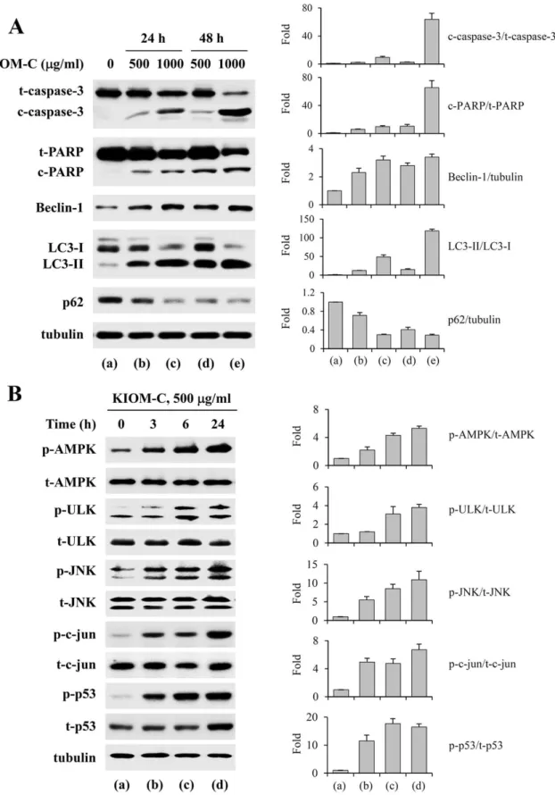
Figure 4. Induction of apoptosis and autophagy by KIOM-C via activation of AMPK, ULK, JNK, c-jun, and p53.(A) Apoptosis- and autophagy-related markers, such as caspase-3 activation, PARP cleavage, Beclin-1 up-regulation, LC3-II conversion, and p62 down-regulation, were assessed by Western blotting of KIOM-C-treated cells. (B) Lysates prepared from cells treated with 500mg/ml KIOM-C for 3, 6, and 24 h were
subjected to Western blotting to determine the levels of AMPK, ULK, JNK, c-jun, p53, and their phosphorylated forms. In (A) and (B), the band intensities relative to those of the untreated ‘‘control’’ cells were determined after normalizing to tubulin expression and represented as the means6
SD of two independent experiments. doi:10.1371/journal.pone.0098703.g004
and ap-value less than 0.05 was considered to indicate a significant result.
Results
KIOM-C decreases cell viability and induces G1arrest in
human cancer cells
To assess the effect of KIOM-C on the inhibition of cell growth, the MTT assay was used. As shown in Figure 1A, exposure of human cancer cells to increasing KIOM-C concentrations, ranging from 100 to 1000mg/ml, for 48 h resulted in a
dose-dependent decrease in cell viability, with IC50 values of 408.22mg/ml (HT1080), 406.59mg/ml (AGS), and 607.86mg/ ml (A431). Of these cell lines, that of the human fibrosarcoma HT1080 was used in all subsequent experiments to examinein vitro
andin vivothe underlying chemotherapeutic mechanisms. Normal
hepatocytes were not affected by KIOM-C treatment even after 48 h with the 1000mg/ml dose, suggesting that KIOM-C is
non-hepatotoxic. Since many individual medicinal herbs have greater pharmacological efficacy when used together with other ingredi-ents as herbal cocktails, we evaluated the potential synergistic KIOM-C anti-cancer activity by comparing the individual activities of each herb. Cells were treated for 48 h with each herb at the individual concentrations present in 500 and 1000mg/ml
doses of KIOM-C, and the cell viability was subsequently determined by MTT assay. At these concentrations, none of the herbs caused more than a 5% reduction in HT1080 cell viability, suggesting synergism among the multiple herbs present in KIOM-C (data not shown). Under a phase contrast microscope, KIOM-C treatment, in a dose-dependent manner, caused the majority of the cells to shrink, round up, and display numerous vacuoles in the cytoplasm (Figure 1B), a typical morphologic appearance induced Figure 5. The effect of KIOM-C on JNK-mediated intracellular ROS generation and ER stress.(A) Cells pre-treated with or without NAC (1 mM) or the JNK inhibitor SP600125 (5mM) for 1 h were treated with KIOM-C for another 3 h. After incubation with DCF-DA (5mM) for 30 min at
37uC, the intracellular ROS levels were measured using flow cytometry. Values are the means6SD of three independent experiments. *p,0.01 vs. untreated control, **p,0.01 vs. KIOM-C treatment. (B) Expression of CHOP mRNA in KIOM-C treated cells was examined by RT-PCR, and the relative values were determined after normalizing to GAPDH expression. (C) Cells were pre-treated with or without SP600125 for 1 h and then incubated with KIOM-C for an additional 24 h. In cell lysates, CHOP protein levels were determined by Western blotting, and the relative values were determined after normalizing to tubulin expression. In (B) and (C), the values are the means6SD of two independent experiments. *p,0.01 vs. untreated control, **p,0.01 vs. KIOM-C treatment
by apoptosis and autophagy. Analysis of the cell cycle revealed that KIOM-C treatment for 12 and 24 h increased the proportion of cells in G1 phase to 57.14 and 55.53%, respectively, compared with that in untreated control cells (36.69%)(Figure 2A). This G1 phase increase was accompanied by a corresponding decrease in the proportion of cells in S and G2/M phases. The apoptotic sub-G0/G1peak was considerably increased with KIOM-C treatment to 7.92 and 13.96% after 12- and 24-h incubations, respectively, compared with the control cells (3.24%), indicating that the KIOM-C-induced G1 cell cycle arrest retarded growth and subsequently induced cell death. We next examined the effect of KIOM-C on the expression of the G1phase regulatory proteins p21, p27, and cyclin D1. Western blotting showed that KIOM-C treatment up-regulated the levels of the cyclin-dependent kinase inhibitors p21 and p27, while it down-regulated the level of cyclin D1, compared with those in control cells (Figure 2B).
KIOM-C induces both apoptotic and autophagic cell death in HT1080 cells
To investigate the ability of KIOM-C to induce programmed cell death in HT1080 cells, we initially assessed the level of
YO-PRO-1 uptake in KIOM-C-treated HT1080 using flow cytometry. YO-PRO-1, DNA-intercalating dye, selectively passes through the plasma membranes of cells that are beginning to undergo apoptosis, and labels them with green fluorescence. As shown in Figure 3A, YO-PRO-1 uptake was moderately increased to 5.9 and 8.9% after 24 h treatment at concentrations of 500 and 1000mg/ml, respectively, compared with control cells (2.0%). KIOM-C treatment for 48 h at 1000mg/ml resulted in an approximately 7.5-fold (26.95%) increase in YO-PRO-1 uptake (3.6% for control). In TUNEL assays, the proportion of TUNEL-positive cells overlapping with the nuclear marker DAPI was increased by 15.53 and 57.33%, compared with control cells (2.62%), in response to KIOM-C treatment at 500 and 1000mg/ ml, respectively, suggesting massive induction of nuclear DNA fragmentation (Figure 3B). Next, we examined LC3 distribution as an autophagy marker in response to KIOM-C treatment in HT1080 cells after transfection with an expression construct for LC3 fused to RFP (RFP-LC3). As shown in Figure 3C, in control cells, RFP-LC3 was weakly expressed in the cytoplasm, whereas KIOM-C treatment remarkably increased punctuate structure of RFP-LC3, indicating the connection of LC3-II with the Figure 6. Blockade of KIOM-C-mediated cell death by the JNK inhibitor SP600125 and JNK-specific siRNA transfection.(A) Cells pre-treated with or without SP600125 (5mM) were treated with KIOM-C (500mg/ml) for 24 h, harvested, and examined for p-JNK, Beclin-1, p62, Bcl-2, and
PARP cleavage by Western blotting. The band intensities relative to those of the untreated ‘‘control’’ cells were determined after normalizing to tubulin expression. Values represent the means6SD of two independent experiments. *p,0.01 vs. untreated control, **p,0.01 vs. KIOM-C treatment. (B) After pre-incubation with SP600125, SB203580, PD98059, and Bafilomycin A1 for 1 h, the cells were treated with KIOM-C (500mg/ml)
for 48 h, and cell viability was assessed using the MTT assay and cell morphology was observed under an inverted microscope. (C) Cells were transfected with scrambled siRNA (Scr siRNA) or JNK siRNA, incubated for additional 72 h, and then mRNA and protein levels of JNK1 and JNK2 were examined by RT-PCR and Western blotting at the indicated times, respectively. (D) Scr siRNA- or JNK siRNA-transfected cells were treated with KIOM-C (500mg/ml) for 24 h and cell morphology was observed. (E) Scr siRNA- or JNK siRNA-transfected cells were treated with KIOM-C (500mg/ml) for 24 h,
and then the levels of Beclin-1, LC3, PARP, and cleaved caspase-3 were detected using Western blotting. The data were obtained from two independent experiments and are expressed as the means6SD. *p,0.01 vs. untreated control, **p,0.01 vs. Scr siRNA transfection
doi:10.1371/journal.pone.0098703.g006
autophagosome membranes. In addition, 500 and 1000mg/ml KIOM-C caused increases of 33.10 and 57.63%, respectively, in the proportion of MDC-positive fluorescent cells, compared with control cells (3.73%), indicating the formation of autophagic vacuoles (Figure 3D). To further confirm the ability of KIOM-C to induce autophagic flux, cells were incubated with KIOM-C in the absence or presence of vacuolar H+-ATPase inhibitor, Bafilomy-cin A1 to prevent lysosomal acidification causing accumulation of autophagosome [23]. As shown in Figure S1A, blockade of lysosomal-mediated protein turnover by Bafilomycin A1 resulted in accumulation of p62 and LC3-II. In addition, pre-treatment with Bafilomycin A1 increased punctuate structure of RFP-LC3 (Figure S1B), indicating that KIOM-C efficiently increases autophagic flux.
KIOM-C regulates the levels of apoptosis- and autophagy-related proteins in HT1080 cells
To further clarify the mechanisms by which KIOM-C induces cell death, we examined the effect of KIOM-C on the expression of apoptosis- and autophagy-related protein by Western blotting. Caspase-3 is a key mediator of apoptosis that cleaves cellular proteins, such as the inhibitor of caspase-activated DNase (ICAD), poly (ADP-ribose) polymerase (PARP), and others [24]. Cleavage from LC3-I into LC3-II occurs during autophagy via proteolytic cleavage and lipidation. Conversion to LC3-II is essential for the formation of autophagosomes and completion of autophagy [25]. p62 selectively incorporated into autophagosome through direct binding to LC3 is efficiently degraded by autophagy [23]. As shown in Figure 4A, KIOM-C treatment increased the cleavages of caspase-3 and PARP, the latter being a downstream target of the former, in a dose- and time-dependent manner. In addition, the protein level of Beclin-1, which is critical for autophagosome formation during autophagy, was markedly increased, the ratio of LC3-II to LC3-I was significantly increased, and p62 expression was efficiently decreased in KIOM-C-treated HT1080 cells
(Figure 4A). These data indicate that KIOM-C induced both apoptosis and autophagy.
KIOM-C activates phosphorylation of AMPK, ULK, JNK, c-jun, and p53
In previous studies, it has been demonstrated that autophagy is regulated by multiple signaling pathways, including those involv-ing class II PI3K, class I PI3K/Akt/mTOR, and MAPKs. AMPK, a repressor of mTOR, interacts with the PS domain of ULK1 and phosphorylates and activates the ULK1 protein kinase to induce autophagy by inhibiting mTORC1 activity via phosphorylation of raptor in the ULK1 autophagic complex [26,27]. In accordance with these reports, the levels of phosphor-ylated AMPK and ULK1 increased gradually with incubation time in response to 500mg/ml KIOM-C treatment (Figure 4B). In
addition, consistent with recent reports that p53 phosphorylation, which is mediated by JNK activation, is critical for autophagic death induction in HT1080 cells [15,28], KIOM-C treatment caused a marked elevation in the levels of p53, phospho-p53, phospho-JNK, and phospho-c-jun in a time-dependent manner, and persisted through the time course of 24 h (Figure 4B). The effect of KIOM-C on the activation of p38 and ERK1/2 was also examined. As shown in Figure S2, KIOM-C slightly increased the level of p-p38 to about 2-fold, but had little effect on the ERK1/2 activation. These results indicate that, in addition to its tumor-suppressing activity, p53 regulated autophagy and that its activation was involved in KIOM-C-induced autophagic and apoptotic cell death in HT1080 cells.
KIOM-C induces ROS generation and ER stress via JNK activation
Under starvation or stress conditions, responses to intracellular ROS production and prolonged ER stress participate in autoph-agy and apoptosis induction [29,30]. Therefore, we evaluated the effect of KIOM-C on ROS production using flow cytometry and
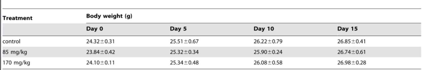
Table 1.Means of body weights of mice administrated with 85 mg/kg or 170 mg/kg of KIOM-C.
Treatment Body weight (g)
Day 0 Day 5 Day 10 Day 15
control 24.3260.31 25.5160.67 26.2260.79 26.8560.41
85 mg/kg 23.8460.42 25.3260.34 25.9060.24 26.7460.61
170 mg/kg 24.1060.11 25.3460.48 26.0860.58 26.9860.28
Data are presented as mean6S.D. Each group of mice (n = 3) were orally administrated with 85 mg/kg or 170 mg/kg of KIOM-C daily and weighed body weight at 0, 5, 10, and 15 days.
doi:10.1371/journal.pone.0098703.t001
Table 2.Organ weights of mice administrated with 85 mg/kg or 170 mg/kg of KIOM-C.
Treatment Weight of organs (g)
Liver Heart Lung Spleen Kidney (L) Kidney (R)
control 1.0960.07 0.1160.01 0.1460.01 0.0760.01 0.1360.01 0.1260.01
85 mg/kg 1.1760.12 0.1160.01 0.1560.01 0.0760.01 0.1360.01 0.1260.01
170 mg/kg 1.1060.08 0.1160.01 0.1560.02 0.0860.03 0.1360.01 0.1260.01
Data are presented as mean6S.D. Each group of mice (n = 3) were orally administrated with 85 mg/kg or 170 mg/kg of KIOM-C daily, sacrificed at 15 days, and weighed organs.
on CHOP expression using RT-PCR and Western blotting. In addition, using the JNK-specific inhibitor SP600125, we examined whether JNK activation by KIOM-C is directly related to ROS production and CHOP expression. As shown in Figure 5A, KIOM-C markedly increased intracellular ROS levels by 2.9- and 4.7-fold at 500 and 1000mg/ml, respectively, while the ROS
scavenger NAC dramatically blocked KIOM-C-enhanced ROS production by,60%, as compared with levels in control cells. In a
parallel experiment, pretreatment with SP600125 blocked KIOM-C-enhanced ROS production almost completely. The levels of CHOP mRNA in response to KIOM-C was dose- and time-dependently increased by ,20-fold (1000mg/ml, 48 h)
(Figure 5B). Similarly, the levels of CHOP protein were also significantly elevated by KIOM-C, while pretreatment with SP600125 dramatically prevented the KIOM-C-enhanced CHOP expression (Figure 5C). These results indicate that JNK activation by KIOM-C is essential for activating oxidative and ER stresses and that it acts as an upstream regulator of KIOM-C-induced cell death.
JNK activation is required for simultaneous induction of autophagy and apoptosis by KIOM-C
To further investigate the role of JNK activation in KIOM-C-mediated cell death, we pre-treated cells with pharmacological inhibitors for 1 h, followed by KIOM-C treatment for an additional 48 h. Western blot analysis showed that pre-incubation
with SP600125 prevented the induction of Beclin-1, reduction of Bcl-2, degradation of p62, and PARP cleavage by KIOM-C almost completely, resulting in levels comparable to those observed in untreated control cells (Figure 6A). In addition, SP600125 pre-treatment blocked cytoplasmic vacuole formation and dramatically protected KIOM-C-treated cells from cell death by ,95% (Figure 6B). Meanwhile, pre-treatment with p38
inhibitor SB203580 and ERK1/2 inhibitor PD98059 failed to prevent KIOM-C-induced cell death, and Bafilomycin A1 exacerbated cell death. To verify the significance of JNK activation for cell death by KIOM-C, we targeted JNK by siRNA that can recognize a common sequence in both JNK1 and JNK2 [31]. In cells transfected with JNK siRNA, mRNA and protein levels of JNK1 and JNK2 were strongly reduced compared with those in cells transfected with Scr siRNA (Figure 6C). As shown in Figure 6D and 6E, knockdown of JNK significantly rescued cells from the cytotoxic effect by C and clearly blocked KIOM-C-mediated cell death as evidenced by the inhibition of Beclin-1 induction, LC3-II conversion, PARP cleavage, and caspase-3 activation. These data collectively indicate that KIOM-C-medi-ated cell death was attributable mainly to JNK activation and subsequent modification of autophagy- and apoptosis-related protein expression. To further establish the anti-cancer effect of KIOM-C, we utilized two additional cell lines including murine melanoma B16F10 cells and human gastric carcinoma AGS cells. As demonstrated in the HT1080 cell system, KIOM-C treatment decreased cell viability in a dose-dependent manner, caused
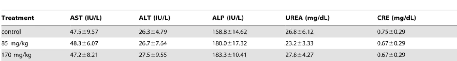
Table 3.Chemical analysis of serums obtained from mice administrated with 85 mg/kg or 170 mg/kg of KIOM-C.
Treatment AST (IU/L) ALT (IU/L) ALP (IU/L) UREA (mg/dL) CRE (mg/dL)
control 47.569.57 26.364.79 158.8614.62 26.866.12 0.7560.29
85 mg/kg 48.366.07 26.767.64 180.0617.32 23.263.33 0.6760.29
170 mg/kg 47.268.21 27.569.55 183.3610.41 27.864.27 0.6760.29
Data are presented as mean6S.D. Each group of mice (n = 3) were orally administrated with 85 or 170 mg/kg of KIOM-C daily, sacrificed at 15 days, and analyzed the levels of AST, ALT, ALP, BUN, and CRE. AST, aspartate aminotransferase; ALT, alanine aminotransferase; ALP, alkaline phosphatase; CRE, creatinine.
doi:10.1371/journal.pone.0098703.t003
Table 4.Hematological analysis of bloods obtained from mice administrated with 85 mg/kg or 170 mg/kg of KIOM-C.
parameter control 85 mg/kg 170 mg/kg
WBCP (6103cells/ml) 1.760.21 1.560.11 1.660.25
WBCB (6103cells/ml) 2.060.16 2.360.32 2.160.26
RBC (6106cells/ml) 9.4
60.20 9.460.10 9.560.23
Means HGB (g/dL) 14.160.20 14.160.22 14.060.10
HCT (%) 50.861.12 50.661.08 51.261.16
MCV (fL) 54.161.10 54.161.21 54.161.11
MCH (pg) 15.060.29 14.960.17 14.760.23
MCHC (g/dL) 27.860.56 27.560.49 27.260.46
PLT (6104cells/ml) 77.669.54 82.9610.11 78.068.52
% NEUT 40.865.24 37.763.14 43.164.12
% LYM 46.963.11 42.761.96 46.162.08
% MONO 2.660.11 2.460.13 360.27
Data are presented as mean6S.D. Each group of mice (n = 3) were orally administrated with 85 or 170 mg/kg of KIOM-C daily, sacrificed at 15 days, and analyzed hematologic parameters. CBC, complete blood cell count; WBCP, white blood cell count peroxidase method; WBCB, white blood cell count basophile method; RBC, red blood cell count; HGB, hemoglobin, HCT, hematocrit; MCV, mean corpuscular volume; MCH, mean corpuscular hemoglobin; MCHC, mean corpuscular hemoglobin concentration; PLT, platelet; NEUT, neutrophil; LYM, lymphocyte; MONO, monocyte.
doi:10.1371/journal.pone.0098703.t004
morphological changes, and increased PARP cleavage, Beclin-1 expression, and conversion to LC3-II. In addition, we also confirmed in these two cell lines that KIOM-C induces cell death via JNK activation (Figure S3).
KIOM-C administration dramatically inhibits tumorigenic growth of HT1080 cellsin vivo without adverse effects
To examine whether repeated administration of KIOM-C elicits systemic toxicity, we compared body weight, organ weight, and serological/hematological parameters in mice after treatment with KIOM-C or saline only (control). The KIOM-C doses used for the mice were based on the amounts used in human adults (49.13 g/day/60 kg of body weight) and on the yield of powdered extraction (20.7%). The administration of KIOM-C at doses of 85 or 170 mg/kg for 15 days did not cause death or abnormal behavior. Body (Table 1) and organ (Table 2) weights of the mice and the ratios of AST/ALT and BUN/CRE were not significantly
altered by KIOM-C administration compared with those of the control group, suggesting that KIOM-C administration did not cause hepatic or renal damage (Table 3). The hematological parameters of the KIOM-C-treated mice were also similar to those of the control mice (Table 4). These serological and hematological findings indicate that KIOM-C caused no adverse effects during the treatment period. To confirm the inhibitory effect of KIOM-C on tumor growth in vivo, HT1080 cells were subcutaneously injected into the femoral region of athymic nude mice, and five days later, the mice were treated with 85 or 170 mg/kg of KIOM-C for 10 days commencing 5 days after tumor inoculation. As shown in Figure 7A and 7B, KIOM-C administration successfully retarded tumor growth and suppressed tumor volume by,80%
compared with the control mice on day 14. Control mice exhibited a mean tumor weight of 0.61960.178 g, while mice treated with 85 and 170 mg/kg KIOM-C had tumor weights of 0.15760.065 and 0.15860.091 g, reflecting 74.6 and 74.5% reductions, Figure 7. The inhibitory effect of KIOM-C onin vivotumor growth in a xenograft model.Athymic nude mice were subcutaneously injected with HT1080 cells (26106), and 5 days after tumor implantation, mice were administered daily with saline (control) or KIOM-C at 85 or 170 mg/kg for
10 days (n = 4 per group). (A) Tumor volume was determined by measuring the two axes of the tumors as described in the ‘‘Materials and Methods’’. (B) Fourteen days after tumor implantation, tumor masses were surgically removed and imaged. Data are representative of three independent experiments. (C) The tumor masses in Figure 7B were weighed and expressed as the means6SD. *p,0.01 vs. control (saline) (D) Serum samples were obtained from mice of each group for measurement of IFN-clevels by ELISA. Data are the means6SD of duplicate samples. *p,0.01 vs. control (saline).
respectively (Figure 7C). Interestingly, in KIOM-C-treated mice, the serum IFN-cconcentration was markedly elevated compared with that in control mice (Figure 7D), indicating that KIOM-C administration affects host defenses against tumors. Altogether, these data suggested that KIOM-C administration effectively suppressed growth of malignant cancer cells without causing side effects.
Discussion
Oriental medicinal herbs have been widely used for cancer adjuvant therapies and are considered to be potential chemopre-ventive and chemotherapeutic agents. Because advanced malig-nancies require potent therapies targeting multiple cellular pathways, properly formulated herbal cocktails have the advan-tages of synergism and improved therapeutic efficacy compared with individual herbs. In this study, we found that KIOM-C at 500 and 1000mg/ml induced G1 cell cycle arrest (Figure 2); it ultimately induced cancer cell death via autophagy and apoptosis in a complementary and cooperative manner by regulating signaling pathways, particularly JNK activation, upstream of both of these processes (Figure 3–6). Meanwhile, none of the individual herb treatments when used at the concentrations present in the
500 and 1000mg/ml doses of KIOM-C inhibited cell viability,
suggesting synergism among herbs present in KIOM-C. In the evaluation of subacute toxicity of KIOM-C after 15 days of daily oral administration, KIOM-C did not cause any systemic toxicity with respect to body weight loss, organ abnormalities, and hematological/serological parameter changes (Tables 1–4), indi-cating that KIOM-C is a safe alternative treatment. The results presented in Figure 7A–C strongly supported the inhibitory effect of KIOM-C on tumor growth in vivo. Moreover, KIOM-C dramatically increased secretion of IFN-c(Figure 7D). IFN-chas been shown to exert potent anti-tumor activity bothin vitroandin vivo [32], and deficiencies in IFN-c (IFN-c-/-) or the IFN-c receptor (IFN-cR-/-) accelerated tumor development in mice [33]. Therefore, it would be reasonable to assume that KIOM-C-stimulated IFN-cproduction may be involved in the mechanisms of host defense against tumors.
Several studies have demonstrated that autophagy inhibits apoptosis by autophagosomal degradation of pro-apoptotic proteins such as caspases, resulting in a pro-survival effect [34]. Inhibition of autophagy by 3-MA and/or chloroquine accelerated apoptotic cell death, and induction of autophagy delayed apoptotic response to DNA damage, suggesting that autophagy and apoptosis play opposite roles in cancer cells [35]. In this study, Bafilomycin A1 treatment efficiently prevented KIOM-C-mediat-ed lysosomal-mKIOM-C-mediat-ediatKIOM-C-mediat-ed protein turnover at early stage (Figure S1), and eventually increased KIOM-C-mediated cell death (Figure 6B). Meanwhile, recent studies have shown that sustained and excessive autophagy leads to apoptotic cell death, autophagy and apoptosis occurs simultaneously by the same stimuli, and ROS acts as upstream signaling molecules [6–10,12]. Cellular redox status plays an essential role in the regulation of cell death pathways, including autophagy and apoptosis, and it is influenced by the balance between the rate of production and the rate of breakdown of reactive oxygen and/or nitrogen species (ROS/ RNS), such as superoxide (O2N
-), the hydroxyl radical (HON), and
hydrogen peroxide (H2O2) [36]. In many studies, it has been demonstrated that an increase in intracellular ROS levels along with MAPK activation participates in cancer cell death [36–38]. In the present study, we observed that intracellular ROS generation and subsequent ER stress were induced by KIOM-C treatment and that JNK activation was critical for ROS generation, ER stress, and decreased cell viability (Figure 5). Unlike previous reports suggesting that ROS act as strong signals for MAPK activation and lead to JNK activation in malignant cells [39], the JNK-specific inhibitor SP600125 almost completely abolished KIOM-C-induced ROS generation and ER stress responses (Figure 5), while the ROS scavenger NAC was incapable of blocking KIOM-C-induced JNK activation (data not shown). These results indicate that JNK activation acts upstream of ROS generation and ER stress responses in KIOM-C-induced cell death.
Beclin-1 as part of the Beclin-1-Vps34-Vps15 core complex has been reported to induce considerable autophagic cell death, inhibit cancer cell growth, and act as an important molecular switch between autophagy and apoptosis [40]. Meanwhile, the BH3 domain of Beclin-1 interacts with certain anti-apoptotic B cell lymphoma 2 (Bcl-2) family members, including Bcl-2 or Bcl-xL; Bcl-2 was shown to inhibit the autophagic function of Beclin-1. Therefore, disruption of the Beclin-1/Bcl-2 interaction by phosphorylation of Bcl-2 and Beclin-1 or by caspase-dependent cleavage of Beclin-1 promoted crosstalk between apoptosis and autophagy pathways [40–41]. Consistent with these results, KIOM-C increased Beclin-1 expression but decreased Bcl-2 expression, which was mediated by JNK activation (Figure 6A). Figure 8. Schematic diagrams of the mechanisms underlying
KIOM-C-mediated cell death.KIOM-C induces both autophagy and apoptosis by induction of Beclin-1 and reduction of Bcl-2 via JNK-mediated oxidative and ER stresses. SP600125: JNK-specific inhibitor; ROS: reactive oxygen species; ER stress: endoplasmic reticulum stress; JNK: c-jun-N-terminal kinase.
doi:10.1371/journal.pone.0098703.g008
Certain KIOM-C components, including Radix Scutellariae, Radix Paeoniae Alba, Radix Angelicae Gigantis, and Platycodon grandiflorum have been shown to elicit anti-cancer effects by inducing apoptosis. However, in this study, none of the individual herbs comprising KIOM-C exhibited anti-cancer effects in HT1080 cells at the concentrations used, suggesting synergistic effects when used together. Currently, we are attempting to isolate the active ingredients in KIOM-C subfractions. In this future study, we will compare the chemotherapeutic effects and underlying mechanisms among the KIOM-C active ingredients. In addition, further elucidation of the mechanisms involved in the immune-potentiating effects of KIOM-C is needed.
In summary, our results clearly demonstrated that KIOM-C simultaneously induced autophagy and apoptosis, primarily through JNK activation, in malignant cancer cells (Figure 8). Moreover, oral administration of KIOM-C considerably sup-pressed in vivo tumor cell growth of subcutaneously injected HT1080 cells, possibly through induction of cell death and potentiation of immune responses, without causing systemic toxicity. Collectively, these results suggest that KIOM-C repre-sents a safe herbal therapy for controlling malignant tumor growth.
Supporting Information
Figure S1 KIOM-C induces autophagic flux.HT1080 cells were treated with 500mg/ml KIOM-C for 24 h with or without pretreatment with Bafilomycin A1 (10 nM) for 1 h. To monitor autophagic flux, Western blotting for p62 and LC3-II/LC3-I expression (A) and fluorescence analysis for RFP-LC3 distribution (B) was performed. *p,0.01 vs. untreated control, **p,0.01 vs. KIOM-C treatment.
(TIF)
Figure S2 KIOM-C slightly induces p38 activation but not ERK phosphorylation.(A) HT1080 cells were treated with 500mg/ml KIOM-C for 3, 6, and 24 h, and cell lysates were subjected to Western blotting to determine the levels of p38, ERK, and their phosphorylated forms. (B) HT1080 cells were treated with 500mg/ml KIOM-C with or without pretreatment with pharmacological inhibitors for 1 h. After 48 h, cells were observed under an inverted microscope.
(TIF)
Figure S3 KIOM-C induces autophagic and apoptotic cell death via JNK activation in murine melanoma B16F10 cells and human gastric carcinoma AGS cells. (A) After B16F10 and AGS cells were treated with KIOM-C (100– 1000mg/ml) for 48 h, cell viability was determined by MTT
assay. (B) After treatment with 500 and 1000mg/ml KIOM-C for
48 h, morphologic changes were observed under an inverted microscope. (C) Apoptosis- and autophagy-related marker pro-teins, such as PARP cleavage, Beclin-1 increase, and LC3-II conversion, were detected by Western blotting in KIOM-C-treated cells at the specified conditions. (D) MAPK activation was determined by Western blotting in lysates prepared from cells treated with KIOM-C (1000mg/ml) for 3, 6, and 24 h. (E) After pre-incubation with SP600125 (5mM), cells were treated with KIOM-C (1000mg/ml) for 48 h and then observed under an inverted microscope.
(TIF)
Author Contributions
Conceived and designed the experiments: AYK JYM. Performed the experiments: AYK MJI NHY. Analyzed the data: AYK TSK. Contributed reagents/materials/analysis tools: AYK JYM. Wrote the paper: AYK.
References
1. Maddika S, Ande SR, Panigrahi S, Paranjothy T, Weglarczyk K, et al. (2007) Cell survival, cell death and cell cycle pathways are interconnected: implications for cancer therapy. Drug Resist Updat 10: 13–29.
2. Petrylak DP (2005) The current role of chemotherapy in metastatic hormone-refractory prostate cancer. Urology 65: 3–7; discussion 7–8.
3. Evans T (2005) Chemotherapy in advanced non-small cell lung cancer. Semin Respir Crit Care Med 26: 304–313.
4. Tan ML, Ooi JP, Ismail N, Moad AI, Muhammad TS (2009) Programmed cell death pathways and current antitumor targets. Pharm Res 26: 1547–1560. 5. Kondo Y, Kanzawa T, Sawaya R, Kondo S (2005) The role of autophagy in
cancer development and response to therapy. Nat Rev Cancer 5: 726–734. 6. Scott RC, Juhasz G, Neufeld TP (2007) Direct induction of autophagy by Atg1
inhibits cell growth and induces apoptotic cell death. Curr Biol 17: 1–11. 7. Liu YL, Yang PM, Shun CT, Wu MS, Weng JR, et al. (2010) Autophagy
potentiates the anti-cancer effects of the histone deacetylase inhibitors in hepatocellular carcinoma. Autophagy 6: 1057–1065.
8. Filippi-Chiela EC, Villodre ES, Zamin LL, Lenz G (2011) Autophagy interplay with apoptosis and cell cycle regulation in the growth inhibiting effect of resveratrol in glioma cells. PLoS One 6: e20849.
9. Song KS, Kim JS, Yun EJ, Kim YR, Seo KS, et al. (2008) Rottlerin induces autophagy and apoptotic cell death through a PKC-delta-independent pathway in HT1080 human fibrosarcoma cells: the protective role of autophagy in apoptosis. Autophagy 4: 650–658.
10. Trejo-Solis C, Jimenez-Farfan D, Rodriguez-Enriquez S, Fernandez-Valverde F, Cruz-Salgado A, et al. (2012) Copper compound induces autophagy and apoptosis of glioma cells by reactive oxygen species and JNK activation. BMC Cancer 12: 156.
11. Schwartzman RA, Cidlowski JA (1993) Apoptosis: the biochemistry and molecular biology of programmed cell death. Endocr Rev 14: 133–151. 12. Duan W, Jin X, Li Q, Tashiro S, Onodera S, et al. (2010) Silibinin induced
autophagic and apoptotic cell death in HT1080 cells through a reactive oxygen species pathway. J Pharmacol Sci 113: 48–56.
13. Torres M, Forman HJ (2003) Redox signaling and the MAP kinase pathways. Biofactors 17: 287–296.
14. Wong CH, Iskandar KB, Yadav SK, Hirpara JL, Loh T, et al. (2010) Simultaneous induction of non-canonical autophagy and apoptosis in cancer cells by ROS-dependent ERK and JNK activation. PLoS One 5: e9996.
15. Duan WJ, Li QS, Xia MY, Tashiro S, Onodera S, et al. (2011) Silibinin activated p53 and induced autophagic death in human fibrosarcoma HT1080 cells via reactive oxygen species-p38 and c-Jun N-terminal kinase pathways. Biol Pharm Bull 34: 47–53.
16. Malhotra JD, Kaufman RJ (2007) The endoplasmic reticulum and the unfolded protein response. Semin Cell Dev Biol 18: 716–731.
17. Park SH, Park HS, Lee JH, Chi GY, Kim GY, et al. (2013) Induction of endoplasmic reticulum stress-mediated apoptosis and non-canonical autophagy by luteolin in NCI-H460 lung carcinoma cells. Food Chem Toxicol 56: 100– 109.
18. Corson TW, Crews CM (2007) Molecular understanding and modern application of traditional medicines: triumphs and trials. Cell 130: 769–774. 19. Kiyohara H, Matsumoto T, Yamada H (2004) Combination Effects of Herbs in
a Multi-herbal Formula: Expression of Juzen-taiho-to’s Immuno-modulatory Activity on the Intestinal Immune System. Evid Based Complement Alternat Med 1: 83–91.
20. Chung TH, Lee GS, Yu KJ, Kim H, Kim DS, et al. (2012) Effects of the novel herbal medicine, KIOM-C, on the growth performance and immune status of porcine circovirus associated disease (PCVAD) affected pigs. J Med Plants Res 6: 4456–4466.
21. Kim EH, Pascua PN, Song MS, Baek YH, Kwon HI, et al. (2013) Immunomodulaton and attenuation of lethal influenza A virus infection by oral administration with KIOM-C. Antiviral Res 98: 386–393.
22. Kim A, Yim NH, Im M, Jung YP, Kim T, et al. (2014) Suppression of the invasive potential of highly malignant tumor cells by KIOM-C, a novel herbal medicine, via inhibition of NF-kappaB activation and MMP-9 expression. Oncol Rep 31: 287–297.
23. Mizushima N, Yoshimorim T, Levine B (2010) Methods in mammalian autophagy research. Cell 140: 313–326.
24. Miller DK (1997) The role of the Caspase family of cysteine proteases in apoptosis. Semin Immunol 9: 35–49.
25. Kabeya Y, Mizushima N, Ueno T, Yamamoto A, Kirisako T, et al. (2000) LC3, a mammalian homologue of yeast Apg8p, is localized in autophagosome membranes after processing. EMBO J 19: 5720–5728.
26. Zhao M, Klionsky DJ (2011) AMPK-dependent phosphorylation of ULK1 induces autophagy. Cell Metab 13: 119–120.
28. Zhang Y, Wu Y, Wu D, Tashiro S, Onodera S, et al. (2009) NF-kappab facilitates oridonin-induced apoptosis and autophagy in HT1080 cells through a p53-mediated pathway. Arch Biochem Biophys 489: 25–33.
29. Haberzettl P, Hill BG (2013) Oxidized lipids activate autophagy in a JNK-dependent manner by stimulating the endoplasmic reticulum stress response. Redox Biol 1: 56–64.
30. Tabas I, Ron D (2011) Integrating the mechanisms of apoptosis induced by endoplasmic reticulum stress. Nat Cell Biol 13: 184–190.
31. Gururajan M, Chui R, Karuppannan AK, Ke J, Jennings CD, et al. (2005) c-Jun N-terminal kinase (JNK) is required for survival and proliferation of B-lymphoma cells. Blood 106: 1382–1391.
32. Giovarelli M, Cofano F, Vecchi A, Forni M, Landolfo S, et al. (1986) Interferon-activated tumor inhibition in vivo. Small amounts of interferon-gamma inhibit tumor growth by eliciting host systemic immunoreactivity. Int J Cancer 37: 141– 148.
33. Kaplan DH, Shankaran V, Dighe AS, Stockert E, Aguet M, et al. (1998) Demonstration of an interferon gamma-dependent tumor surveillance system in immunocompetent mice. Proc Natl Acad Sci U S A 95: 7556–7561. 34. Gordy C, He YW (2012) The crosstalk between autophagy and apoptosis: where
does this lead? Protein Cell 3: 17–27.
35. Abedin MJ, Wang D, Lehmann U, Kelekar A (2007) Autophagy delays apoptotic death in breast cancer cells following DNA damage. Cell Death Differ 14: 500–510.
36. Kamata H, Honda S, Maeda S, Chang L, Hirata H, et al. (2005) Reactive oxygen species promote TNFalpha-induced death and sustained JNK activation by inhibiting MAP kinase phosphatases. Cell 120: 649–661.
37. Scherz-Shouval R, Shvets E, Fass E, Shorer H, Gil L, et al. (2007) Reactive oxygen species are essential for autophagy and specifically regulate the activity of Atg4. EMBO J 26: 1749–1760.
38. Mates JM, Sanchez-Jimenez FM (2000) Role of reactive oxygen species in apoptosis: implications for cancer therapy. Int J Biochem Cell Biol 32: 157–170. 39. Xie CM, Chan WY, Yu S, Zhao J, Cheng CH (2011) Bufalin induces autophagy-mediated cell death in human colon cancer cells through reactive oxygen species generation and JNK activation. Free Radic Biol Med 51: 1365– 1375.
40. Kang R, Zeh HJ, Lotze MT, Tang D (2011) The Beclin 1 network regulates autophagy and apoptosis. Cell Death Differ 18: 571–580.
41. Zhou F, Yang Y, Xing D (2011) Bcl-2 and Bcl-xL play important roles in the crosstalk between autophagy and apoptosis. FEBS J 278: 403–413.