Prevalence of the Prefoldin Subunit 5 Gene
Deletion in Canine Mammary Tumors
Silvia Hennecke1☯, Julia Beck2☯*, Kirsten Bornemann-Kolatzki2, Stephan Neumann1, Hugo Murua Escobar3,6, Ingo Nolte3, Susanne Conradine Hammer3,6, Marion Hewicker-Trautwein4, Johannes Junginger4, Franz-Josef Kaup5, Bertram Brenig1,
Ekkehard Schütz1,2
1Institute of Veterinary Medicine, Georg-August-University Goettingen, Goettingen, Germany,2Chronix Biomedical, Goettingen, Germany,3Small Animal Clinic, University of Veterinary Medicine Hannover, Hannover, Germany,4Department of Pathology, University of Veterinary Medicine Hannover, Hannover, Germany,5German Primate Center, Goettingen, Germany,6Hematology Oncology and Palliative Medicine, ClinicIII, University of Rostock, Rostock, Germany
☯These authors contributed equally to this work. *[email protected]
Abstract
Background
A somatic deletion at the proximal end of canine chromosome 27 (CFA27) was recently reported in 50% of malignant mammary tumors. This region harbours the tumor suppressor gene prefoldin subunit 5 (PFDN5) and the deletion correlated with a higher Ki-67 score. PFDN5has been described to repress c-MYC and is, therefore, a candidate
tumor-suppres-sor and cancer-driver gene in canine mammary cancer. Aim of this study was to confirm the recurrent deletion in a larger number of tumors.
Methods
Droplet digital PCR forPFDN5was performed in DNA from 102 malignant, 40 benign
mam-mary tumors/dysplasias, 11 non-neoplastic mammam-mary tissues and each corresponding genomic DNA from leukocytes. The copy number ofPFDN5was normalized to a reference
amplicon on canine chromosome 32 (CFA32). Z-scores were calculated, based on Gauss-ian distributed normalizedPFDN5copy numbers of the leukocyte DNA. Z-scores-3.0 in tissue were considered as being indicative of thePFDN5deletion and called as such. The
Ki-67 proliferation index was assessed in a subset of 79 tissue samples by immunohistochemistry.
Results
The deletion was confirmed in 24% of all malignant tumors, detected in only 7.5% of the benign tumors and was not present in any normal mammary tissue sample. The subgroup of solid carcinomas (n = 9) showed the highest frequency of the deletion (67%) and those malignomas without microscopical high fraction of benign tissue (n = 71) had a 32%
OPEN ACCESS
Citation:Hennecke S, Beck J, Bornemann-Kolatzki K, Neumann S, Escobar HM, Nolte I, et al. (2015) Prevalence of the Prefoldin Subunit 5 Gene Deletion in Canine Mammary Tumors. PLoS ONE 10(7): e0131280. doi:10.1371/journal.pone.0131280
Editor:Xuefeng Liu, Georgetown University, UNITED STATES
Received:February 19, 2015
Accepted:June 1, 2015
Published:July 1, 2015
Copyright:© 2015 Hennecke et al. This is an open access article distributed under the terms of the Creative Commons Attribution License, which permits unrestricted use, distribution, and reproduction in any medium, provided the original author and source are credited.
Data Availability Statement:All relevant data are within the paper.
frequency (p<0.01 vs. benign samples). The Ki-67 score was found to be significantly higher (p<0.05) in thePFDN5-deleted group compared to malignant tumors without the deletion.
Conclusions
A somatic deletion of thePFDN5gene is recurrently present in canine mammary cancer,
supporting a potential role in carcinogenesis. The association of this deletion with higher Ki-67 indicates an increased proliferation rate and thus a link to tumor aggressiveness can be hypothesized. The confirmation of earlier results warrants further studies onPFDN5as
can-cer-driver gene.
Introduction
Mammary tumors represent the majority of neoplastic diseases in female dogs, constituting
over 40% of all female canine tumors [1]. Of these approximately 50% are malignant and in
majority being of epithelial origin (simple adenoma/carcinoma). In contrast to human breast cancer 20% of the canine tumors exhibit myoepithelial cell proliferation (complex carcinoma). Canine mammary tumors (CMT) have been proposed as a model for human breast cancer and increasing knowledge about the molecular differences and similarities will support the use of
CMT in breast cancer research [2–4]. However, molecular research in canine mammary gland
tumors in the recent years seemed to focus primarily on genes with well established roles in
human carcinogenesis (for review see: [2]). In order to gain a deeper understanding of
molecu-lar processes that can lead to neoplastic transformation of the canine mammary gland a broader spectrum of investigated genes is required.
Recently, our group identified a recurrent deletion of the proximal part of cfa27 by high-throughput sequencing analyses of five canine malignant mammary tumors (MMTs). Within
this regionPFDN5appeared as the most plausible candidate with cancer-driving functional
rel-evance. [5].
The Prefoldin Subunit 5 gene (PFDN5) maps to the proximal end of the canine
chromo-some (cfa) 27 (1,906,133–1,909,613 bp, Broad CanFam 3.1). The encoded protein is part of the
prefoldin complex that functions as chaperone and is involved in the posttranslational folding
of high-order protein structures such as actin and microtubules [6]. Furthermore, prefoldin
and its subunits can localize to the nucleus and act as transcriptional regulators by binding to
the DNA or transcription factors [6]. PFDN5 in particular is known to repress the c-MYC
tran-scription factor [7]. Three different c-MYC repression mechanisms are described that are
mediated by PFDN5: i) repression of the transcriptional activity by binding to the N-terminal region of c-MYC ii) recruiting of the ubiquitin ligase complex to c-MYC that drives protea-some dependent degradation iii) repression of the Wnt4 expression that leads to the repression
of c-MYC expression [6–11]. Therefore,PFDN5has the functional potential to act as a tumor
suppressor gene. Indeed, a frequent amino acid substitution (A157R) that led to a loss of its
c-MYC transcriptional repression activity was found in 50–60% of leukemia/lymphoma cells and
in more than 75% of human tongue cancers [8]. Furthermore, tumor-specific alternative
splic-ing variants were detected in thyroid and head/neck cancer [12,13]. One variant was generated
by the retention of intron 2 that generated a premature stop-codon and another detected splice
isoform led to the insertion of 101 amino acids [12,13]. Both splice-variants are expressed at
higher levels in tumor as compared to normal tissues or benign lesions [12,13].
decision to publish, or preparation of the manuscript. The specific roles of these authors are articulated in the author contributions section.
In human breast cancerPFDN5was amongst 102 genes for which differential expression in tumor versus normal tissue was detected; 69% of the analysed tumor samples showed a
decreased expression [14].
However,PFDN5cannot be considered as a well-known established cancer-driver gene,
nei-ther in human nor in canine cancers. Nevertheless, significantly higher Ki-67 scores, indicative
of a higher proliferative activity, were detected in the tumors withPFDN5deletion [5], which
can serve as hint for a functional association. Since the sample number in our first explorative study was small the primary goal of this study was to confirm the deletion of PFDN5 in a larger number of canine MMTs compared to benign tissue.
Material and Methods
Clinical samples
A total of 153 mammary tissues (CMT n = 142 and non-neoplastic controls n = 11) were inves-tigated, of which 82 were from patients of the Clinic for Small Animals, Institute of Veterinary Medicine, Georg-August-University Göttingen and 71 mammary tissues were sampled at the Clinic for Small Animals, University of Veterinary Medicine Hannover. The CMTs and the non-neoplastic mammary tissues were collected from 127 bitches with a median age of 9 years
(range 3–15 years), of which most were intact (16.3% surgically castrated). Cross breeds
(n = 35) were the most common breed represented, followed by Cocker spaniels (n = 7), Ger-man shepherds (n = 5), Dachshund (n = 5), Golden Retriever (n = 5) and Labrador Retriever (n = 5). A total of 40 different other breeds were included in the study. Considering the compo-sition of the herein analysed samples the median age of occurrence, the affected breeds and
dis-tribution of different tumor types in this study were consistent with other reports [1,15,16].
Tumors were removed by mastectomy and the diagnosis was confirmed by histopatholog-ical evaluation. All tumors and the corresponding peripheral blood mononuclear cells (PBMC) were obtained as part of routine diagnostic procedures with informed consent of the owner. Therefore the study was exempt of ethical approval according to German regulations.
The 153 samples were classified histological using formalin fixed paraffin embedded (FFPE) tumor tissue with haematoxylin and eosin staining according to the published criteria of the
World Health Organization [17]. The samples consisted of 102 (67%) MMT (1 non-infiltrating
carcinoma, 27 complex carcinomas, 36 simple carcinomas, 5 spindle cell carcinomas, 1 osteo-sarcoma, 4 carcinosarcomas, 28 carcinomas in benign tumors) and 32 (21%) BMT (17 adeno-mas, 4 fibroadenoadeno-mas, 11 benign mixed tumors), 6 (4%) MH, 2 (1%) duct ectasias (DE) and 11 (6%) samples with normal histopathological findings.
In order to separate cancerous and adenomatous parts of 12 carcinomas in benign tumors (6 pre-dominantly carcinomas and 6 prepre-dominantly adenomas) the FFPE specimens were macrodissected (malignant and benign parts), by using a 3mm dermal punch and were then examined separately.
DNA extraction from tissue, blood sampling and FFPE
DNA was extracted from 25mg of homogenized tumor and non-neoplastic tissue and 200μL of
the corresponding PBMC using the DNeasy Blood & Tissue Kit (Qiagen, Hilden, Germany). DNA from macrodissected carcinoma in benign tumor FFPE tissues was extracted using the Gene Read DNA FFPE Kit (Qiagen, Hilden, Germany) following the manufacturers instructions.
Droplet Digital PCR assays
A ddPCR assay was designed to evaluate a deletion at the proximal end of chromosome
(GeneID: 607260; location CFA27: 1,906,133–1,909,613bp on CanFam3.1 reference genome;
NCBI; NW_006609.3). Primers and probes were designed using the Primer3 program (http://
Frodo.wi.mit.edu/primer3/). All sequences were cross-checked against the canine reference
genome to ensure unambiguous results. The sequences of the primers and probe were: CFA27. F: 5´-GCCTTAGAACTATACCTATACATTCCG-3´, 5´-CFA27.R: GTAGCATATGTCATTT TCTCCATAAGG-3´ and CFA27-Probe:
5´-6-Fam-ACCGACTTCTCTGTTCCTTCCCATTC-BHQ1-3´ yielding an 80bp amplified fragment (CFA27:1,908,080–1,908,159; Broad CanFam3.1)
in the intronic region. A 79bp fragment on CFA32 (location CFA32: 2,342,421–2,342,499; Broad
CanFam3.1) served as control amplicon; this region had equal copy-numbers in PBMC/tumor pairs of the included dogs. The sequences of the primers and probe for CFA32 were: CFA32.F: 5´-AAAAGCCTCCAATCCCCGAG-3´, CFA32.R: 5´-CCTGACAGAAAAAGCAGCCC-3´ and CFA32-Probe: 5´-HEX-CTCCGTGACAAGTCAAGCTCAATAGCCT-BHQ1-3´.
1μg of genomic DNA of the tumors and corresponding PBMCs was digested with 5 UApoI
restriction endonuclease in a total reaction volume of 25μL. 60ng digested DNA, 10μL 2x
ddPCR Master Mix (Bio-Rad, Munich, Germany), 900nmol/L of each primer and 250nmol/L of hydrolysis probes were supplemented with nuclease- and protease free water to a final
vol-ume of 20μL. Approximately 23,000 droplets per reaction were generated using the QX200
Droplet Generator (Bio-Rad, Munich, Germany) and PCR amplification was performed in a thermal cycler (Biometra, Göttingen, Germany) at 95°C for 10min followed by 50 cycles at 95°C for 30sec and 60°C for 1min, 98°C for 10min and 12°C hold.
The droplets were analysed using the QX200 Droplet Reader (Bio-Rad, Munich, Germany). On average ddPCR yielded 11,252 (SD:1,505) detected usable droplets in PBMC DNA and 11,653 (SD:1,471) detected droplets in the tissue DNA. Using the QuantaSoft software the con-centration of the respective template and the relative standard uncertainty was calculated based
on the Poisson distribution. The obtained copy number of the target amplicon in thePFDN5
gene locus was calculated relative to the CFA32 reference amplicon (diploid).
Immunohistochemistry
Immunohistochemistry was performed on a subset of 79 paraffin-embedded samples using the avidin-biotin complex (ABC) method (VECTASTAIN Elite ABC kit, Vector Laboratories,
Bur-lingame, California, USA) and 3,3’-diaminobenzidine (DAB, Sigma-Aldrich, Munich,
Ger-many) to produce a brown colour reaction as described elsewhere [18].
Briefly, antigen retrieval was performed in citrate buffer (pH 6.0) for 20min at 95°C followed by blocking of endogenous peroxidase activity with 0.5% hydrogen peroxide in 70% ethanol for 30min. Subsequent to the application of normal goat serum (20% in PBS, 30 min), primary mouse anti-Ki-67 antibodies (1:100 in PBS containing 1.0% BSA: clone MIB-1, Dako, Hamburg, Germany) were added and incubated over night at 4°C. Biotinylated goat anti-mouse IgG secondary antibodies (1:200 in PBS, Vector Laboratories) were applied for 30min at room temperature. Sections were
counter-stained with Mayer’s hematoxylin solution. Specimens from normal canine lymph node
were used as positive control. Primary antibodies were replaced by ascites fluid from non-immu-nized BALB/cJ mice (Cedarlane, Ontario, Canada) in equal concentrations for negative controls. Sections were photographed at 200x magnification in five randomly selected areas of the highest nuclear labelling. A total of at least 350 proliferating and non-proliferating cells were counted manually to calculate the percentage of Ki-67 expressing cells.
Statistical analysis
measured in the PBMCs of all animals (μ= 2.02, SD = 0.09). For comparisons ofPFDN5
dele-tion a T-test assuming equal variance was used. A deviadele-tion of z-3 was regarded as
signifi-cant aberration, and differences in the group frequencies were compared using Fisher’s exact
test (one-tailed). Since not Gaussian distributed (KS-test), the Ki-67 scores were compared using the Mann-Whitney U-test. Samples from BMT, MH, DE and normal tissue where grouped as being of benign origin.
Results
Overall, the relative copy numbers ofPFDN5in malignant tumors (n = 102) was significantly
lower (p<0.05) compared to benign tissue (n = 51), while, no significant difference was present
in PBMC genomic DNA for the same groups (p = 0.16). If those tumors with high amount of benign tissue (carcinoma in benign tumors) and the carcinoma in situ were omitted from this analysis, the remaining 74 cases showed a highly significant deviation from benign controls (p<0.01).
Hence, by using a z-score delimiter of -3.0 the frequency of thePFDN5deletion was higher
in malignancies (n = 102), yielding an odds ratio of 4.9 (95% Cl: 1.4–17.2, p = 0.004 Fisher’s
exact test).
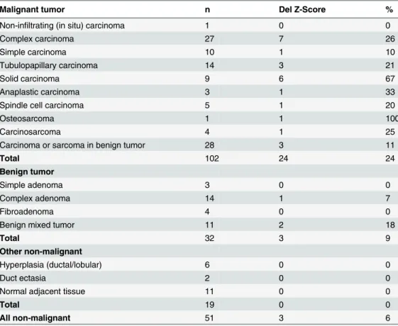
At that delimiter of z = -3.0, the deletion was detected in 26% of complex carcinomas (7|27), in 21% of tubulopapillary carcinomas (3|14) and in 67% of solid carcinomas (6|9). Furthermore the deletion was detected in one simple carcinoma (1|10), one anaplastic carcinoma (1|3), one spindle cell carcinoma (1|5), one osteosarcoma (1|1) and in one carcinosarcoma (1|4). In the most frequently occurring carcinoma/sarcoma in benign tumors (3|28) the deletion was detect-able in 11%. In 9% (3|32) of the BMTs the deletion was detected, which were one complex ade-noma and two benign mixed tumors. In the 19 non-neoplastic mammary tissues, including the
six MH and the two DE the deletion was not detected (Table 1).
In samples that were histopathologically categorized as malignancy within benign tumors, the content of DNA from malignant cells is usually low in total tissue, which may result in fail-ure to detect the deletion, even if present in malignant cells. To prove this, in a set of 6 tumors, the malignant and the benign parts were macrodissected from FFPE samples, which were
scored negative of for thePFDN5deletion, when homogenized total tissue was used. In contrast
to the results in tissue, the deletion was found in three of the predominantly malignant parts (50%) but not in the areas, which were defined as predominantly adenomas (0|6).
Immunolabelling with Ki-67 antibodies was performed in a subset of 100 tissue samples and revealed a clear nuclear labelling that showed a variable intensity. Due to the low content of malignant cells in the so called carcinomas in benign tumors (n = 21), the KI-67 was reduced, congruent with the low abundance of the PFDN5 deletion in the whole tissue of that tumor type. This type was therefore excluded from statistical analysis to avoid a bias in favour of an association between KI-67 and the somatic deletion. The percentage of Ki-67 expressing cells was evaluated in 79 tumor samples (MMT n = 50; BMT n = 21; MH n = 6 and DE n = 2) and ranged between 4% and 81%. Ki-67 expression was significantly higher (p = 0.0001;
Mann-Whitney U-test) in MMT (median: 27%, 25th-75thpercentile: 21.3%-33%) compared to
benign neoplasms (median: 15%, 25th-75thpercentile: 13%-24%) (Table 2).
In the MMTs a significant higher Ki-67 expression between thePFDN5-deleted (median:
29%, 25th-75thpercentile: 25%-35%) and non-deleted group (median: 26%, 25th-75thpercentile:
Discussion
This study was intended to validate our earlier finding [5] on the presence of a recurrent
somatic deletion of thePFDN5gene on CFA27. The results confirm that this somatic deletion
is present in canine MMT and represents one potential new tumor marker. Furthermore, the
somatic presence of the deletion in 23% of the analysed malignant tumors supportsPFDN5as
being a potential tumor driver gene. Cancer drivers are defined as genes that when somatically
altered contribute to the development of cancer [19,20]. It is thought that several different
mutations are required to drive oncogenesis, the actual number, however, can only be
esti-mated and is variable between different cancers [20]. Cancer driver genes are commonly
thought to be altered at a certain level of recurrence [20]. An amplification of the c-MYC
onco-gene for example is found in 15.7% of human breast cancers [21].
In the current study the highest deletion frequency (67%) of caninePFDN5was detected in
the group of solid carcinomas followed by 26% in complex carcinomas. On the other hand, only 10% of the analysed simple carcinomas carried the deletion. These differences might be indicative of different carcinogenic mechanisms that give rise to the different carcinoma types,
as recently suggested [3].
The very low percentage of detected deletions in the special types of carcinoma or sarcoma
in benign tumors is most likely due to the "dilution”of the often low numbers of malignant
cells by the benign fraction of the tumor. In these tumors foci of malignant-appearing cells or
distinct nodules of malignant cells reside within complex adenomas or benign tumors [22].
Our results in macrodissected malignant areas, where the deletion was found in 3 of 6 samples Table 1. PFDN5deletion in different tumor groups.
Malignant tumor n Del Z-Score %
Non-infiltrating (in situ) carcinoma 1 0 0
Complex carcinoma 27 7 26
Simple carcinoma 10 1 10
Tubulopapillary carcinoma 14 3 21
Solid carcinoma 9 6 67
Anaplastic carcinoma 3 1 33
Spindle cell carcinoma 5 1 20
Osteosarcoma 1 1 100
Carcinosarcoma 4 1 25
Carcinoma or sarcoma in benign tumor 28 3 11
Total 102 24 24
Benign tumor
Simple adenoma 3 0 0
Complex adenoma 14 1 7
Fibroadenoma 4 0 0
Benign mixed tumor 11 2 18
Total 32 3 9
Other non-malignant
Hyperplasia (ductal/lobular) 6 0 0
Duct ectasia 2 0 0
Normal adjacent tissue 11 0 0
Total 19 0 0
All non-malignant 51 3 6
that were not detectable when homogenized total tissue was used, confirmed such a dilution effect of the DNA from malignant cells by the benign fraction.
Interestingly, the deletion was also found in a few BMTs, though they did not show histo-pathological evidence of malignancy at the time of surgery. However, those three cases were in complex and mixed tumors, where the histopathological evaluation is extremely difficult, a
recent survey unveiled a considerable false negative rate [23], furthermore focal differences
between the section used for microscopy and for molecular analyses can never be excluded. On the other hand, benign mammary tumors (BMT) have been considered as precancerous
lesions and carry an increased risk of malignant transformation [24]. In this line it could also
be hypothesized that a deletion of the c-Myc repressor genePFDN5may be a risk indicator for
precancerosis.
Further indication for the proposed role ofPFDN5as tumor-suppressor is the detected
associa-tion between CFA27 deleassocia-tion and higher Ki-67 scores. Ki-67 antigen is a non-histone protein, which is only expressed in proliferating cells and enabling to determine the growth fraction of a
cell population [25] and cell proliferation in mammalian tumors e.g. humans and dogs [4]. In
CMT increased cell proliferation may serve as an indicator of malignancy since Ki-67 was reported
to be significantly different between BMT and MMT [2]. High Ki-67 scores were associated with
increased tumor grade, larger tumor size, lymph node involvement and metastasis and the survival
time was significantly decreased for bitches with high Ki-67 score tumor [2]. Even though a
statis-tical significant difference between Ki-67 scores of malignant and benign tumors was seen in our
sample set, the large overlap (cf.Fig 1) seems to compromise a general use as prognostic marker in
dogs. Similarly, Ki-67 scores in malignant tumors are significantly higher in those with the deletion compared to tumors without the deletion, but again both groups largely overlap.
Due to the location ofPFDN5close to the centromere it can not be excluded that its
fre-quent deletion in canine mammary tumors is solely related to its position. Centric fusions that result from reciprocal translocation appear frequently in canine tumor cells and are thought to occur randomly and with greater frequency at later stages of tumor progression Table 2. Ki-67 score and SD in different tumor groups.
Malignant Mammary Tumors n median (%) min (%) max (%)
In situ carcinoma 1 26.6
Complex carcinoma 17 23.7 4.1 42.9
Simple carcinoma 2 47.1 13.5 80.7
Tubulopapillary carcinoma 10 26.7 11.1 48.9
Solid carcinoma 8 33.4 21.8 57.1
Anaplastic carcinoma 2 25.4 23.7 27.1
Spindle cell carcinoma 5 27.7 17.6 30.9
Osteosarcoma 1 22.6
Carcinosarcoma 4 29.3 22.3 46
Total 50 27 4 81
Non-malignant Mammary Tissue
Simple adenoma 2 14.9 14.3 15.4
Complex adenoma 11 15.4 7.4 49.4
Fibroadenoma 3 29.4 22.6 34
Benign mixed tumor 5 14 6.9 27.1
Duct ectasia 2 15.2 15 15.4
Hyperplasia 6 15.6 4.0 33
Total 29 15 4 49
[26]. Reciprocal translocations often lead to large deletions at the breakpoints [27]. Therefore, it is possible that the recurrent deletion of PFDN5 in canine tumors is attributable to its posi-tion rather than to its funcposi-tion during carcinogenesis. Nevertheless, the fact that such a somatic telomeric deletion was not found for other chromosomes and that the PFDN5 deletion has a substantial frequency in malignant tumors makes a functional role well conceivable.
Conclusion
The present study confirms and extends our previous findings of the abundance of a deletion
of the proximal part of CFA27 harbouring thePFDN5gene in canine mammary tumors. This
confirmation warrants further studies including the assessment of biochemical and in vitro
data that evidently prove the functional link toPFDN5and oncogenic transformation. With
the current knowledge, the question as to whether the observedPFDN5deletion is a primary or
secondary driver event during tumorgenesis cannot be answered.
Acknowledgments
The authors acknowledge Sarah Bierau, Stefan Balzer (Chronix Biomedical GmbH, Göttingen), Klaus-Peter Kuhlmann, Bettina Buck and Kerstin Schöne (Department of Pathology, Univer-sity of Veterinary Medicine Hannover) for their excellent technical assistance.
Fig 1. Box plot of Ki-67 scores.Boxplots of Ki-67 score in all malignant mammary tumors (MMT), all benign mammary tumors (BMT) (***p<0.0001), in malignant mammary tumors with (w/) and without (w/o) thePFDN5deletion and without the carcinoma in benign tumor (*p<0.05)
Author Contributions
Conceived and designed the experiments: JB BB ES. Performed the experiments: SH KBK JJ FJK. Analyzed the data: SH JB KBK ES. Contributed reagents/materials/analysis tools: SH JB SN IN SCH MHT JJ FJK BB ES. Wrote the paper: SH JB KBK HME IN JJ BB ES.
References
1. Sleeckx N, de Rooster H, Veldhuis Kroeze EJ, Van Ginneken C, Van Brantegem L. Canine mammary tumours, an overview. Reproduction in domestic animals = Zuchthygiene. 2011; 46(6):1112–31. doi: 10.1111/j.1439-0531.2011.01816.xPMID:21645126.
2. Klopfleisch R, von Euler H, Sarli G, Pinho SS, Gartner F, Gruber AD. Molecular carcinogenesis of canine mammary tumors: news from an old disease. Veterinary pathology. 2011; 48(1):98–116. doi: 10.1177/0300985810390826PMID:21149845.
3. Liu D, Xiong H, Ellis AE, Northrup NC, Rodriguez CO Jr, O'Regan RM, et al. Molecular homology and difference between spontaneous canine mammary cancer and human breast cancer. Cancer research. 2014; 74(18):5045–56. doi:10.1158/0008-5472.CAN-14-0392PMID:25082814; PubMed Central PMCID: PMC4167563.
4. Queiroga FL, Raposo T, Carvalho MI, Prada J, Pires I. Canine mammary tumours as a model to study human breast cancer: most recent findings. In vivo. 2011; 25(3):455–65. PMID:21576423.
5. Beck J, Hennecke S, Bornemann-Kolatzki K, Urnovitz HB, Neumann S, Strobel P, et al. Genome aber-rations in canine mammary carcinomas and their detection in cell-free plasma DNA. PloS one. 2013; 8 (9):e75485. doi:10.1371/journal.pone.0075485PMID:24098698; PubMed Central PMCID:
PMC3787092.
6. Millan-Zambrano G, Chavez S. Nuclear functions of prefoldin. Open biology. 2014;4(7). doi:10.1098/ rsob.140085PMID:25008233; PubMed Central PMCID: PMC4118604.
7. Mori K, Maeda Y, Kitaura H, Taira T, Iguchi-Ariga SM, Ariga H. MM-1, a novel c-Myc-associating protein that represses transcriptional activity of c-Myc. The Journal of biological chemistry. 1998; 273
(45):29794–800. PMID:9792694.
8. Fujioka Y, Taira T, Maeda Y, Tanaka S, Nishihara H, Iguchi-Ariga SM, et al. MM-1, a c-Myc-binding pro-tein, is a candidate for a tumor suppressor in leukemia/lymphoma and tongue cancer. J Biol Chem. 2001; 276(48):45137–44. Epub 2001/09/22. doi:10.1074/jbc.M106127200M106127200[pii]. PMID: 11567024.
9. Kimura Y, Nagao A, Fujioka Y, Satou A, Taira T, Iguchi-Ariga SM, et al. MM-1 facilitates degradation of c-Myc by recruiting proteasome and a novel ubiquitin E3 ligase. International journal of oncology. 2007; 31(4):829–36. PMID:17786314.
10. Satou A, Taira T, Iguchi-Ariga SM, Ariga H. A novel transrepression pathway of c-Myc. Recruitment of a transcriptional corepressor complex to c-Myc by MM-1, a c-Myc-binding protein. The Journal of bio-logical chemistry. 2001; 276(49):46562–7. doi:10.1074/jbc.M104937200PMID:11585818.
11. Yoshida T, Kitaura H, Hagio Y, Sato T, Iguchi-Ariga SM, Ariga H. Negative regulation of the Wnt signal by MM-1 through inhibiting expression of the wnt4 gene. Experimental cell research. 2008; 314 (6):1217–28. doi:10.1016/j.yexcr.2008.01.002PMID:18281035.
12. Guimaraes GS, Latini FR, Camacho CP, Maciel RM, Dias-Neto E, Cerutti JM. Identification of candi-dates for tumor-specific alternative splicing in the thyroid. Genes, chromosomes & cancer. 2006; 45 (6):540–53. doi:10.1002/gcc.20316PMID:16493598.
13. Reis EM, Ojopi EP, Alberto FL, Rahal P, Tsukumo F, Mancini UM, et al. Large-scale transcriptome analyses reveal new genetic marker candidates of head, neck, and thyroid cancer. Cancer research. 2005; 65(5):1693–9. doi:10.1158/0008-5472.CAN-04-3506PMID:15753364.
14. Schummer M, Green A, Beatty JD, Karlan BY, Karlan S, Gross J, et al. Comparison of breast cancer to healthy control tissue discovers novel markers with potential for prognosis and early detection. PloS one. 2010; 5(2):e9122. doi:10.1371/journal.pone.0009122PMID:20161755; PubMed Central PMCID: PMC2817747.
15. Stratmann N, Failing K, Richter A, Wehrend A. Mammary tumor recurrence in bitches after regional mastectomy. Vet Surg. 2008; 37(1):82–6. Epub 2008/01/18. doi: VSU00351 [pii]doi: 10.1111/j.1532-950X.2007.00351.xPMID:18199060.
16. Sorenmo KU, Rasotto R, Zappulli V, Goldschmidt MH. Development, anatomy, histology, lymphatic drainage, clinical features, and cell differentiation markers of canine mammary gland neoplasms. Vet Pathol. 2011; 48(1):85–97. Epub 2010/12/15. doi: 0300985810389480 [pii]doi:10.1177/
17. Misdorp RWE W., Hellmén E., Limscomb T.P. Histological Classification of Mammary Tumors of the Dog and the Cat. Schulmann FY, editor. Washington D.C.: Armed Forces Institute of Pathology; 1999. 18. Junginger J, Schwittlick U, Lemensieck F, Nolte I, Hewicker-Trautwein M. Immunohistochemical inves-tigation of Foxp3 expression in the intestine in healthy and diseased dogs. Vet Res. 2012; 43:23. Epub 2012/03/24. doi: 1297-9716-43-23 [pii]doi:10.1186/1297-9716-43-23PMID:22440243.
19. Haber DA, Settleman J. Cancer: drivers and passengers. Nature. 2007; 446(7132):145–6. doi:10. 1038/446145aPMID:17344839.
20. Stratton MR, Campbell PJ, Futreal PA. The cancer genome. Nature. 2009; 458(7239):719–24. doi:10. 1038/nature07943PMID:19360079; PubMed Central PMCID: PMC2821689.
21. Xu J, Chen Y, Olopade OI. MYC and Breast Cancer. Genes & cancer. 2010; 1(6):629–40. doi:10.1177/ 1947601910378691PMID:21779462; PubMed Central PMCID: PMC3092228.
22. Misdorp W, Meuten D. Tumors of Mammary Gland. 4th ed2002.
23. Elmore JG, Longton GM, Carney PA, Geller BM, Onega T, Tosteson AN, et al. Diagnostic concordance among pathologists interpreting breast biopsy specimens. Jama. 2015; 313(11):1122–32. doi:10. 1001/jama.2015.1405PMID:25781441.
24. Sorenmo KU, Kristiansen VM, Cofone MA, Shofer FS, Breen AM, Langeland M, et al. Canine mammary gland tumours; a histological continuum from benign to malignant; clinical and histopathological evi-dence. Veterinary and comparative oncology. 2009; 7(3):162–72. doi:10.1111/j.1476-5829.2009. 00184.xPMID:19691645.
25. Gerdes J, Li L, Schlueter C, Duchrow M, Wohlenberg C, Gerlach C, et al. Immunobiochemical and molecular biologic characterization of the cell proliferation-associated nuclear antigen that is defined by monoclonal antibody Ki-67. Am J Pathol. 1991; 138(4):867–73. Epub 1991/04/01. PMID:2012175. 26. Hahn KA. Chromosomal changes associated with Cancer. In: Morrison WB, editor. Cancer in Dogs &
Cats: Medical & Surgical Management. Jackson WY: Teton New Media; 2002. p. 49–54.