1
INTRODUCTION
Revista da Sociedade Brasileira de Medicina Tropical 43(1):1-3, jan-fev, 2010
Article/Artigo
Evaluation of the histopathological hepatic lesions and opportunistic
agents in Brazilian HIV patients
Avaliação das alterações histológicas hepáticas e infecções oportunistas em pacientes brasileiros
infectados pelo HIV
Graziella Hanna Pereira
1,
Diva Carvalho Collarile Yamaguti
2and João Silva de Mendonça
31. Department of Infectious Diseases and Infection Control, Hospital Brigadeiro, São Paulo, SP, Brazil. 2. Department of Pathology, Hospital Servidor Público Estadual, São Paulo, SP, Brazil. 3. Department of Infectious Diseases and Infection Control, Hospital Servidor Público Estadual, São Paulo, SP, Brazil.
Address to: Dra. Graziella Hanna Pereira. CCIH/Hospital Brigadeiro. Av. Jandira 79/231 Bloco A2, Moema, 04080-000 São Paulo, SP.
Tel: 55 11 9915-8008/55 11 3170-6112 e-mail: [email protected]
Received in 05/08/2009
Accepted in 09/12/2009
ABSTACT
Introduction: to evaluated the type histopathological hepatic lesions and opportunistic agents in Brazilian HIV-infected patients. Methods: we examined 52 percutaneous liver biopsies of 50 HIV-infected patients who had at least two of the following conditions: fever of unknown origin, unexplained severe emaciation, hepatomegaly or abnormal liver chemistry. he specimens were cultured for mycobacteria and fungi and stained by standard procedures. Results: reactive paterns, granulomatous hepatitis and chronic active hepatitis were veriied in 28 (54%), 11 (21%) and 8 (15%) of the patients respectively. Opportunistic infections were diagnosed in 18 (36%) patients: mycobacteria in 12 (24%), Cryptococcus neoformans in 5 (10%) patients and mycobacteria and yeast was isolated from the same liver fragment in one patient. Conclusions: mycobacteriosis was the most common opportunistic infection and liver tissue culture is an important method to detect opportunistic agents, even in the absence of histological lesions.
Key-words: Liver biopsy. Mycobacteriosis. AIDS. Liver histology.
RESUMO
Introdução: avaliar os tipos de lesões histopatológicas e infecções oportunistas de Brasileiros infectados pelo HIV. Métodos: Foram analisadas 52 biópsias hepáticas percutâneas de 50 pacientes que apresentavam pelo menos duas das alterações: febre de origem indeterminada, emagrecimento inexplicado, hepatomegalia ou anormalidades na bioquímica hepática. O fragmento de tecido hepático foi submetido a histopatologia por métodos habituais e cultura para micobacteria e fungo. Resultados: padrão reacional, hepatite granulomatosa e hepatite crônica ativa foram encontrados em 28 (54%), 11 (21%) e 8 (15%) dos pacientes respectivamente. Infecções oportunistas foram diagnosticadas em 18 (36%) dos pacientes: micobacteria em 12 (24%), Cryptococcus neoformans em 5 (10%) pacientes e micobacteria e fungo foram isolados no mesmo fragmento em um paciente. Conclusões: micobacteriose foi a infecção oportunista mais comum e a cultura de tecido hepático foi um importante método para detecção de infecções, mesmo na ausência de lesões histológicas.
Palavras-chaves: Biópsia hepática. Micobacteriose. SIDA. Histologia hepática.
Research involv ing AIDS patients has determined a high prevalence of underlying hepatic abnormalities. The spectrum and prevalence of diferent diseases among patients with AIDS varies between countries¹.
Hepatic manifestations can be a harbinger of disseminated opportunistic infections in AIDS patients². Liver biopsy is a useful technique for the diagnosis of fever of unknown origin (FUO) in HIV-infected patients. Early biopsy should be considered in patients with hepatosplenomegaly and above-normal alkaline phosphatase levels³. Many liver and pancreatic lesions caused by opportunistic agents have been diagnosed based on autopsy results4.
Studies in underdeveloped countries suggest that hepatic tuberculosis is the most common liver disease in AIDS patients¹, followed by Mycobacterium avium complex infection, cytomegalovirus, toxoplasmosis, fungal infections, malignant tumors5 and concomitant pathologies².
Few studies of liver complications in Brazilian patients have been conducted, some involving case reports of hepatic pathology secondary to opportunistic diseases in HIV-infected patients, associated with tuberculosis6, histoplasmosis7, and Salmonella-Schistosoma mansoni8. Histopathological exams of liver biopsies were not performed in these Brazilian AIDS studies.
2
METHODS
RESULTS Pereira GH et al - Histopathological hepatic lesions in HIV
he State Public Servants’ Hospital (Hospital Servidor Público Estadual) is a public, tertiary care, teaching hospital that belongs to the Brazilian National Healthcare System (SUS). We analyzed 50 cases in which 52 percutaneous liver biopsy (PLB) had been performed. In two patients, the procedure was repeated due to clinical indications. Patients that presented at least two clinical or laboratory alterations including: prolonged fever, accentuated weight loss, hepatomegaly and liver function test abnormalities were selected for PLB. he following data was recorded for these patients: age, sex, alcohol abuse, use of medicines, as well as clinical symptoms, including fever, weight loss, hepatomegaly, signs of hepatic deiciency and abnormal laboratory parameters. Liver function test abnormalities were deined as above normal levels of serum transaminases, alanine aminotransferase (ALT) and aspartate aminotransferase (AST), alkaline phosphatase (Aph) and glutamic-transferase (Gt). he normal values considered were: ALT 5-35U/l, AST 8-40U/l, Aph 30-40U/l and Gt up to 30U/l.
All percutaneous liver biopsies were performed with a Menghini needle, using standard techniques.
Unfixed tissue was cultivated for Mycobacterium and fungi. Formalin-ixed tissue sections were stained with hematoxylin and eosin, Masson’s trichrome, Prussian blue, Gomori-methenamine silver, periodic acid-Schif and Ziehl-Neelsen stains. Histology was analyzed by a single pathologist (DCCY). he Chi square and Fisher exact tests were used to compare categorical variables (α = 0.05).
Among the 50 patients, 48 were male; median age was 35 years of age (range 16-60 years-old). Nine (18%) patients had alcohol abuse problems and 30% were illicit drug users.
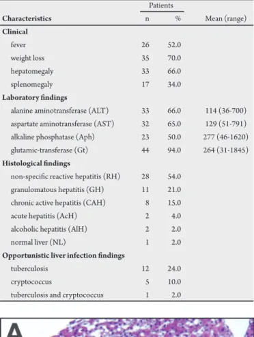
hirty (60%) patients presented opportunist infections, ive (10%) had Kaposi’s sarcoma, two (4%) had Hodgkin’s lymphoma and one (2%) had idiopathic thrombocytopenic purpura. he clinical characteristics, laboratorial and histological indings of the patients are described in Table 1.
Liver biopsy showed histological alterations in 98% of the patients; the only patient who apparently had a normal liver (2%) was infected with mycobacteria. Two patients were submited to two biopsies each, once ater the initial diagnosis of acute hepatitis and the other 10 months later, ater the disease had evolved to chronic active hepatitis in one patient and two months later in a patient with nonspeciic reactive hepatitis that had evolved to acute cholestatic hepatitis.
Nonspecific reactive hepatitis (RH) involved hyperplasia and hypertrophy of the Kupffer cells (100%), hepatocelular degeneration (76%), inlammatory iniltration in the portal and periportal area (70%) and lobular (42%), sinusoidal dilatation (60%) and focal steatosis (56%) were observed in the liver biopsies. Granulomatous hepatitis (GH) presented lobular and portal granulomas, rarely formed by Langhans’ cells, with minimal central necrosis. Mycobacteria were veriied in three patients and yeast in ive patients with RH. Among the 11 patients with GH, nine presented opportunistic infections (eight mycobacteria and one yeast, Figure 1). In one patient, extensive portal inlammation with Hodgkin’s lymphoma was veriied, associated with granulomas, with no infectious agents.
TABLE 1 - Description of the clinical characteristics, laboratory test results and histological indings of Brazilian HIV patients with liver problems.
Patients
Characteristics n % Mean (range)
Clinical
fever 26 52.0 weight loss 35 70.0 hepatomegaly 33 66.0 splenomegaly 17 34.0
Laboratory indings
alanine aminotransferase (ALT) 33 66.0 114 (36-700) aspartate aminotransferase (AST) 32 65.0 129 (51-791) alkaline phosphatase (Aph) 23 50.0 277 (46-1620) glutamic-transferase (Gt) 44 94.0 264 (31-1845)
Histological indings
non-speciic reactive hepatitis (RH) 28 54.0 granulomatous hepatitis (GH) 11 21.0 chronic active hepatitis (CAH) 8 15.0 acute hepatitis (AcH) 2 4.0 alcoholic hepatitis (AlH) 2 2.0 normal liver (NL) 1 2.0
Opportunistic liver infection indings
tuberculosis 12 24.0 cryptococcus 5 10.0 tuberculosis and cryptococcus 1 2.0
Figure 1 - A: Granulomatous hepatitis of tuberculosis (HE stain x 100): B.
3
DISCUSSION
Rev Soc Bras Med Trop 43(1):1-3, jan-fev, 2010
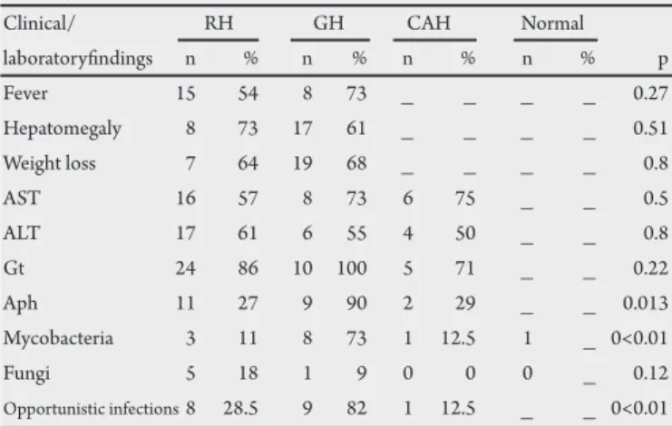
TABLE 2- Correlation of clinical, laboratory and opportunistic indings with histological indings.
Clinical/ RH GH CAH Normal laboratoryindings n % n % n % n % p Fever 15 54 8 73 _ _ _ _ 0.27 Hepatomegaly 8 73 17 61 _ _ _ _ 0.51 Weight loss 7 64 19 68 _ _ _ _ 0.8 AST 16 57 8 73 6 75 _ _ 0.5 ALT 17 61 6 55 4 50 _ _ 0.8 Gt 24 86 10 100 5 71 _ _ 0.22 Aph 11 27 9 90 2 29 _ _ 0.013 Mycobacteria 3 11 8 73 1 12.5 1 _ 0<0.01 Fungi 5 18 1 9 0 0 0 _ 0.12
Opportunistic infections 8 28.5 9 82 1 12.5 _ _ 0<0.01 RH: non-speciic reactive hepatitis, GH: granulomatous hepatitis, CAH: chronic active hepatitis, AST: aspartate aminotransferase, ALT: alanine aminotransferase, Gt: glutamic-transferase, Aph: alkaline phosphatase.
Mycobacteria were observed in 13 liver biopsies and yeast in six (ive Cryptococcus neoformans and one unidentiied yeast). One patient presented Cryptococcus and Mycobacteria in the same liver fragment. Liver biopsy permited the diagnosis of opportunistic infections in nine (18%) patients, these included eight with mycobacteriosis and one with Cryptococcus infection.
he clinical alterations were not signiicantly correlated with the presence of opportunistic infections. Alkaline phosphatase was signiicantly altered in liver granuloma tissue. Levels of other liver enzymes, such as AST, ALT and Gt, were not correlated with histological indings. here was a signiicant relation between GH and opportunistic infections, speciically those caused by Mycobacteria. here was no correlation between histological indings and fungus infection (Table 2).
Among the 13 patients with a diagnosis of Mycobacteria, Ziehl-Neelsen staining was positive in six out of 12 (50%) patients, cultures were positive in all nine patients tested; Mycobacterium tuberculosis was identiied in four of these. Due to a lack of hepatic tissue, histopathology was not performed for one patient and no cultures were made from samples of four patients. Among the patients diagnosed with fungi, two out of six were determined by staining and four out of ive by speciic culture.
Hepatic histological abnormalities were observed in all the patients; this frequency was higher than reported in other studies. Lanjewar et al1 reported 58% patients with signiicant pathological lesions; the most common pathological processes involving the liver appeared to be secondary to infections¹. hese indings could be explained by the phase of illness in these patients, with opportunistic disease occurring in 76%, along with drug abuse and frequent use of medicines.
Opportunistic infections were diagnosed in 36% of the patients, half of these by liver biopsy. Altered alkaline phosphatase levels were related to granulomatous hepatitis and mycobacteria infection. Garcia-Ordonez et al3 studied 58 HIV-infected patients who underwent PLB for evaluation of FUO in Spain. he diagnosis was established in 51 (87.9%) patients; tuberculosis (50%) and leishmaniasis (20.7%) were the most common. PLB was diagnostic in 25 (43.1%) cases, helpful in 13 (22.4%), and normal or nonspeciic in the remaining 20 (34.5%); they concluded that PLB is a useful technique for the diagnosis of FUO in HIV-infected persons and that
early PLB should be considered in patients with hepatosplenomegaly and increased alkaline phosphatase levels³. Other studies have reported similar conclusions2,9.
Echejoh et al10 evaluated postmortem hepatic histopathological indings in HIV patients in Nigeria; most (65%) patients had clinical tuberculosis. Granulomatous hepatitis, chronic hepatitis, nonspeciic reactive hepatitis and steatosis were the most common hepatic histopathological lesions, occurring in 34, 20, 15 and 12% of patients, respectively. Seven percent had normal histological features10.
In our study, the histopathological findings were often nonspeciic. A correlation was determined between granulomatous hepatitis and opportunistic hepatic infections, with prevalence of Mycobacterium tuberculosis, and Cryptococcus neoformans among the fungi. Culture for mycobacteriosis and of the deep mycoses was an important approach for the detection of opportunists, even when Ziehl-Neelsen and PAS staining of the liver was negative.
In conclusion, Mycobacteriosis was the most prevalent hepatic infection in Brazilian patients and culture of liver fragmentsis important for the diagnosis of opportunistic infection.
ACKNOWLEDGMENTS
CONFLICT OF INTEREST
he authors declare that there is no conlict of interest.
REFERENCES
We would like to thank Maria de Fátima Beu for preparing the cultures of the hepatic fragments.
1. Lanjewar DN, Rao RJ, Kulkarni SB, Hira SK. Hepatic pathology in AIDS: a pathological study from Mumbai, India. HIV Medicine 2004;5:253-257. 2. Chang YG, Chen PJ, Hung CC, Chen MY, Lai MY, Chen DS. Opportunistic
hepatic infections in AIDS patients with fever of unknown origin. Journal of the Formosan Medical Association 1999;98:5-10.
3. Garcia-Ordonez MA, Colmenero JD, Jimenez-Onate F, Martos F, Martinez J, Juarez C. Diagnostic usefulness of percutaneous liver biopsy in HIV-infected patients with fever of unknown origin. Journal of Infection 1999;38:94-98. 4. Danesi G, Pianta P, Mastroianni A, Cicognani C, Cristoni L, Sama C. Hepatic
and pancreatic disease in patients with acquired immunodeiciency syndrome (AIDS). Minerva Medica 1999;90:123-131.
5. Shakhgil’dian VI., Kravchenko AV, Parkhomenko Iu G, Tishkevich OA, Serova VV, Gruzdev B M. Liver involvement in secondary infections in HIV-infected patients. Terapevticheskiî Arkhiv 2002;74:40-43.
6. Barone B, Kreuzig PL,Gusmão PM, Chamie D, Bezerra S, Pinheiro P, et al. Case report of lymph nodal, hepatic and splenic tuberculosis in an HIV-positive patient. Brazilian Journal Infectious Disease 2006;10:149-153.
7. Borges AS, Ferreira MS, Silvestre MT, Nishioka SA, Rocha A. Histoplasmosis in immunodepressed patients: study of 18 cases seen in Uberlandia, MG. Revista da Sociedade Brasileira de Medicina Tropical 1997;30:119-124.
8. Lambertucci JR, Rayes AA, Gerspacher-Lara R. Salmonella-S.mansoni association in patients with acquired immunodeiciency syndrome. Revista do Instituto de Medicina Tropical de São Paulo 1998;40:233-235.
9. Piratvisuth T, Siripaitoon P, Sriplug H, Ovartlarnporn B. Findings and beneit of liver biopsies in 46 patients infected with human immunodeiciency virus. Journal of Gastroenterology and Hepatology 1999;14:146-149.