100
INTRODUCTION Revista da Sociedade Brasileira de Medicina Tropical 43(1):100-101, jan-fev, 2010
Communication/Comunicação
PCR-RFLP of 16S ribosomal DNA to confirm the identification of
Enterococcus gallinarum
and
Enterococcus casseliflavus
isolated from
clinical and food samples
PCR-RFLP do 16S DNA ribossomal para conirmar a identiicação de
Enterococcus gallinarum
e
Enterococcus casselilavus
isolados de amostras clínicas e alimentares
Aline Weber Medeiros¹, Pedro d’Azevedo
2, Rebeca Inhoque Pereira³, Ana Paula Cassenego¹, Sueli Van Der Sand³,
Jeverson Frazzon
4and Ana Paula Guedes Frazzon³
1. Graduate Program of Agricultural and Environmental Microbiology, Federal University of Rio Grande do Sul, Porto Alegre, RS, Brazil. 2. Department of Basic Health Sciences, Microbiology Department, Federal Health Sciences University of Porto Alegre, Porto Alegre, RS, Brazil. 3. Microbiology Department, Federal University of Rio Grande do Sul, Porto Alegre, RS, Brazil. 4. Science and Food Technology Institute, Federal University of Rio Grande do Sul, Porto Alegre, RS, Brazil.
Address to: Dr. Ana Paula Guedes Frazzon. ICBS/UFRS. Campus do Centro. Av. Sarmento Leite 500/ sala 158, 90050-170 Porto Alegre, RS.
Tel: 55 51 3308-4111; Fax: 55 51 3308-4111 e-mail: [email protected]
Received in 16/11/2009 Accepted in 13/01/2010
ABSTACT
Introduction: his study aimed to conirm the identiication of Enterococcus gallinarum and
Enterococcus casselilavus isolated from clinical and food samples by PCR-RFLP. Methods:
Fity-two strains identiied by conventional biochemical exams were submited to PCR ampliication and digested with HinfI. Only 20 (38.5%) of the 52 strains showed a DNA patern expected for E. gallinarum and E. casselilavus. Results: Analysis of the results of this study showed that E. gallinarum and E. casselilavus are occasionally erroneously identiied and conirmed the potential application of 16S rDNA analysis for accurate identiication of these species. Conclusions: A correct identiication is important to distinguish between intrinsic and acquired vancomycin resistance.
Key-words: PCR-RFLP of 16S rDNA. Enterococcus gallinarum.Enterococcus casselilavus.
RESUMO
Introdução: O objetivo deste estudo foi conirmar a identiicação de amostras clínicas e alimentos de Enterococcus gallinarum e Enterococcus casselilavus por PCR-RFLP. Métodos: Cinquenta e duas cepas identiicadas por exames bioquímicos convencionais foram submetidos a ampliicação por PCR e digestão com HinfI. Apenas 20 (38,5%) das 52 amostras apresentaram um padrão de DNA esperado E. gallinarum e E. casselilavus. Resultados: Analise dos resultados deste estudo demonstraram que, algumas vezes E. gallinarum e E. casselilavus são erroneamente identiicados e conirmaram a potencial aplicação da análise do 16S rDNA para identiicação exata destas espécies. Conclusões: A correta identiicação é importante a im de distinguir entre resistência intrínseca e adquirida à vancomicina.
Palavras-chaves: PCR-RFLP de 16S rDNA. Enterococcus gallinarum.Enterococcus casselilavus.
Enterococci are opportunistic pathogens and well known as the principal microorganisms associated with the development of infections, especially in immunosuppressed patients. Furthermore, strains have been recognized as emerging human pathogens mostly associated with nosocomial infections1. he
emergence of enterococci in nosocomial infections has grown in parallel with the rise in strains resistant to a large number of antimicrobial drugs used in the treatment of human infections. Enterococcus gallinarum and Enterococcus casseliflavus exhibit low-level intrinsic resistance to vancomycin, conferred by the vanC-1 gene2. Commercial kits
for species identiication of Enterococcus are unable to distinguish E. gallinarum and E. casselilavus from other enterococci3. Rapid and reliable diferentiation
of these species in patients infected with vancomycin resistant enterococci (VRE) is essential for an infection control program. he aim of this work was to conirm the identiication of E. gallinarum and E. casselilavus using the PCR-restriction fragment length polymorphism (PCR-RFLP) technique.
METHODS
In the current study, E. gallinarum (n=32) and E. casselilavus (n=20) isolated from clinical samples and food identified by conventional biochemical were analyzed. Two references strains E. gallinarum (PAD 262) and E. casselilavus (PAD 71) were obtained from the culture collection at the laboratory of microbiology of the Federal University of Health Sciences (Universidade Federal de Ciências da Saúde) of Porto Alegre and used as controls (Table 1). Extraction of total DNA from cells followed the method described by Riboldi et al4
101
RESULTS AND DISCUSSIONMedeiros AW et al - PCR-RFLP conirmation Enterococcus gallinarum and Enterococcus casselilavus
cycler (Eppendorf Mastercycler Personal). he primers 16Sent-F (5’-CTGACGCTGAGGCTCGAAAGCG-3’) and 16Sent-R (5’-TGTGACGGGCGGTGTGTACAAGGGGG-3’) corresponded to nucleotide sequences of 16SrDNA of the genus Enterococcus. he PCR product of 661 bp ampliied was submited to digestion with the restriction enzyme Hinf I ( Jena Bioscience GmbH, Germany), according to the manufacturer’s instructions. he DNA fragments obtained were resolved by electrophoresis on 2% agarose gel stained by ethidium bromide.
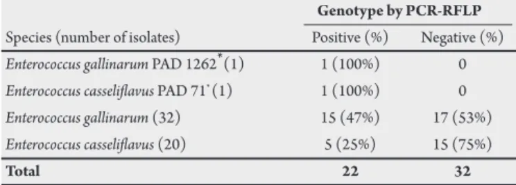
Table 1. Enterococcus gallinarum and Enterococcus casselilavus isolated from clinical and food samples in South Brazil.
Genotype by PCR-RFLP Species (number of isolates) Positive (%) Negative (%) Enterococcus gallinarum PAD 1262* (1) 1 (100%) 0 Enterococcus casselilavus PAD 71* (1) 1 (100%) 0
Enterococcus gallinarum (32) 15 (47%) 17 (53%) Enterococcus casselilavus (20) 5 (25%) 15 (75%)
Total 22 32
* PAD culture collection at the Laboratory of Microbiology of Federal University of Health Sciences of Porto Alegre.
he PCR-RFLP results from reference strains of E. gallinarum and E. casselilavus showed two distinguishable DNA fragments of 589bp and 72bp (Figure 1). PCR-RFLP from the 52 strains tested demonstrated that 47% (15/32) of E. gallinarum and 25% (5/20) showed the expected PCR-RFLP paterns (Figure 1). Two PCR-RFLP positive E. gallinarum and one E. casselilavus were analyzed
FINANCIAL SUPPORT
he authors grateful to the Conselho Nacional de Desenvolvimento Cientíico e Tecnológico, the Coordenação de Aperfeiçoamento de Pessoal de Nivel Superior and the Fundação de Amparo a Pesquisa do Rio Grande do Sul for their inancial support.
CONFLICT OF INTEREST
he authors declare that there is no conlict of interest.
FIGURE 1 - Restriction DNA fragments obtained with the digestion of PCR ampliication products with the enzyme HinfI. (1) Ladder 100 bp; (2) PCR product from E. gallinarum non digested; (3) E. galllinarum PAD 1262; (4) E. casselilavus PAD 71; (5) E. galllinarum clinical isolate; (6) misidentiied E. galllinarum clinical isolate; (7) E. casselilavus clinical isolate; (8) Negative control; (9) misidentiied E casselilavus clinical isolate; (10-11) E. casselilavus isolated from food; (12) misidentiied E. casselilavus isolated from food; (14) E. galllinarum isolated from food; (15-19) misidentiied E. galllinarum isolated from food.
by the SDS-PAGE method and conirmed the results obtained. On the other hand, 53% (17/32) of E. gallinarum and 75% (15/20) of E. casselilavus strains showed three DNA fragments of 504, 85 and 72bp (Figure 1). hese strains were resubmited to a new set of biochemical tests and reclassiied as: E. faecium, E. faecalis and Enterococcus sp. he 16S rDNA gene has been useful for the identiication of Enterococcus genus and species5,6. All 16S rDNA sequences deposited in GenBank
of the NCBI of E. gallinanum and E.casselilavus have a conserved thymidine (T) at position 1248, while other species of enterococci predominantly present a cytosine (C) or T at the equivalent position. A single conserved base substitution in this position in E. gallinarum and E. casselilavus eliminates the restriction endonuclease site for HinfI. he present results demonstrate that E. gallinarum and E. casselilavus are occasionally erroneously identiied and conirmed the potential application of 16S rDNA analysis to accurately identify these species. Correct identiication is very important to discriminate between natural and VRE strains.
REFERENCES
1. d'Azevedo PA, Dias CA, Teixeira LM. Genetic diversity and antimicrobial resistance of enterococcal isolates from Southern region of Brazil. Rev Inst Med Trop São Paulo 2006; 48,11-16.
2. Clark NC, Teixeira LM, Facklam RR, Tenover FC. Detection and diferentiation
of van C-1, van C-2and van C-3, glycopeptide resistance genes in enterococci. J
Clin Microbiol 1998; 36: 2294-2297.
3. Facklam RR, Carvalho MGS, Teixeira LM. History, taxonomy, biochemical characteristics, and antibiotic susceptibility testing of enterococci. In The
enterococci: pathogenesis, molecular biology, and antibiotic resistance. Eds,
Washington: ASM Press; 2002. p 1-54.
4. Riboldi GP, Frazzon J, d'Azevedo PA, Frazzon APG. Antimicrobial resistance proile of Enterococcus spp isolated from food in Southern Brazil. Braz J Microbiol 2009; 40:125-128.
5. Fortina MG, Ricci G, Borgo F, Manachini PL. Rapid identiication of Enterococcus
italicus by PCR with primers targeted to 16S rRNA gene. LetAppl Microbiol
2007; 44:443-446