○ ○ ○ ○ ○ ○ ○ ○ ○ ○ ○ ○ABST RAC T○ ○ ○ ○ ○ ○ ○ ○ ○ ○ ○ ○ ○ ○ ○ ○IN T RO D U C T IO N○ ○ ○ ○ ○ ○ ○ ○ ○ ○
The ABO system is the most investigated erythrocyte antigen system for all populations and, due to the ease of identifying its phenotypes, it has been used as a genetic marker in studies of associations with infectious and non-infectious diseases.1,2 On the other hand, the Lewis blood group system and the secretor and non-secretor phenotypes of ABH antigens have been less intensively studied.1
A compilation by Lima et al. of results from different studies among Brazilians showed that the frequencies of the ABO and Lewis blood group systems and the secretor and non-secretor phenotypes are similar to those observed in European populations.3 Recently, Mattos et al. demonstrated that the ABO blood groups among Brazilians are highly polymorphic when analyzed by molecular methods.4
Among the first epidemiological studies to establish associations between blood groups and diseases, there were some demonstrations of high frequencies of the O blood group and non-secretor phenotype of ABH antigens among patients suffering from peptic ulcers.5,6 It was later shown that the Helicobacter pylori bacillus is the main etiologic agent associated with gastric ulceration, being present in more than 80% of patients with this disease.7,8 Subsequently, it was demonstrated that infection by this bacillus affects more than 90% of the adult population around the world, especially in underdeveloped countries.9 In Brazil, investigations of the presence of H. pylori among children, youths and blood donors have demonstrated that the frequency of this
infection ranges from 34% to 66%.10,11 In the early 90s, Borén et al. reported that this bacillus chooses to attach itself to the Lewis b antigen (Leb), which is rich in fucose and is expressed on the surface of the epithelial cells of the gastric mucosa.12 With this obser-vation, the authors tried to establish connec-tions between the associaconnec-tions of the ABO blood groups and the secretor phenotypes of ABH antigens with peptic ulcers that had been observed in previous decades.
T he ABH and Lewis antigens on the gastric and duodenal mucosae are synthesized through a specific glycosyl transferase, which incorporates molecules of fucose in common type I oligosaccharide precursor.13,14 T he O and Le(a-b+) phenotypes express a greater quantity of these fucosylated antigens in comparison with other groups, and Borén et al. believed that this difference predisposed these carriers to H. pylori infection.12, 15
Various studies have evaluated the associations of the ABO and Lewis blood groups and the secretor and non-secretor phenotypes with H. pylori infection. Hooke-Nikanne et al. did not find an association between the secretor phenotype and positive serology for this bacillus among 271 blood donors.16 Clyne & Drum observed that the Leb antigen expression did not influence the adherence of H. pylori to the gastric epithe-lium.17 On the other hand, Alkout et al. showed that the O blood group had great susceptibility towards peptic ulcers and that the H and Leb antigens had an influence on H. pylori infection.18 Lin et al. demonstrated a high frequency of infection with this bacteria in 90.3% of O blood group patients suffering
O
ri
gi
n
a
l
A
rt
ic
le
• Luiz Carlos de M attos
• Juliana Rodrigues Cintra
• Fábio Eduardo Sanches
• Rita de Cássia M artins Alves
da Silva
• M ilton Artur Ruiz
• Haroldo W ilson M oreira
ABO, Lewis, secretor and
non-secretor phenotypes in
patients infected or
uninfected by the
Helicobacter pylori
bacillus
Department of Molecular Biology, Faculty of Medicine of São José do Rio
Preto, São José do Rio Preto, São Paulo, Brazil
CON TEX T: Epidemio lo g ical studies have demo nstrated hig her frequencies o f the O blo o d g ro up and the no n-secreto r pheno type o f ABH antig ens amo ng p a tie nts suffe ring fro m p e p tic ulc e rs. Sinc e Helico bacter pylo ri has been established as the ma in etio lo g ic a l fa c to r in this disea se, c o ntro -versies abo ut the asso ciatio ns o f the ABO and Lewis blo o d g ro up pheno types and secreto r and no n-secreto r pheno types in relatio n to susceptibility to w a rd s infe c tio n b y this b a c illus ha ve b e e n presented.
O BJECTIVE: To verify the frequencies o f ABO , Lewis blo o d gro up pheno types, secreto r and no n-secreto r pheno types in patients infected o r uninfected by H. pylo ri.
DESIGN : Cro ss-sectio nal study.
SETTIN G: O utpatient clinic.
PARTICIPAN TS: O ne hundred and twenty patients with dyspeptic sympto ms who underwent endo sco py.
M AIN M EASUREM EN TS: ABO a nd Le wis b lo o d g ro up pheno types were determined by a standard hemag g lutinatio n test and the secreto r and no n-sec reto r pheno types were eva lua ted b y sa liva samples using the inhibito r hemag g lutinatio n test.
RESULTS: The diag no sis o f infectio n, made via breath and urea tests and co nfirmed using po lymerase chain reactio n (PCR) in g astric bio psy frag ments, sho wed the presence o f H. pylo ri in 6 1 .7 % o f the patients and absence in 3 8 .3 %. The differences between the frequencies o f the ABO blo o d g ro up pheno types amo ng infected (A 2 7 .0 %; B 1 2 .2 %; AB 4 .0 % and O 5 6 .8 %) and uninfected patients (A 5 8 .7 %; B 1 3 .0 %; AB 4 .3 % and O 2 4 .0 %) were sig nificant. The Lewis blo o d type, secreto r a nd no nse c re to r p he no typ e s sho w e d ho mo -g e ne o us d istrib utio n b e tw e e n the -g ro up s o f patients analyz ed.
CO N CLUSIO N S: O ur results sug g est that the infectio n o f H. pylo ri can be related to ABO blo o d g ro ups but no t to the Lewis blo o d g ro up no r to secreto r and no n-secreto r pheno types.
KEY W O RDS: Blo o d g ro ups. ABO . Lewis. Secreto r pheno type. N o n-secreto r pheno type. Helico bacter pylo ri.
São Paulo M edical Journal - Revista Paulista de M edicina
56
from gastroduodenal diseases.19
T hese data show that the associations of AB O , Lewis, secretor and non-secretor phenotypes with H. pylori infection are controversial because of the discordance between clinical observations and laboratory evidence.12,15-19
T he aim of this study was to verify the frequencies of ABO, Lewis, secretor and non-secretor phenotypes in patients with symptoms of dyspepsia who underwent upper gastrointestinal endoscopy and to cross-check whether they were infected by H. pylori or not.
○ ○ ○ ○ ○ ○ ○ ○ ○ ○ ○ ○ ○ ○M ET H O D○ ○ ○ ○ ○ ○
One hundred and twenty adult patients who were seen over a period of one year in the D epartment of Endoscopy and/or the Clinical Gastroenterology outpatient service of the Medical School (FAMERP) were evaluated. Patients were included in the study if they had symptoms of dyspepsia and if upper gastrointestinal endoscopy was indicated. T his group included sufferers of gastric and duodenal ulcers.
T he study had prior approval from the Research Ethics Committee of the Institution (case 4657/97) and informed consent was obtained from all the participants.
Patients who were less than 18 years old or pregnant, those who had gastrointestinal tract hemorrhage or acute gastritis, and those who had used a proton-pump inhibitor in the previous week or had used an H2 receptor antagonist in the previous 24 hours, were excluded from the study.
A sample of 5 ml of peripheral blood was obtained using EDTA to type the ABO and Lewis blood groups and 2 ml of saliva was collected for the determination of secretor and non-secretor phenotypes.
ABO blood groups were determined using standardized hemagglutination methods20 and the Lewis phenotypes were ascertained using the gel method.21 Secretor and non-secretor phe-notypes were identified using the hemagglu-tination inhibition test.22 Investigation of H. pylori was done in a routine manner by a specialized gastroenterology laboratory using urea breath and urease tests. In all cases, whether positive, negative or discordant, the presence of the infection was confirmed using the polymerase chain reaction (PCR), in accordance with the protocol of Valentine et al.23 The χ2 test and dependence analysis were used to calculate associations among the results.
○ ○ ○ ○ ○ ○ ○ ○ ○ ○ ○ ○ ○ ○ ○R ESU LT S○ ○ ○ ○ ○ One hundred and twenty patients, both male and female, including Caucasians and non-Caucasians, with a mean age of 42 years (ranging from 18 to 74 years old), were assessed and divided into groups infected and uninfected by H. pylori.
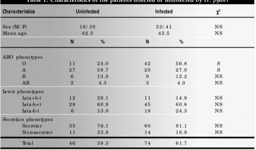
Of the 120 patients enrolled, 40.8% (49/ 120) were male and 59.2% (71/120) were female; 77.5% (93/120) were Caucasians and 22.5% (27/120) were non-Caucasians. H. pylori infection was present in 61.7% (74/120) and absent in 38.3% (46/120) of the patients. No significant differences were observed when comparing gender and racial group with H. pylori infection.
Table 1 shows the characteristics of in-fected and uninin-fected patients. Analysis of the whole data set demonstrated that the distribution of the ABO blood groups among the patients, independent of infection by H. pylori, followed the same proportions as the blood groups found in the general Brazilian population.3,4
When the frequencies of these phenotypes were analyzed separately in accordance with infection and non-infection, it was possible to verify that the O and A blood groups were distinct. Of the 74 infected patients, 56.8% (42/74) were type O and 27% (20/74) were type A; of the 46 uninfected patients, only 24% (11/46) were type O and 58.7% (27/ 46) were type A. The χ2 test demonstrated that the difference observed between the higher prevalence of type O in the infected group and the higher prevalence of blood group A in the uninfected group is significant, giving a p-value of 0.003. D ependence analysis applied to these results shows that there is an association between the blood group and infection by H. pylori, in which type O has a greater tendency towards infection and type A to non-infection.
The distribution of the Lewis, secretor and non-secretor phenotypes did not exhibit significant differences between infected and uninfected patients.
○ ○ ○ ○ ○ ○ ○ ○ ○ ○ ○ ○ D ISC U SSIO N○ ○ ○ ○ ○ ○ ○ ○
Studies of associations between blood groups and diseases have interested researchers since the 1950s, when the first results showing a high prevalence of the O blood group among patients with peptic ulcer disease were published.5,6
T hese associations remained relatively unstudied until Borén et al. suggested that the Leb antigen of the Lewis blood group system acts as a receptor for H. pylori on the gastric mucosa.12 The Leb antigen is a oligosaccharide rich in fucose molecules and is expressed in greater quantities in the O blood group.13,14 Borén et al. investigated the hypothesis that, due to the presence of fucose molecules in the Leb antigen, the carriers of the O blood group would have a greater tendency towards infection by H. pylori.12,15 These researchers believed that this host characteristic justified the observation made during previous decades that persons with blood group O have a greater tendency towards peptic ulcers in comparison with those with the A, B and AB blood groups, which continues to be reported in current publications.24,25 Table 1. Characteristics of the patients infected or uninfected by H. pylori
Characteristics Uninfected Infected χχχχχ2
Sex (M/ F) 1 6 / 3 0 3 3 / 4 1 N S
Mean ag e 4 2 .3 4 3 .5 N S
N % N %
ABO pheno types
O 1 1 2 4 .0 4 2 5 6 .8 S
A 2 7 5 8 .7 2 0 2 7 .0 S
B 6 1 3 .0 9 1 2 .2 N S
AB 2 4 .3 3 4 .0 N S
Lewis pheno types
Le(a+b-) 1 2 2 6 .1 1 1 1 4 .9 N S
Le(a-b+) 2 8 6 0 .9 4 5 6 0 .8 N S
Le(a-b-) 6 1 3 .0 1 8 2 4 .3 N S
Secretio n pheno types
Secreto r 3 5 7 6 .1 6 0 8 1 .1 N S
N o n-secreto r 1 1 2 3 .9 1 4 1 8 .9 N S
To tal 4 6 3 8 .3 7 4 6 1 .7
S = sig nificant; N S = no t sig nificant.
São Paulo M edical Journal - Revista Paulista de M edicina
57
With the demonstration that H. pylori is the agent in the majority of cases of peptic ulcer disease,several studies have tried to establish a relationship between the presence of this bacillus and the ABO and Lewis blood groups and the secretor and non-secretor phenotypes.12, 15-19
T he results of this current study show a strong association between the O blood group and infection caused by H. pylori, which is reinforced by data obtained from other scientific papers.18,19 Significant differences observed in the distribution of the frequencies of infected patients in comparison with uninfected patients support the epide-miological view of the greater susceptibility of blood group O to infection by H. pylori.
T hese observations support the conclu-sions of Alkout et al., who demonstrated that the H antigen represents an important receptor expressed in the gastroduodenal mucosa cells to which H. pylori adheres.18 Mollicone et al. showed that the H antigen expression in the duodenal mucosa is controlled by FUT 1 (H ) using type I I oligosaccharide precursor.26 As this fucosylated
antigen is not modified to A or B antigens in the O blood group, its greatest potential expression can be a important factor in establishing a connection between this blood group, the H. pylori infection and the diseases resulting from its presence, independent of Lewis phenotypes and the secretory or non-secretory condition of the ABH antigens.
Our results disagree with some previous reports in which it was demonstrated that the O blood group did not represent a risk factor for H. pylori infection.16,27-29 In these studies, the authors used other methods to detect infection by H. pylori, including ELISA,16,28,29 urease27,28 and histological27,28 tests. In our work, the confirmation of infection by this pathogen was based on PCR tests. T here is strong evidence that other tests employed in the diagnosis of H. pylori infection differ in specificity and sensitivity when compared to the PCR.23,30-34 Atherton et al. used PCR to show the presence of H. pylori in patients treated with amoxicillin whose urease and histological test results were negative.34 T his characteristic of PCR may have an influence on the different infection frequencies observed
within distinct populations, by improving the diagnosis even in cases where there is low bacterial density on the gastric mucosa.30,31,33
Variations in the percentages between infected and uninfected patients, in relation to gender, Lewis phenotypes, secretor and non-secretor phenotypes did not result in significant differences. However, we observed an elevated frequency of the Le(a-b-) phenotype among infected patients. Our observations do not support the hypothesis that patients with the Le(a-b+) phenotype present a higher susceptibility to H. pylori infection.
○ ○ ○ ○ ○ ○ ○ ○ ○ ○ ○C O N C LU SIO N S○ ○ ○ ○ ○ ○ ○ ○ ○
Our results suggest that H. pylori infection is strongly associated with the O blood group, which is in agreement with other published data, but there is no association with the Lewis, secretor and non-secretor phenotypes. Nevertheless, the high frequency of the O blood group among patients infected by this bacillus and in those with gastroduodenal diseases caused by the presence of H. pylori remains unexplained.
1. Mourant AE, Kopec AC. D omaniewska-Sobczak, K. T he distribution of the human blood groups and other polymorphisms. London: Oxford University Press; 1976. 2. Mourant AE, Kopec AC, Domaniewska-Sobczak K. Blood groups
and diseases: a study of associations of diseases with blood groups and other polymorphisms. London: Oxford University Press; 1978. 3. Lima LM A, C allado M R M , Santos J A. Curso de Imunohematologia da Divisão Hemocentro da Faculdade de Medicina de Botucatu — Campus Unesp. Apostila. Botucatu (SP): Universidade Estadual de São Paulo; 1992. 4. Mattos LC, Sanchez FE, Cintra JR, et al. Genotipagem do locus
ABO (9q34.1) em doadores de sangue da região noroeste do Estado de São Paulo. Rev Bras Hematol Hemoter 2001;1:15-22. 5. Clarke CA, Edwards JW, Haddock DRW, et al. ABO blood groups and secretor character in duodenal ulcer. B M J 1956;2:725-31.
6. Clark CA, Evand DAP, McConnell RB, Sheppard PM. Secretion of blood group antigens and peptic ulcer.BMJ 1959;1:603-7. 7. Warren JR. Unidentified curved bacilli on gastric epithelium in
active chronic gastritis. Lancet 1983;4:1273-5.
8. NIH Consensus Conference. Helicobacter pylori in peptic ulcer. JAMA 1994;272(1):65-9.
9. Pounder RE, Ng D. T he prevalence of Helicobacter pylori
infection in different countries. Alim Pharmac T herap 1995;9:33-40.
10. O liveira AMR, Queiroz D MM, Rocha GA, Mendes EN. Seroprevalence of Helicobacter pylori infection in children of low socioeconomic level in Belo Horizonte, Brazil. Am J Gastroenterol 1994;89:2201-4.
11. Rocha GA, Q ueiroz D M, Mendes EN, et al. Source of
Helicobacter pylori infection: studies in abattoir workers and pigs. Am J Gastroenterol 1992;87:1525.
12. Borén T, Falk P, Roth KA. Attachment of Helicobacter pylori to
○ ○ ○ ○ ○ ○ ○ ○ ○ ○ ○ ○ ○ ○ ○ ○ ○ ○ ○ ○ ○ ○ ○ ○ ○ ○ ○ ○ ○ ○ ○ ○ ○ ○ ○ ○ ○ ○ ○ ○ ○ ○ ○ ○ ○ ○ ○ ○ ○ ○ ○ ○ ○ ○ ○ ○ R EFER EN C ES○ ○ ○ ○ ○ ○ ○ ○
human gastric epithelium mediated by blood group antigens. Science 1993;262:1892-5.
13. Watkins WM. Biochemistry and genetics of the ABO, Lewis and P blood group systems. Adv Hum Genet 1980;10:1-136. 14. Mollicone R, Bara J, Le Pendu J. Immunohistologic pattern of type 1 (Lea, Leb) and type 2 (X, Y, H) blood group-related
antigens in the human pyloric and duodenal mucosae. Lab Invest 1985;53:219-27.
15. Borén T, Normark S, Falk P. Helicobacter pylori: molecular basis for host recognition and bacterial adherence. Trends in Microbiol 1994;2(7):221-8.
16. Hooke-Nikanne J, Sistonen P, Kosunen T U. Effect of ABO blood group and secretor status on the frequency of Helicobacter pylori antibodies. Scan J Gastroenterol 1990;25:815-8. 17. Clyne M, Drum B. Absence of effect of Lea and Leb expression
on adherence of Helicobacter pylori to human gastric cell. Gastroenterology 1997;113:72-80.
18. Alkout AM, Blackwell CC, Weir DM, et al. Isolation of cell surface component of Helicobacter pylori that binds H type 2, Lewis A and Lewis B antigens. Gastroenterology 1997;112:1179-87. 19. Lin CW, Chang YS, Wu SC, Cheng KS. Helicobacter pylori in
gastric biopsies of Taiwanese patients with gastroduodenal diseases. Jpn J Med Sci Biol 1998;51(1):13.
20. Vengelen-Tyler V. Technical manual, 12th ed. American
Association of Blood Banks; 1996.
21. Lapierre Y. T he gel test: a new way to detect red cell antigen-antibody reactions. Transfusion 1990;30:109-13. 22. Oliveira MCVC. Práticas em imunologia eritrocitária. Rio de
Janeiro: Editora Médica e Científica; 1999.
23. Valentine JL, Arthur RR, Mobley HLT, Dick JD. Detection of
Helicobacter pylori by using polymerase chain reaction. J Clin Microbiol 1991;29:689-5.
24. Hallstone AE, Perez EA. Blood type and the risk of gastric
disease. Science 1994;264:1386-7.
25. Anderson RP, Street A, Gibson PR. Blood group O predicts risk and severity for bleeding peptic ulcers and von Willebrand disease. J Gastrol Hepatol 1997;12(suppl):26.
26. Mollicone R, Bara J, Le Pendu J, Oriol R. Immunohistologic pattern of type 1 (Lea, Leb) and type 2 (X, Y, H) blood
group-related antigens in the human pyloric and duodenal mucosae. Lab Invest 1985;53:219-27.
27. Dickey W, Collins JSA, Watson RGP, Sloan JM, Porter KG. Secretor status and Helicobacter pylori infection are independent risk factors for gastroduodenal disease. Gut 1993;34:351-3. 28. Loffeld RJF, Stobberingh E. Helicobacter pylori and ABO blood
groups. J Clin Pathol 1991;44:516-7.
29. Niv Y, Fraser G, Delpre G, Neeman A, et al. Helicobacter pylori
infection and blood groups. Am J Gastroenterol 1996;91(1):101-4. 30. Fabre R, Sobhani I, Laurent-Puig P, et al. Polymerase chain reaction assay for the detection of Helicobacter pylori in gastric biopsy specimens: comparison with culture, rapid urease test and histopathological tests. Gut 1994;35(7):905-8. 31. Clayton CL, Kleanthous H, Coates PJ, Morgan DD, Tabaqchali
S. Sensitive detection of Helicobacter pylori by using polymerase chain reaction. J Clin Microbiol 1992;30:192-200. 32. Kawamata O, Yoshida H, Hirota K, et al. Nested-polymerase
chain reaction for the detection of Helicobacter pylori infection with novel primers designed by sequence analysis of urease A gene in clinically isolated bacterial strains. Bioch Biophys Res Commun 1996;219:266-72.
33. Li C, Ha T, Ferguson Jr DA, et al. A newly developed PCR assay of Helicobacterpylori in gastric biopsy, saliva and feces. Dig Dis Sci 1996;41(11):2142-9.
34. Atherton JC, Cockayne A, Balsitis M, et al. Detection of intragastric sites at which Helicobacter pylori evades treatment with amoxicillin and cimetidine. Gut 1995;36(5):670-4.
São Paulo M edical Journal - Revista Paulista de M edicina
58
C O NT EX T O : Estudos epidemiológicos evidenciaram alta freqüência do tipo O e do fenótipo não-secretor dos antígenos ABH em pacientes com úlceras pépticas. Desde que o
Helicobacter pylori foi estabelecido como o
principal agente etiológico destas doenças, controvérsias sobre as associações dos grupos sangüíneos ABO , Lewis e dos fenótipos secretor e não-secretor com a suscetibilidade à infecção por esse bacilo foram apresentadas.
OBJETIVO: Verificar as freqüências dos fenótipos ABO , Lewis, secretor e não-secretor em pacientes infectados ou não infectados pelo H. pylori.
TIPO DE ESTUDO: Estudo transversal.
LOCAL: Ambulatório de Gastroenterologia do Hospital de Base de São José do Rio Preto e Laboratório de Imunogenética Molecular do Hemocentro de São José do Rio Preto.
PARTICIPANTES: Foram avaliados 120 pa-cientes com sintomas de dispepsia submetidos a endoscopia digestiva alta.
VARIÁVEIS EST U DADAS: Fenótipos eritrocitários ABO e Lewis determinados pelos métodos de hemaglutinação
padro-○ ○ ○ ○ ○ ○ ○ ○ ○ ○ ○ ○ ○ ○ ○ ○ ○ ○ ○ ○ ○ ○ ○ ○ ○ ○ ○ ○ ○ ○ ○ ○ ○ ○ ○ ○R ESU M O○ ○ ○ ○ ○ ○ Ack now ledgements: The autho rs wish to express their
appreciatio n to Fabiana Lo ng hi Seg antini, Arlete Aparecida Alexandre Camacho and Patrícia Martins Pinto fo r their helpful technical assistance. Special g ratitude to Pro f. Dr. Jo sé Anto nio Co rdeiro o f the Department o f Epidemio lo g y and Public Health, Faculty o f Medicine o f São Jo sé do Rio Preto , fo r his statistical assistance.
Luiz Ca rlos de M a ttos, M D, PhD. Asso ciate Pro fesso r, Department o f Mo lecular Bio lo g y, Faculty o f Medicine o f Sã o Jo sé d o Rio Pre to ; Dire c to r o f the M o le c ula r Immuno g enetic Labo rato ry, Hemo center o f São Jo sé do Rio Preto , São Jo sé do Rio Preto , Braz il.
Julia na Rodrigues Cintra . Bio lo g ist a t the Mo lec ula r Immuno g enetic Labo rato ry, Hemo center o f São Jo sé do Rio Preto , São Jo sé do Rio Preto , Braz il.
Fá b io Ed ua r d o Sa nches. M e d ic a l Bio lo g ist a t the Mo lecular Immuno g enetic Labo rato ry, Hemo center o f São Jo sé do Rio Preto , São Jo sé do Rio Preto , Braz il.
Rita de Cá ssia M a rtins Alves da Silva , M D, PhD.
Asso ciate Pro fesso r, Department o f Internal Medicine o f the Faculty o f Medicine o f São Jo sé do Rio Preto ; Directo r o f the spe c ia liz e d G a stro e nte ro lo g y La b o ra to ry o f the Ho spital o f Base/ FUN FARME, São Jo sé do Rio Preto , Brazil.
M ilton Ar tur Ruiz, M D. PhD. Asso c ia te Pro fe sso r, Department o f Internal Medicine, Faculty o f Medicine o f São Jo sé do Rio Preto ; Co o rdinato r o f the Bo ne Marro w Transplant Unit, Ho spital o f Base/ FUN FARME, São Jo sé do Rio Preto , Braz il.
H a r o ld o W ilso n M o r e ir a , M D, Ph D. Pro fe sso r, Department o f Clinical Analyses, Faculty o f Pharmaceutical Sciences, São Paulo State University, Araraquara Campus, São Paulo , Braz il.
Sources of funding: BAP FAMERP (case no . 0 4 1 2 3 / 9 8 ).
Conflict of interest: N o ne
Da te of first submission: 1 0 Aug ust 2 0 0 1
La st received: 2 8 N o vember 2 0 0 1
Accepted: 1 7 December 2 0 0 1
Address for correspondence
Luiz Carlo s de Matto s
Departamento de Bio lo g ia Mo lecular Faculdade de Medicina de São Jo sé do Rio Preto Avenida Brig adeiro Faria Lima, 5 4 1 6
São Jo sé do Rio Preto / SP – Brasil – CEP 1 5 0 9 0 -0 0 0 Tel. (+5 5 1 7 ) 2 2 7 -5 7 3 3 - R.1 5 4
E-mail:lumatto s@ famerp.br
CO PYRIG HT© 2 0 0 2 , Asso ciação Paulista de Medicina ○ ○ Pu b l i sh i n g i n f o r m a t i o n○ ○ ○ ○ ○ ○ ○ ○ ○ ○ ○ ○ ○ ○ ○ ○ ○ ○
nizados e fenótipos secretor e não-secretor pelo método de inibição de hemaglutinação.
RESULTADOS: O diagnóstico de infecção e não-infecção, realizado pelos testes respiratório e da urease, e confirmado pelo método da reação de polimerase em cadeia (PCR) em fragmentos de biópsias gástricas demonstrou
a presença do H. pylori em 61,7% dos
pa-cientes e ausência em 38,3%. As diferenças nas freqüências dos grupos sangüíneos ABO entre os pacientes infectados (A 27,0%; B 12,2%; AB 4,0%; O 56,8%) e não-infectados (A 58,7%; B 13,0%; AB 4,3%; O 24,0%) foram significantes. O s tipos sangüíneos Lewis e os fenótipos secretor e não-secretor mostraram distribuição semelhante entre os grupos de pacientes analisados.
CONCLUSÕES: Os resultados sugerem que a infecção pelo H. pylori parece ser influenciada pelos fenótipos ABO, mas não pelos fenótipos Lewis, secretor e não-secretor.
PALAVRAS-C H AVE: G rupos sangüíneos. Sistema ABO . Sistema Lewis. Fenótipo secretor. Fenótipo não-secretor. Helicobacter
pylori.