○ ○ ○ ○ ○ ○ ○ ○ ○ ○ ○ ○ABSTRACT○ ○ ○ ○ ○ ○ ○ ○ ○ ○ ○ ○ ○ ○ ○ ○ ○ ○ ○ ○ ○ ○ ○ ○ ○ ○
INTRODUCTION
Endometrial adenocarcinoma represents 90% of tumors in the uterine body and is the fourth commonest neoplasm in Western women and the fifth among Brazilians. Among gynecological cancers it ranks second as the cause of death.1-3 The incidence of this
type of cancer has been increasing in the Bra-zilian population.4
The classical prognostic factors are his-tological type and grading, depth of myo-metrial invasion and extent in the cervix.5-7
It has been suggested that the expression of p53 protein, as well as estrogen and proges-terone receptors, may have prognostic sig-nificance.8,9 p53 protein is expressed more in
aggressive tumors, which suggests that mu-tation of the gene renders the neoplastic cells resistant to apoptosis.10 On the other hand,
estrogen and progesterone receptors are usu-ally associated with well-differentiated en-dometrial tumors.8,10-12
Our aims were: 1) to compare histologi-cal type and grading of endometrial carcinoma in curettage and hysterectomy samples; 2) to assess the expression of p53, estrogen and pro-gesterone receptors in curettage specimens; and 3) to correlate these data with morphol-ogy and staging of the disease in the hysterec-tomy specimens.
○ ○ ○ ○ ○ ○ ○ ○ ○ ○ ○ ○ ○ ○ ○ ○ ○ ○ ○ ○
METHODS
A review was made of 51 consecutive cases of endometrioid carcinoma from January 1994 to December 1998. For all patients, di-agnostic curettage and hysterectomy
speci-mens, intraoperative frozen sections and par-affin sections, were available.
Representative 4 µm sections from for-malin-fixed, paraffin-embedded curettage specimens were histologically classified, ac-cording to the World Health Organization guidelines,13 as endometrioid or
non-endome-trioid types. Based on the International Fed-eration of Gynecology and Obstetrics (FIGO) criteria,14 the histological grades for
endome-trial carcinoma are: well-differentiated or grade I; moderately differentiated or grade II; and poorly differentiated or grade III. In accord-ance with the disease stage, the histological parameters evaluated were depth of myome-trial invasion, extent in the cervix and lymph node metastasis.
Further sections from the same tissue were submitted to immunohistochemical reactions for p53, estrogen and progesterone receptors, via the avidin-biotin-peroxidase method, us-ing antigen recovery with microwave oven (15 minutes, citrate buffer 0.01N, pH 6.0). The primary antibodies for detecting p53 and pro-gesterone receptors were, respectively: Dako D07 (code M7001), dilution 1:100; and Dako 1AE (code M3529-1), dilution 1:100. For estrogen, non-commercial antibody from a private source (University of Toulouse), was used at dilution 1:2000. Positive and nega-tive controls for all procedures were obtained from duct breast carcinoma for estrogen and progesterone receptors and colon adenocarci-noma for p53. Immunohistochemical reac-tions occur in the nucleus, which acquires a brownish color; when more than 10% of tumor cell nuclei became stained, the reac-tion was considered positive.
Original Ar
ticle
• Vera Lúcia Leite Bonfitto • Liliana Aparecida Lucci
de Angelo Andrade
p53, estrogen and progesterone
receptors in diagnostic curettage
for endometrial adenocarcinoma
and their correlation with
morphological data and disease
stage at hysterectomy
Department of Pathological Anatomy, Faculdade de Ciências Médicas,
Universidade Estadual de Campinas, Campinas, Brazil
CONTEXT: Diagnostic staging is an important prognos-tic factor for endometrial adenocarcinoma. Apart from the histological type and histological grade, some markers seem to be associated with the stage and biological behavior of the disease. Among these are p53, estrogen and progesterone receptors.
OBJECTIVE: The objectives of the present study were: to compare histological type and grading of en-dometrial carcinoma in curettage and hysterectomy samples; to assess expression of p53, estrogen and progesterone receptors in curettage specimens; and to correlate these data with morphology and stag-ing of the disease in hysterectomy specimens.
TYPE OF STUDY: Retrospective.
SETTING: Department of Pathology, Faculdade de Ciências Médicas, Universidade Estadual de Campinas.
SAMPLE: Histological diagnosis from 51 consecutive files.
PROCEDURES: Immunohistochemical reactions for p53, estrogen and progesterone receptors via the avidin-biotin-peroxidase method in 51 curettage samples endometrial carcinoma were compared with the mor-phological data and disease stage in hysterectomy. Marker expression was correlated with histological type and grade and the final stage of the disease.
RESULTS: According to the histological type:
44 cases (86%) were of endometrioid and 7 (14%) non-endometrioid carcinoma. p53 expression was observed in 16% of endometrioid and 71% of non-endometrioid cases (p < 0.05). Although estrogen expression was more evident in endo-metrioid (54%) than non-endoendo-metrioid cases (29%), this was not statistically significant. Proges-terone receptor expression was significantly higher in endometrioid than non-endometrioid cases (70% vs. 14%, p < 0.05). According tothe histo-logical grade: Estrogen and progesterone receptors were expressed more frequently in grade I endometrioid carcinoma, while p53 was mainly reported in tumor grades II and III. According to final disease stage: p53 and estrogen ex-pression in curettage specimens was not related to stage; progesterone receptors, however, were expressed significantly less in advanced disease.
CONCLUSION: p53 was observed in the majority of non-endometrioid and in high-grade non-endometrioid carci-noma, but was not related to stage. Estrogen and progesterone receptors were mainly found in grade I endometrioid carcinoma. The markers studied in cu-rettage were no more valuable for predicting the dis-ease stage than classical histological criteria.
KEY WORDS: Endometrial carcinoma. p53. Estrogen receptor. Progesterone receptor.
São Paulo Medical Journal — Revista Paulista de Medicina
164
The Fisher Exact test was used for statis-tical analysis of the positivity of the tests, in relation to histological type and grade and disease stage. The association was considered significant when p < 0.05.
○ ○ ○ ○ ○ ○ ○ ○ ○ ○ ○ ○ ○ ○ ○ ○ ○ ○ ○ ○
RESULTS
The sample consisted of 51 women with endometrioid carcinoma, with ages ranging from 43 to 83 years (mean of 64 years); 44 cases (86%) were of endometrioid and 7 (14%) non-endometrioid carcinoma (Figures 1 and 2). The 44 endometrioid carcinoma cases were graded as follows: grade I = 26/44 (59.1%), grade II = 13/44 (29.5%) and grade III = 5/44 (11.4%).
The curettage diagnoses were compared to those from the hysterectomy. Diagnostic agreement regarding histological type and grade was observed in 80%.
According to the stage of the disease the majority of the patients were diagnosed in stage I. However, non-endometrioid
carci-noma was mainly diagnosed in more advanced stages (71%, Table 1).
Markers and histological types: p53 ex-pression was found in 16% of the endo-metrioid and in 71% of the non-endometrial carcinomas (Figure 3); the estrogen and pro-gesterone receptors were more frequent in the endometrioid type (Table 2).
Markers and histological grade: In the 44 cases of endometrioid carcinoma, those with grade I expressed estrogen (15 out of 26 cases = 63%) and progesterone receptors (21 out of 26 cases = 68%) more frequently than those with grades II and III. p53 was expressed in only 16% of endometrioid carcinomas, the ma-jority of which were grade II or III (Table 3).
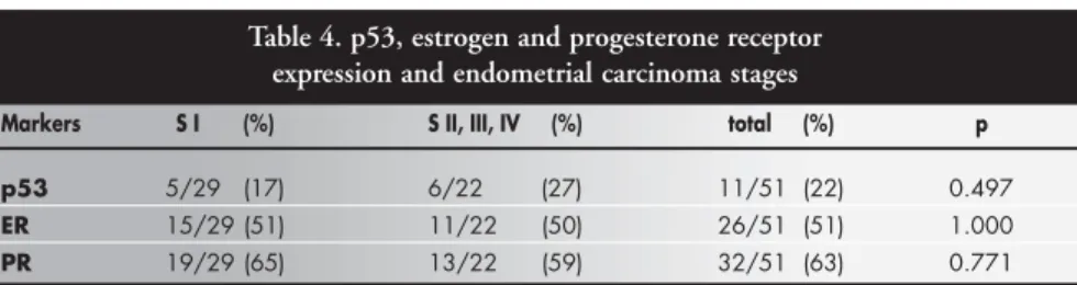
Markers and final disease stage: The ex-pression of the markers in curettage mate-rial did not bear a significant relationship to the final disease stage as evaluated in the hysterectomy specimens (Table 4). We ob-served a slight increase in p53 positivity in the advanced stages.
○ ○ ○ ○ ○ ○ ○ ○ ○ ○ ○ ○ ○ ○ ○ ○ ○ ○ ○ ○
DISCUSSION
Our results agree with the literature with regard to the higher frequencies of the endo-metrioid type and histological grade I (well-dif-ferentiated) carcinomas.5-7,15-17
Non-endome-trioid carcinoma predominated in elderly women, as expected, although the frequency of 14% was higher than found in other studies.5,6
This difference may be attributable to the rela-tively small size of the population studied and the fact that our service is a reference center in which complex cases are concentrated.
The 80% diagnostic agreement between the two procedures can be explained by the small size of the fragments obtained at curettage. Most patients were in stage I at diagnosis: this corre-lates with the predominance of the endometrioid type, which has slower progression. On the other hand, most non-endometrioid carcinomas were in stages II or III, although this was not statisti-cally significant due to the small size of the sam-ple. All these observations are in accordance with the literature.5,6,15,18
p53 was significantly more expressed in non-endometrioid (71%) than in endome-trioid carcinoma (16%), and was mostly in histological grades II and III. This indicates possible mutation of p53 as an early event, which is typical of the carcinogenic pathway for non-endometrioid carcinomas.19-21
Con-versely, estrogen and progesterone receptors are more frequently positive in endometrioid carcinoma, particularly if well-differentiated, as reported by others.9,18,21-23 However, in our
results, only progesterone receptor expression was statistically significant, which could pos-sibly be explained by the antibodies coming from different sources or having different sen-sitivity and specificity, as reported by others.23
We were able to confirm that p53 ex-pression is inversely related to that of estrogen and progesterone receptors, thus indicating a dual theory of carcinogenesis in the en-dometrium.11,23-26
Expression of the markers studied was not significantly associated with the disease stage. It has been reported that p53 expression correlates with more advanced stages.23,25,27,28 Although we
found slightly greater p53 expression in stages II and III, this was not statistically significant.
○ ○ ○ ○ ○ ○ ○ ○ ○ ○ ○ ○ ○ ○ ○ ○ ○ ○ ○ ○
CONCLUSION
We conclude that the expression of p53, estrogen and progesterone receptors in curettage material was no more valuable for predicting the disease stage than the classical morphological criteria of histological type and grading. Table 1. Stages and histological types of endometrial carcinoma specimens
EC (%) NEC (%) TOTAL
S I 27/44 (61) 2/7 (29) 29
S II, III, IV 17/44 (39) 5/7 (71) 22
TOTAL 44 7 51
S I, II, III, IV: stages I, II, III, IV; EC: endometrioid carcinoma; NEC: non-endometrioid carcinoma; (p = 0.216).
Table 2. p53, estrogen and progesterone receptor expression and histological types of endometrial carcinoma specimens
Markers EC (%) NEC (%) p
p53 7/44 (16) 5/7 71) 0.005
ER 24/44 (54) 2/7 (29) 0.248
PR 31/44 (70) 1/7 (14) 0.007
EC: endometrioid carcinoma; NEC: non-endometrioid carcinoma; ER: estrogen receptor; PR: progesterone receptor; p: significance by Fisher Exact test.
Table 3. p53, estrogen and progesterone receptor expression and histological grades of endometrioid carcinoma
Markers EC G I EC G II EC G III total (%) p
p53 2/26 3/13 2/5 7/44 (16) 0.103
ER 15/26 6/13 3/5 24/44 (54) 0.829
PR 21/26 6/13 4/5 31/44 (70) 0.079
EC G I, G II, G III: endometrioid carcinoma grades I, II and III; ER: estrogen receptor; PR: progesterone receptor; p: significance by Fisher Exact test.
Table 4. p53, estrogen and progesterone receptor expression and endometrial carcinoma stages
Markers S I (%) S II, III, IV (%) total (%) p
p53 5/29 (17) 6/22 (27) 11/51 (22) 0.497
ER 15/29 (51) 11/22 (50) 26/51 (51) 1.000
PR 19/29 (65) 13/22 (59) 32/51 (63) 0.771
S I, II, III, IV: stages I, II, III, IV; ER: estrogen receptor; PR: progesterone receptor; p: significance by Fisher Exact test.
São Paulo Medical Journal — Revista Paulista de Medicina
165
1. Ball HG, Blessing JA, Lentz SS, Mutch DG. A phase II trial of paclitaxel in patients with advanced or recurrent adenocarci-noma of the endometrium: a Gynecologic Oncology Group study. Gynecol Oncol 1996;62(2):278-81.
2. Parker SL, Tong T, Bolden S, Wingo PA. Cancer statistics, 1996. CA Cancer J Clin 1996;46(1):5-27.
3. Brasil. Ministério da Saúde, Instituto Nacional do Câncer/Inca, Coordenação de Programas de Controle do Câncer. [Coordi-nation Office for Cancer Control Programs]. Pró-câncer no Brasil: dados dos registros de base hospitalar. Rio de Janeiro: Pró-onco; 1993.
4. Pedro AO, Pinto-Neto AM, Costa-Paiva LHS, Borges MH, Lane E. A neoplasia maligna como causa de óbito feminino no Complexo Hospitalar da Unicamp: um aspecto de transição epidemiológica. Rev Bras Ginecol Obstet 1996;18(4):349-53. 5. Kurman RJ, Zaino RJ, Norris HJ. Endometrial carcinoma. In: Kurman RJ, editor. Blaustein’s pathology of the female genital tract. 4th ed. Berlin: Springer-Verlag; 1994. p.439-86.
6. Gompel C, Silverberg SG. The Corpus Uteri. In: Gompel C, Silverberg SG, editors. Pathology in Gynecology and Obstetrics. 4th ed. Philadelphia: JB Lippincott Company; 1994. p.163-283.
7. Hendrickson MR, Kempson RL. The Uterine Corpus. In: Stern-berg SS, Antonioli DA, Mills SE, Carter D, Oberman HA, edi-tors. Diagnostic Surgical Pathology. 2nd ed. Philadelphia:
Lippincott-Raven Publishers; 1994. p.2091-193. 8. Berchuck A, Kohler MF, Marks JR, Wiseman R, Boyd J, Bast
RC. The p53 tumor suppressor gene frequently is altered in gynecologic cancers. Am J Obstet Gynecol 1994;170(1 Pt 1):246-52.
9. Geisler JP, Wiemann MC, Zhou Z, Miller GA, Geisler HE. p53 as a prognostic indicator in endometrial cancer. Gynecol
○ ○ ○ ○ ○ ○ ○ ○ ○ ○ ○ ○ ○ ○ ○ ○ ○ ○ ○ ○ ○ ○ ○ ○ ○ ○ ○ ○ ○ ○ ○ ○ ○ ○ ○ ○ ○ ○ ○ ○ ○ ○ ○ ○ ○ ○ ○ ○ ○ ○ ○ ○ ○ ○ ○ ○ ○ ○ ○ ○ ○ ○ ○ ○
REFERENCES
Oncol 1996;61(2):245-8.
10. Burton JL, Stewart RL, Healtley MK, Royds JA, Wells M. p53 expression, p21 expression and the apoptotic index in endome-trial endomeendome-trial adenocarcinoma. Histopathology 1999;35(3):221-9.
11. Bokhman JV. Two pathogenetic types of endometrial carcinoma. Gynecol Oncol 1983;15(1):10-7.
12 - Honda T, Kato H, Imamura T, et al. Involvement of p53 gene mutations in human endometrial carcinomas. Int J Cancer 1993;53(6):963-7.
13. Scully RE, Bonfiglio TA, Kurman RJ, Silverberg SG, Wilkinson EJ. Histological Typing of Female Genital Tract Tumours: in collaboration with Pathologists in 10 Countries. 2nd ed.
Heibelberg: Springer-Verlag; 1994.
14. Shepherd JH. Revised FIGO staging for gynaecological cancer. Br J Obstet Gynaecol 1989;96(8):889-92.
15. Burton JL, Wells M. Recent advances in the histopathology and molecular pathology of carcinoma of the endometrium. Histopathology 1998;33(4):297-303.
16. Ioffe OB, Papadimitriou JC, Drachenberg CB. Correlation of proliferation indices, apoptosis, and related oncogene expression (bcl-2 and c-erbB-2) and p53 in proliferative, hyperplastic, and malignant endometrium. Hum Pathol 1998;29(10):1150-9. 17. Noumoff JS, Menzin A, Mikuta J, et al. The ability to evaluate
prognostic variables on frozen section in hysterectomies per-formed for endometrial carcinoma. Gynecol Oncol 1991;42(3):202-8.
18. Kounelis S, Kapranos N, Kouri E, Coppola D, Papadaki H, Jones MW. Immunohistochemical profile of endometrial ad-enocarcinoma: a study of 61 cases and review of the literature. Mod Pathol 2000;13(4):379-88.
19. Mutter GL. Endometrial intraepithelial neoplasia (EIN): will it bring order to chaos? The Endometrial Collaborative Group. Gynecol Oncol 2000;76(3):287-90.
20. Lax SF, Kendall B, Tashiro H, Slebos RJ, Hedrick L. The fre-quency of p53, K-ras mutations, and microsatellite instability differs in uterine endometrial and serous carcinoma evidence of distinct molecular genetic pathway. Cancer 2000;88(4):814-24. 21. Fernando SSE, Wu X, Perera LS. p53 Overexpression and Ster-oid Hormone Receptor Status in Endometrial Carcinoma. Int J Surg Pathol 2000;8(3):213-22.
22. Hamel NW, Sebo TJ, Wilson TO, et al. Prognostic value of p53 and proliferating cell nuclear antigen expression in endome-trial carcinoma. Gynecol Oncol 1996;62(2):192-8. 23. Ito K, Watanabe K, Nasim S, et al. Prognostic significance of
p53 overexpression in endometrial cancer. Cancer Res 1994;54(17):4667-70.
24. Taskin M, Lallas TA, Barber HR, Shevchuk MM. bcl-2 and p53 in endometrial adenocarcinoma. Mod Pathol 1997;10(7):728-34.
25. Ozsaran AA Tüker S, Dikmen Y, et al. p53 staining as a prog-nostic indicator in endometrial carcinoma. Eur J Gynaecol Oncol 1999;20(2):156-9.
26. Elhafey AS, Papadimitriou JC, El-Hakim MS, et al. Compu-terized image analysis of p53 and proliferating cell nuclear an-tigen expression in benign, hyperplastic, and malignant en-dometrium. Arch Pathol Lab Med 2001;125(7):872-9. 27. Ambros RA, Vigna PA, Figge J, et al. Observations on tumor
and metastatic suppressor gene status in endometrial carcinoma with particular emphasis on p53. Cancer 1994;73(6):1686-92. 28. Rose PG. Endometrial carcinoma. N Engl J Med
1996;335(9):640-9.
São Paulo Medical Journal — Revista Paulista de Medicina
166
p53, receptores de estrógeno e progesterona e sua relação com dados morfológicos no car-cinoma endometrial
CONTEXTO: O estádio do diagnóstico é
im-portante fator prognóstico no carcinoma do endométrio. Além do tipo histológico e do grau de diferenciação da neoplasia, alguns marcadores parecem estar associados ao está-dio e comportamento biológico da doença, entre eles, o p53 e os receptores de estrógeno e de progesterona.
OBJETIVO: O objetivo foi comparar a
expres-são do p53 e os receptores de estrógeno e pro-gesterona no material de curetagem diagnóstica para carcinoma, relacionando-os com o tipo e grau de diferenciação histoló-gicos e com o estádio final da doença avalia-do após em histerectomias.
TIPO DE ESTUDO: Retrospectivo.
LOCAL: Departamento de Anatomia
Patológi-ca/Faculdade de Ciências Médicas/Universi-dade Estadual de Campinas.
PARTICIPANTES (AMOSTRA):
Levantamen-to de diagnósticos hisLevantamen-tológicos de 51 casos consectivos do arquivo.
PROCEDIMENTOS: 51 curetagens de
carcino-ma foram estudadas através da reação imu-noistoquímica para os marcadores p53, re-ceptores de estrógeno e progesterona pelo método da avidina-biotina-peroxidase e os resultados foram comparados aos dados de histerectomia e estádio final da doença.
VARIÁVEIS ESTUDADAS: Relação da
expres-são dos marcadores com o tipo e grau histo-lógicos e o estádio final da doença.
○ ○ ○ ○ ○ ○ ○ ○ ○ ○ ○ ○ ○ ○ ○ ○ ○ ○ ○ ○ ○ ○ ○ ○ ○ ○ ○ ○ ○ ○ ○ ○ ○ ○ ○ ○ ○ ○ ○ ○ ○ ○
RESUMO
Vera Lúcia Leite Bonfitto, MD. Faculdade de Ciências Médicas, Universidade Estadual de Campinas, Campinas, São Paulo, Brazil.
Liliana Aparecida Lucci De Angelo Andrade, MD.
Associate professor, Department of Pathological Anatomy, Faculdade de Ciências Médicas, Universidade Estadual de Campinas, Campinas, São Paulo, Brazil.
Sources of funding: Fundo de Apoio ao Ensino e à
Pesquisa/Universidade Estadual de Campinas – 1128/97
Conflict of interest: None
Date of first submission: April 17, 2002
Last received: November 27, 2002
Accepted: February 14, 2003
Address for correspondence
Vera Lúcia Leite Bonfitto
Departamento de Anatomia Patológica Faculdade de Ciências Médicas Universidade Estadual de Campinas Caixa Postal 6111
Campinas/SP – Brasil – CEP 13083-970 Tel. (+55 19) 3243-3031
E-mail: [email protected]
COPYRIGHT © 2003, Associação Paulista de Medicina
○ ○ ○ ○ ○ ○ ○ ○ ○ ○ ○ ○ ○ ○ ○ ○ ○ ○ ○ ○
Publishing information
RESULTADOS: Quanto ao tipo histológico: 44
casos (86%) eram do tipo endometrióide e 7 (14%), não-endometrióide. A expressão do p53 foi observada em 16% dos carcinomas endometrióides e em 71% dos não-endometrióides (p < 0,05). Embora a expres-são do receptor de estrógeno fosse mais evi-dente nos endometrióides (54%) do que nos não-endometrióides (29%), isto não foi es-tatisticamente significativo. A expressão do receptor de progesterona foi significativamen-te maior no carcinoma endometrióide (70% X 14%, p < 0,05). Quanto ao grau
histo-lógico: o carcinoma endometrióide grau I
expressou melhor os receptores de estrógeno e progesterona, enquanto p53 foi demons-trado principalmente nos tumores de graus II e III. Estádio da doença na peça de
histerectomia: o p53 e o receptor de estrógeno
na curetagem não foram relacionados com o estádio da doença. Já a expressão do receptor de progesterona foi significantemente menor nos estádios avançados.
CONCLUSÃO: O p53 foi observado na maioria
dos carcinomas não-endometrióides e nos endometrióides de alto grau, não estando lacionado com o estádio da doença. Os re-ceptores de estrógeno e de progesterona são encontrados principalmente nos carcinomas endometrióides G I. Os marcadores estuda-dos não superaram os critérios histológicos clássicos para predizer o estádio da doença.
PALAVRAS-CHAVE: Carcinoma endometrial.
p53. Receptor de estrógeno. Receptor de progesterona.