Original article
Randomized clinical trial on the preservation of the medial
pectoral nerve following mastectomy due to breast cancer:
impact on upper limb rehabilitation
Estudo clínico aleatório sobre a preservação do nervo peitoral medial em mastectomia por
câncer de mama: impacto na reabilitação do membro superior
Andrea de Vasconcelos Gonçalves
1, Luiz Carlos Teixeira
2, Renato Torresan
3, César Alvarenga
3, César Cabello
2Department of Obstetrics and Gynecology, School of Medical Sciences, Universidade Estadual de Campinas (Unicamp), Campinas, São Paulo, Brazil
1PT, MSc. Physiotherapist, Department of Obstetrics and Gynecology, School of Medical Sciences, Universidade Estadual de Campinas (Unicamp), Campinas, São Paulo, Brazil. 2MD, PhD. Associate professor, Department of Obstetrics and Gynecology, School of Medical Sciences, Universidade Estadual de Campinas (Unicamp), Campinas, São Paulo, Brazil.
ABSTRACT
CONTEXT AND OBJECTIVE: Systematic modiications to the surgical technique of mastectomy have been proposed with the objective of minimizing injuries to the pectoral nerves and their effects. The aim of this study was to compare muscle strength and mass of the pectoralis major muscle (PMM) and
abduction and lexion of the homolateral upper limb following mastectomy among women with breast cancer undergoing either preservation or sectioning of the medial pectoral nerve (MPN).
DESIGN AND SETTING: Randomized, double-blind, clinical trial on 30 women with breast cancer who underwent mastectomy between July 2002 and May 2003 in Campinas, Brazil.
METHODS: The women were allocated to a group, in which the MPN was preserved, or to another group in which it was sectioned. Fisher’s exact and Wilcoxon tests were used to analyze the data, along with Friedman and ANOVA analysis of variance.
RESULTS: In the MPN preserved group, 81% of the women did not lose any PMM strength, compared with 31% in the sectioned MPN group (conidence interval, CI = 1.21; relative risk, RR = 2.14; P < 0.03). There were no differences between the groups regarding muscle mass (CI = 0.32; RR = 0.89;
P = 0.8), shoulder abduction (CI = 1.36; RR = 0.89; P = 0.28) and shoulder lexion (CI = 1.36; RR = 1.93; P = 0.8).
CONCLUSIONS: Preservation of the MPN was signiicantly associated with maintenance of PMM strength, compared with nerve sectioning. No differences in muscle mass or in abduction and lexion of the homolateral shoulder were found between the groups.
CLINICAL TRIAL REGISTRATION NUMBER: ANZCTR - 00082622
RESUMO
CONTEXTO E OBJETIVO: Modiicações sistemáticas técnica cirúrgica das mastectomias têm sido propostas com o objetivo de minimizar lesões dos nervos peitorais e seus efeitos. O objetivo deste artigo foi comparar força e troismo do músculo peitoral maior (MPM) e amplitude de movimento do membro
superior, homolaterais à mastectomia em mulheres com carcinoma de mama submetidas à preservação ou não do nervo peitoral medial (NPM).
TIPO DE ESTUDO E LOCAL: Ensaio clínico aleatório, duplo-cego, com 30 mulheres com carcinoma de mama submetidas a mastectomias entre julho de 2002 e maio de 2003, em Campinas, Brasil.
MÉTODOS: As mulheres foram alocadas em um grupo em que o NPM foi preservado e em outro no qual o NPM foi seccionado. Para análise de dados, foram utilizados os testes exato de Fisher e Wilcoxon, além das análises de variância de Friedman e Anova (análise de variância).
RESULTADOS: No grupo com NPM preservado, 81% das mulheres não sofreram perda de força do MPM comparado a 31% no outro grupo (intervalo de coniança, IC = 1.21 e risco relativo, RR = 2.14, P < 0,03). Em relação a troismo muscular (IC = 0.32 e RR = 0.89, P = 0.8), a abdução (IC = 1.36, RR = 0.89, P = 0.28) e lexão (IC = 1.36, RR = 1.93 e P = 0.8) do ombro homolateral, não houve diferenças entre os grupos.
CONCLUSÃO: A preservação do MPM foi signiicativamente associada a manutenção da força do MPM comparada com a secção do nervo. Não foram encontradas diferenças no troismo muscular ou na amplitude de movimento do ombro entre os grupos.
REGISTRO DE ENSAIO CLÍNICO NÚMERO: ANZCTR – 00082622
KEY WORDS:
Breast cancer. Mastectomy.
Pectoral nerves. Rehabilitation.
Physical therapy (specialty).
PALAVRAS-CHAVE:
Neoplasias da mama.
Mastectomia. Nervos torácicos.
Reabilitação.
INTRODUCTION
In breast cancer surgery, axillary lymphadenectomy and manipu-lation of the pectoral muscles carries a risk of tissue lesions and gen-eral complications in up to 70% of cases, thereby negatively afecting the quality of life of patients.1-4 he complications resulting from
ax-illary lymphadenectomy include chronic pain, limitations to shoul-der lexion range of motion, winged scapula, atrophy of the thoracic muscles and shoulder, and paresthesia of the upper limb due to nerve lesions.2,4,5 In most cases, the pectoral nerves are sectioned during
Pat-ey’s surgery.
here are two ways of classifying the pectoral nerves: according to their origin in the brachial plexus or according to the surgeon’s perspec-tive during the procedure of axillary lymphadenectomy.6-10 For the
pres-ent study, the nomenclature based on their origin in the brachial plexus was used. he lateral pectoral nerve is thus described because it origi-nates in the lateral fascicle of the brachial plexus, while the medial pec-toral nerve is so named because it originates in the medial fascicle of the brachial plexus.9-13 he peripheral branches of these nerves are routinely
damaged, together with the pectoral muscles, during radical mastecto-my. Injury to these nerves during axillary dissection may theoretically result in denervation of the pectoralis major even if this muscle has been preserved during the mastectomy. his denervation might provoke atro-phy of the pectoralis major muscle and infraclavicular depression of the thoracic wall, which hampers normal shoulder function and hinders the implantation of a silicone prosthesis or myocutaneous patches during breast reconstruction.1,6,7,13,14,15
With the objective of minimizing or eliminating these efects, sys-tematic modiications to this surgical technique have been proposed, including preservation of the pectoral nerves to avoid atrophy of the pectoralis major muscle. Some investigators have proposed perform-ing axillary dissection by separatperform-ing the pectoral muscles.15 Other
au-thors have defended the technique of axillary lymphadenectomy us-ing the transpectoral anterior approach from the chest wall.3
How-ever, there are very few studies in which pectoral nerve lesions have been correlated with morphofunctional changes in upper limbs. hus, there is little evidence of advantages in preserving the pectoral nerves. Merson et al.14 studied the preservation of the medial pectoral nerve
by preserving the pectoralis minor muscle in a modiied mastectomy procedure. hey reported an atrophy rate of up to 6% in the pectoralis major muscle, compared with a rate of 54% in this muscle in women who underwent removal of the pectoralis minor muscle and the me-dial pectoral nerve.14
OBJECTIVE
herefore, based on these concepts, the objective of this study was to evaluate the efect of preservation of the medial pectoral nerve during modiied mastectomy procedures, in comparison with its resection, in relation to the strength and mass of the pectoralis major muscle and the range of movement of the homolateral upper limb, among women un-dergoing a standardized physiotherapeutic rehabilitation program.
PATIENTS AND METHODS
A randomized, controlled, double-blind clinical trial was carried out involving 30 women with a histological diagnosis of breast cancer and an indication for modiied mastectomy. hese patients were seen between July 2002 and May 2003, at the Department of Obstetrics and Gynecology, School of Medical Sciences, Universidade Estadual de Campinas (Unicamp), Campinas, Brazil. he protocol was approved by the Institutional Review Board and all of the women signed an in-formed consent statement before entering the study.
All of the women underwent modiied mastectomy. he alloca-tion of the patients was done using sealed envelopes prepared by the computing section of the hospital. In 16 of these women, the medi-al pectormedi-al nerve was preserved during the mastectomy, while in the remaining 14, this nerve was sectioned during surgery. he routine mastectomy procedure at this service is to section the medial pecto-ral nerve. To avoid contamination of the groups during the rehabili-tation, the incision and consequently the scar was the same in both groups. he physiotherapist was blinded to the kind of surgery that had been performed and the patients were also not informed about the technique.
All of the patients were included in the rehabilitation program at the Physiotherapy Department, which comprised six weeks of follow-up with thrice-weekly sessions and reevaluations 15 and 43 days after sur-gery. he physiotherapeutic technique used was kinesiotherapy, which consisted of 19 exercises: lexion, extension, abduction, adduction and internal rotation or external rotation of the upper limbs, separately or in combination. he physiotherapeutic procedures also involved tech-niques and tests to evaluate homolateral upper limb function. Goniom-etry was used to measure the range of movement of the upper limb; pal-pation of the pectoralis major muscle compared with sternal insertion was used to evaluate its trophism; and an overload test on the pectoralis major muscle was used to evaluate its strength.
he exclusion criteria were indings of limitations of movement in the homolateral upper limb at the preoperative evaluation; inabil-ity to understand the proposed exercises; and occurrences of accidental damage to the medial pectoral nerve during the surgical procedure in women who had been allocated to the preservation group. Women who failed to attend three consecutive physiotherapy sessions were discon-tinued from the study.
he sample size was estimated as 15 patients in each branch of the study. his was based on the study by Merson et al.,14 which found that
Table 1. Distribution of the control variables studied, according to whether the medial pectoral nerve was preserved or sectioned
n Mean SD Range P-value (a)
Age (years)
MPN preserved 16 50.3 11.9 31-74
0.59
MPN sectioned 14 53.1 14.2 35-76
Body mass index (kg/m2)
MPN preserved 16 27.4 5.5 20.6-40.1
0.66
MPN sectioned 12 27.3 3.7 22.8-36.1
Total number of lymph nodes dissected
MPN preserved 16 18.9 8.8 4.0-39.0
0.69
MPN sectioned 14 18.1 8.7 4.0-35
Number of positive lymph nodes
MPN preserved 11 6.2 9.0 1.0-30.0
0.61
MPN sectioned 9 7.7 10.9 1.0-35.0
Duration of surgery (minutes)
MPN preserved 11 101.4 51.0 50-195.0
0.70
MPN sectioned 10 83.5 25.5 60-150
Number of physiotherapy sessions
MPN preserved 16 20.0 1.3 17-21.0
0.96
MPN sectioned 14 20.0 1.5 17-21.0
SD = standard deviation; MPN = medial pectoral nerve. (a) P-value, according to the Wilcoxon test.
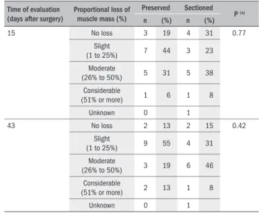
Table 2. Physiotherapeutic evaluation of the proportional loss of muscle mass according to whether the medial pectoral nerve was preserved or sectioned (n = 30)
Time of evaluation (days after surgery)
Proportional loss of muscle mass (%)
Preserved Sectioned
P (a)
n (%) n (%)
15 No loss 3 19 4 31 0.77
Slight
(1 to 25%) 7 44 3 23
Moderate
(26% to 50%) 5 31 5 38
Considerable
(51% or more) 1 6 1 8
Unknown 0 1
43 No loss 2 13 2 15 0.42
Slight
(1 to 25%) 9 55 4 31
Moderate
(26% to 50%) 3 19 6 46
Considerable
(51% or more) 2 13 1 8
Unknown 0 1
P-value, according to Fisher’s exact test.
Time of evaluation (days after surgery)
Proportional loss of strength (%)
Preserved Sectioned
P (a)
n (%) n (%)
15 No loss 7 43 1 7 0.15
Slight (1 to 25%) 1 6 2 14
Moderate
(26% to 50%) 2 13 2 14
Considerable
(51% or more) 6 38 9 64
Unknown 0 0
43 No loss 13 81 4 31 0.03
Slight (1 to 25%) 0 0 2 15
Moderate
(26% to 50%) 1 6 3 23
Considerable
(51% or more) 2 13 4 31
Unknown 0 1
(a) P-value, according to Fisher’s exact test.
Table 3. Physiotherapeutic evaluation of the proportional loss of muscle strength in the pectoralis major muscle at two different postsurgical times, according to whether the medial pectoral nerve was preserved or sectioned (n = 30)
RESULTS
All of the women completed the physiotherapy sessions up to 43 days after surgery. he two groups of women were comparable with re-gard to age, body mass index (BMI; kg/m2), total lymph nodes,
posi-tive lymph nodes, duration of surgery and number of physiotherapy sessions (Table 1).
More women in the group with preservation of the medial pecto-ral nerve sufered slight loss of muscle mass in the pectopecto-ralis major, as evaluated 15 and 43 days after mastectomy, compared with the group of women in whom this nerve was sectioned. However, these diferences were not statistically signiicant (Table 2).
Preservation of the medial pectoral nerve was signiicantly associat-ed with no loss of strength in the pectoralis major muscle 43 days after mastectomy, compared with the group of women in whom this nerve was sectioned. However, this diference had not been observed 15 days after surgery (Table 3). here were no signiicant diferences with regard to loss of range of shoulder lexion and abduction between the 15th and
43rd days following surgery, between the groups of women in whom the
medial pectoral nerve was preserved or sectioned (Tables 4 and 5).
DISCUSSION
he patients who underwent modiied radical mastectomy with preservation of the medial pectoral nerve presented signiicant reduc-tion in the loss of strength in the ipsilateral pectoralis major compared with the patients who underwent mastectomy with nerve sectioning. However, no diferences were found regarding atrophy of the pectora-lis major muscle or loss of range of movement in the homolateral up-per limb.
All the patients completed the physiotherapy program over the 43 days scheduled, making a total of 21 sessions. he results from the study
sessions and there were no losses from the follow-up. One woman alone, who was in the group of patients whose medial pectoral nerve was sec-tioned, failed to appear for the inal 43-day reevaluation visit, for per-sonal reasons.
he range of movement in the upper limb and the strength of the pectoralis major muscle were measured in accordance with the def-initions of good postural alignment and pure lexion and abduction, in which the patients were not allowed to compensate through other movements. his clearly hampered the procedure, particularly during the evaluation carried out on the 15th day following surgery, when the
recent scar tissue provoked signiicant pain and discomfort during the assessment.
sur-in which the muscle mass sur-in the pectoralis major muscle should be eval-uated in women in whom the medial pectoral nerve has been damaged has not yet been well established. A previous study that evaluated this relationship failed to describe how atrophy of the pectoral muscle was evaluated, although it was accepted that this occurs frequently when the pectoral nerve is sectioned during mastectomy.14
In our study, 4 out of the 20 women who had been randomized to the preservation group were excluded because of accidental injury of the medial pectoral nerve and the presence of lymph nodes adhering to it. It is important to emphasize that preservation of the medial pectoral nerve is not a simple procedure to perform. Although recommended by Patey,11
cases of accidental injury of this nerve must be taken into consideration. We were unable to ind any data in the literature on the frequency of such injuries, in order to make comparisons with the indings of the pres-ent study. he same type of lesion, whether accidpres-ental or not, may also occur in Madden’s mastectomy, in which both pectoral muscles are pre-served, and in quadrantectomy, during level II axillary dissection. Mer-son et al.14 indirectly evaluated the sectioning of the medial pectoral nerve
by sectioning the pectoralis minor muscle, and observing the associated complications. hey obtained signiicant data regarding the diferences in muscle mass between the two types of surgical technique studied.
here was no diference in the number of lymph nodes dissected be-tween the group in which the medial pectoral nerve was preserved and the group in which it was sectioned. Similar results were reported from the study carried out by Merson et al.,14 and these indings
con-irm the notion that preservation of the medial pectoral nerve is safe in oncological terms.
CONCLUSION
Preservation of the medial pectoral nerve, compared with section-ing the nerve, among women undergosection-ing modiied mastectomy, was feasible. Forty-three days after surgery, this technique resulted in small-er loss of muscle strength in the pectoralis major muscle, had no efect on muscle mass in the pectoralis major muscle, and did not cause any change in the range of movement of abduction or lexion of the homo-lateral upper limb.
REFERENCES
1. Aitken DR, Minton JP. Complications associated with mastectomy. Surg Clin North Am. 1983;63(6):1331-52.
2. Warmuth MA, Bowen G, Prosnitz LR, et al. Complications of axillary lymph node dissection for carcinoma of the breast: a report based on a patient survey. Cancer. 1998;83(7):1362-8. 3. Dasgupta S, Sanyal S, Sengupta SP. Transpectoral anterior approach to the axilla for lymph
node dissection in association with mastectomy preserving both pectoral muscles and their neurovascular bundles. Tumori. 1999;85(6):498-502.
4. Ververs JM, Roumen RM, Vingerhoets AJ, et al. Risk, severity and predictors of physical and psychological morbidity after axillary lymph node dissection for breast cancer. Eur J Cancer. 2001;37(8):991-9.
5. Torresan RZ, Cabello C, Conde DM, Brenelli HB. Impact of the preservation of the intercos-tobrachial nerve in axillary lymphadenectomy due to breast cancer. Breast J. 2003;9(5): 389-92.
6. Moosman DA. Anatomy of the pectoral nerves and their preservation in modiied mastec-tomy. Am J Surg. 1980;139(6):883-6.
7. Serra GE, Maccarone GB, Ibarra PE, de la Fuente R. Lateral pectoralis nerve: the need to preserve it in the modiied radical mastectomy. J Surg Oncol. 1984;26(4):278-81.
Time of evaluation (days after surgery)
Proportional loss of abduction (%)
Preserved Sectioned
P (a)
n (%) n (%)
15 No loss 1 6 0 0 0.28
Slight
(1% to 25%) 2 13 5 36
Moderate
(26% to 50%) 12 75 7 50
Considerable
(51% or more) 1 6 2 14
Unknown 0 0
43 No loss 1 6 0 0 0.43
Slight
(1% to 25%) 7 44 8 61
Moderate
(26% to 50%) 8 50 4 31
Considerable
(51% or more) 0 0 1 8
Unknown 0 1
P-value, according to Fisher’s exact test.
Table 4. Physiotherapeutic evaluation of the proportional loss of range of movement of the shoulder in abduction at two different postsurgical times, according to whether the medial pectoral nerve was preserved or sectioned (n = 30)
Time of evaluation (days after surgery)
Proportional loss of lexion (%)
Preserved Sectioned
P (a)
n (%) n (%)
15 No loss 1 6 0 0 0.80
Slight
(1% to 25%) 9 56 9 64
Moderate
(26% to 50%) 5 31 3 21
Considerable
(51% or more) 1 6 2 15
Unknown 0 1
43 No loss 0 0 1 8 0.67
Slight
(1% to 25%) 11 69 9 69
Moderate
(26% to 50%) 5 31 3 23
Considerable
(51% or more) 0 0 0 0
Unknown 0 1
P-value, according to Fisher’s exact test.
Table 5. Evaluation of the proportional loss of range of lexion of the shoulder at two different postsurgical times, according to whether the medial pectoral nerve was preserved or sectioned (n = 30)
day after surgery, due to scar dehiscence. In two cases, a silicone prosthe-sis was used, which also technically hampered palpation of the pectoralis major muscle. Moreover, these forms of palpation did not allow evalua-tion of the thickness of the pectoralis major muscle. Such measurement would have provided a better indication of loss of muscle mass. In fu-ture studies, the use of other methods such as magnetic resonance imag-ing should be considered, in order to be able to objectively evaluate the changes in muscle mass in these patients.
8. Lopchinsky RA. Locating the axillary vein and preserving the medial pectoral nerve. Am J Surg. 2004;188(2):193-4.
9. Loukas M, Louis RG Jr, Fitzsimmons J, Colborn G. The surgical anatomy of the ansa pectora-lis. Clin Anat. 2006;19(8):685-93.
10. Macchi V, Tiengo C, Porzionato A, et al. Medial and lateral pectoral nerves: course and branches. Clin Anat. 2007;20(2):157-62.
11. Patey DH. A review of 146 cases of carcinoma of the breast operated on between 1930 and 1943. Br J Cancer. 1967;21(2):260-9.
12. Goss CM. O sistema nervoso periférico. In: Goss CM, editor. Gray anatomia. Rio de Janeiro: Guanabara Koogan; 1977. p. 791-3.
13. Hoffman GW, Elliott LF. The anatomy of the pectoral nerves and its signiicance to the general and plastic surgeon. Ann Surg. 1987;205(5):504-7.
14. Merson M, Pirovano C, Balzarini A, et al. The preservation of minor pectoralis muscle in axillary dissection for breast cancer: functional and cosmetic evaluation. Eur J Surg Oncol. 1992;18(3):215-8.
15. Muscolino G, Leo E, Sacchini V, Bedini AV, Luini A. Resectable breast cancer: axillary
dissec-tion sparing pectoralis muscles and nerves. Eur J Surg Oncol. 1998;14(5):429-33.
Sources of funding: None
Conlict of interest: None
Date of irst submission: January 3, 2008
Last received: July 2, 2009
Accepted: July 13, 2009
Address for correspondence:
Cesar Cabello dos Santos Av. Eng. Carlos Stevenson, 885 Campinas (SP) — Brasil CEP 13092-132