erythrophagocytic particles in primary macrophages
Ines B. Santarino and Ot^ ılia V. VieiraCEDOC, NOVA Medical School, Faculdade de Ci^encias Medicas, Universidade NOVA de Lisboa, Portugal
Keywords
complement-opsonized red blood cells, aged red blood cells; erythrophagocytosis; IgG-opsonized Red Blood Cells; phagosomal maturation
Correspondence
O. V. Vieira, CEDOC, NOVA Medical School, Faculdade de Ciencias M^ edicas,
Universidade NOVA de Lisboa, 1169-056 Lisboa, Portugal
Fax: +351 218 803 010 Tel: +351 218 803 100 E-mail: [email protected]
(Received 31 January 2017, revised 19 June 2017, accepted 25 June 2017)
doi:10.1002/2211-5463.12262
Erythrophagocytosis is a physiological process that aims to remove dam-aged red blood cells from the circulation in order to avoid hemolysis and uncontrolled liberation of iron. Many efforts have been made to under-stand heme trafficking inside macrophages, but little is known about the maturation of phagosomes containing different types of erythrophagocytic particles with different signals at their surfaces. Therefore, we performed a comparative study on the maturation of phagosomes containing three dif-ferent models of red blood cells (RBC): aged/senescent, complement-opso-nized, and IgG-opsonized. We also used two types of professional phagocytes: bone marrow-derived and peritoneal macrophages. By compar-ing markers from different stages of phagosomal maturation, we found that phagosomes carrying aged RBC reach lysosomes with a delay com-pared to those containing IgG- or complement-opsonized RBC, in both types of macrophages. These findings contribute to understanding the importance of the different signals at the RBC surface in phagolysosome biogenesis, as well as in the dynamics of RBC removal.
Red blood cells/erythrocytes (RBC) undergo a matura-tion process in which the precursor cells lose their nuclei and organelles while accumulating hemoglobin. Mature normal human RBC remain in the circulation for roughly 120 days before removal. Aged/senescent RBC are phagocytized by macrophages of the reticu-loendothelial system, found in the spleen, liver, and bone marrow, through a process termed erythrophago-cytosis (EP) [1]. This highly controlled and coordi-nated process is responsible for RBC degradation within the phagolysosome resulting in the breakdown of hemoglobin and the release of heme into the cytosol where its catabolism takes place, leading to iron release [1]. The rapid removal of RBC from the circu-lation is vital for maintaining iron/heme homeostasis, as the majority of iron required to sustain erythro-poiesis is derived from senescent RBC and free iron is a strong pro-oxidant [2].
During aging, RBC undergo several metabolic and physical modifications such as (a) membrane vesicula-tion, disposing nonfunctional membrane patches [3]; (b) oxidation-induced modifications of hemoglobin and membrane protein band 3 [4]; (c) energy depletion [5]; (d) progressive cell shape transformation [6]; and (e) membrane remodeling, such as the exposure of sur-face removal markers like phosphatidylserine (PS) [7]. Directly or indirectly, these modifications trigger erythrophagocytosis.
It has been shown that the central step in the clear-ance of RBC relies on the interaction between macro-phage receptors and the protein band 3 that upon hemoglobin oxidation undergoes clustering, promoting the generation of epitopes on the RBC surface. This provides a signal favoring recognition of redistributed band 3 by autologous IgG [8,9]. Despite this, the immunoglobulins formed are not efficient opsonins,
Abbreviations
agRBC, aged red blood cells; BMDM, bone marrow-derived macrophages; EP, erythrophagocytosis; PM, peritoneal macrophages; PS, phosphatidylserine; RBC, red blood cells; shRBC, sheep red blood cell.
due to their low affinity and low circulation numbers. For this reason, it has been hypothesized that phago-cytosis of RBC can be enhanced by the activation of the classical complement pathway after IgG binding [10]. These immunoglobulins preferentially generate C3b2–IgG complexes in the presence of active comple-ment [11]. Furthermore, prior to senescence, RBC may enter eryptosis, a form of stress-inducing programmed cell death resembling apoptosis in nucleated cells. It is characterized by RBC shrinkage and translocation of PS from the inner leaflet to the outer leaflet of the membrane [12]. Despite some in vivo data showing PS exposure upon RBC aging, there is no convincing evi-dence for the involvement of PS in the physiological removal of aged RBC [13,14]. However, this mecha-nism remains inadequately defined. Recently, it was shown that senescent RBC are more prone to PS expo-sure than young RBC upon oxidation [15].
The bulk of the iron required to synthesize new hemoglobin is derived from senescent RBC that have undergone erythrophagocytosis; it is important to understand this process better. Furthermore, in some pathologies such as chronic kidney disease and sepsis, there is a decreased RBC lifespan and there is an increased eryptosis [16,17], making a detailed under-standing of the kinetics of erythrophagocytosis under different conditions important. Thus, it is fundamen-tal to define the mechanism of RBC clearance as well as the role of the different signals at the RBC sur-face, such as IgG, complement, and PS, in this pro-cess.
In this study, we aimed to address the interaction of different erythrophagosomes containing IgG- or com-plement-opsonized RBC or PS-enriched RBC (aged RBC) with components of the endocytic pathway, namely early endosomes and lysosomes. To do this, we used primary bone marrow-derived mouse macro-phages (BMDM) and peritoneal cavity macromacro-phages (PM). In both BMDM and PM, phagosomes contain-ing aged RBC mature slower than phagosomes con-taining the other RBC models and this information could offer an improved understanding of pathologies with changes in RBC clearance.
Materials and methods
Cell culture
The L929 cell line (kindly provided by Ira Tabas, Columbia University, NY, USA) was routinely cultured to produce L-cell conditioned media (LCCM) enriched in mouse col-ony-stimulating factor (M-CSF) to differentiate monocytes into macrophages, as previously described [18].
Bone marrow-derived macrophages (BMDM) and resi-dent peritoneal macrophages (PM) were obtained from eight- to ten-week-old C57BL/6 wild-type mice. BMDM were maintained for 7 days as described [19] but in RPMI-1640 medium containing 10% FBS, 1% P/S (Gibco, Gaitherburg, MD, USA), and 30% LCCM. PM were col-lected from the mice peritoneal cavity and were maintained in DMEM high glucose (Gibco) supplemented with 20% FBS and 1% P/S and then plated in glass coverslips. After 2-h incubation, nonadherent cells were removed and the adherent cells were washed with warm sterile PBS followed by replacement with fresh DMEM [19].
C57BL/6 mice were purchased from Charles River and were maintained according to the protocols approved by the national (Portuguese Official Veterinary Department; Direccß~ao Geral de Veterinaria) ethics committees according to the Portuguese (Decreto-Lei 113/2013) and European (Directive 2010/63/EU) legislations.
Preparation of the different phagocytic particles
Human blood was collected into sodium heparin tubes (Greiner Bio-One, Kremsm€unster, Austria) from healthy volunteers. Each volunteer signed a consent form approved by the Ethical Review Board of the NOVA Medical School, New University of Lisbon. Sheep blood was col-lected from healthy sheep in a slaughterhouse. All proto-cols were authorized by the national (Portuguese Official Veterinary Department, Direccß~ao Geral de Veterinaria) ethics committees according to Portuguese (Decreto-Lei 113/2013) and European (Directive 2010/63/EU) legislation. Both human and sheep RBC (shRBC) were isolated using the same protocol [20]. Briefly, RBCs were isolated using a Ficoll-Paque (GE Healthcare Life Sciences, Uppsala, Sweden) gradient. After centrifugation at 400gfor 30 min
at 4°C, RBC were located at the bottom of the centrifuge tube. Then, RBC were washed twice with PBS (137 mM NaCl, 2.7 mM KCl, 1.8 mM KH2PO4, 10 mM
NaH-PO4.2H2O, pH 7.4) and finally resuspended in PBS
supple-mented with 0.1% glucose (20% V:V). RBC in suspension were kept at 4°C and used as native RBC.
Aged RBC (agRBC) were prepared as described previ-ously [20]. Briefly, native RBC in PBS without glucose were incubated at 37 °C for 4 days.
IgG-opsonized RBC were prepared according to the fol-lowing protocol: Fresh shRBC were fixed with 4% paraformaldehyde/sucrose at room temperature (RT) for 2 h 30 min in an orbital rotator before IgG opsonization. IgG opsonization was performed overnight at 4°C with a rabbit anti-sheep RBC antibody (Sigma-Aldrich, Darmstadt, Germany) at a ratio of 1 : 50.
C3bi-opsonized shRBC were prepared according to the protocol previously described [21]. Briefly, a 10% shRBC suspension was mixed with PBS and 180 ngmL 1 rabbit
incubated at RT for 1 h in an orbital rotator. Next, IgM-shRBC were incubated with C5-deficient human serum (Sigma-Aldrich) at a ratio of 1 : 10 (v/v). The mixture was incubated for 20 min at 37°C in a thermomixer at 300 rpm. Under these conditions, the Fc region of the IgM pentamer activates the classical pathway of the complement cascade and deposits C3b to the IgM-shRBC where it is rapidly converted to C3bi.
Before phagocytosis and to allow visualization under the microscope, agRBC, IgG-opsonized RBC, and C3bi-RBC were labeled with the vital dye carboxyfluorescein succin-imidyl ester (CFSE) according to the CellTracerTM CFSE
Cell Proliferation Kit instructions. With IgG-opsonized RBC, these particles were incubated with CFSE prior to fixation with PFA.
Phagocytosis and phagosomal maturation assays
Before internalization of the different RBC models, phago-cytic cells were incubated at 4°C for 7 min with agRBC or IgG-opsonized particles and at 37°C for 20 min with C3bi-opsonized particles. This procedure is important because it allows different types of erythrophagocytic parti-cles to bind to receptors on the plasma membrane of phagocytes. Furthermore, it allows the synchronization of the phagocytic process, increasing the number of phago-somes formed during the pulse (engulfment) and then a more accurate analysis of phagolysosome formation. Next, macrophages incubated with agRBC and IgG-opsonized particles were transferred to 37°C for engulfment to occur. For complement-opsonized RBC, phorbol 12-myristate 13-acetate at a final concentration of 150 ngmL 1was added to allow engulfment.
Phagocytosis was performed during 15- and 20-min incu-bations (pulse time, 0-min chase time) for BMDM and PM, respectively. The duration of phagocytosis in the two types of macrophages is different because the phagocytic capacity of BMDM is higher than that of PM [22]. In all phagocytosis assays, four RBC were added per macro-phage. After phagocytosis, noninternalized RBC were removed by extensively washing or by hemolysis with deionized water. Then, the cells were incubated at 37°C for the time points referenced in the graphs abscissa (chase), to follow phagosomal maturation. Next, phagoso-mal maturation was assessed by following the interaction of the phagosomes formed only during the pulse (0-min chase time) with components of the endocytic pathway at different time points (chase times).
Immunofluorescence and microscopy
After the pulse-chase experiments, cells were fixed with 4% PFA for 30 min at RT. Early endosome antigen 1 (EEA-1) staining was performed with mouse anti-EEA-1 antibody (Sigma-Aldrich). The antibody was used at 1 : 50 dilution
for 1 h at RT in cells permeabilized with 0.1% Triton X-100/200 mM glycine in PBS for 30 min. Lysosomal-asso-ciated membrane protein 1 (LAMP-1) was visualized with a rat anti-LAMP-1 antibody (Developmental Studies Hybri-doma Bank, USA). The antibody was used at 1 : 50 dilu-tion for 2 h at RT, in cells fixed and permeabilized with methanol for 10 min. In both cases, after permeabilization and washing with PBS, blocking was performed with gela-tin from cold water fish (Sigma-Aldrich) for 30 min fol-lowed by incubation with the respective primary antibodies. Antibody-bound cells were finally incubated with Cy3 anti-mouse or anti-rat secondary antibodies for 1 h at RT at 1 : 500 dilution (Jackson ImmunoResearch, West Grove, PA, USA).
Stained samples were mounted with Mowiol/DABCO (Calbiochem, Darmstadt, Germany) and analyzed by using a laser scanning confocal microscope (Carl Zeiss, Jena, Ger-many,LSM510 software) with a 63x oil immersion objective N/A=1.30. Digital images were analyzed by LSM IMAGE BROWSERorIMAGE-Jsoftware. A phagosome was considered positive for a given marker when a fluorescent ring or a dot-ted patch was observed around the engulfed particle.
Statistical analysis
Statistical analysis (two-way ANOVA followed by Bonfer-roni post-test) was performed using the GRAPHPAD PRISM software version (GraphPad Software, Inc., La Jolla, San Diego, CA, USA). 5.0. P<0.05 (*) and P<0.001 (***) were considered to be statistically significant.
Results and Discussion
In this study, human and sheep RBCs were used to create different models of nonopsonized and opso-nized RBC. agRBC were used as a model of senes-cent/eryptotic cells for which the main signal for removal by primary phagocytes is the exposure of PS, as described before [20]. RBC were also opso-nized with C3bi to mimic complement-mediated ery-throphagocytosis and IgG to mimic Fc-mediated erythrophagocytosis, both opsonins being involved in the in vivo removal of senescent erythrocytes [23–25].
isolation procedures, the majority of in vitro studies of erythrophagocytosis have been performed in pri-mary cultures of mouse BMDM and PM [31–36].
Despite the fact that PM do not have a physiological role in the removal of senescent/damaged RBC, these phagocytic cells are among the best studied macro-phage populations in terms of cell biology and also in the identification of the molecular mechanisms involved in the removal of senescent RBC in vitro [31,33,37]. Furthermore, in this work, PM were also used as a model of resident macrophages. The peri-toneal cavity has two PM subsets: large periperi-toneal macrophages and small peritoneal macrophages (in-flammatory monocyte population) [38]. In our experi-mental conditions, the PM population was composed mainly of large PM as the experiments were not per-formed in elicited peritoneal macrophages.
Erythrophagocytosis is one of the main physiologi-cal processes responsible for the maintenance of iron homeostasis. In this process, after recognition by
specific receptors on the macrophage membrane sur-face, senescent RBC are internalized leading to the for-mation of a membrane-bound vacuole termed the erythrophagosome. It resembles the plasma membrane in terms of lipid and protein composition and its fluid-phase contents are a reflection of the extracellular milieu. These nascent phagosomes are not able to degrade cargo. However, soon after vacuole scission, there are a series of biochemical modifications that convert the nascent phagosome into a degradative organelle through an ordered choreographed succes-sion of events. These include membrane fissucces-sion and fusion of the nascent phagosomes with components of the endocytic pathway—a process known as
phago-some maturation [39–41].
To assess the initial steps of phagosomal matura-tion, specifically the interaction between the nascent phagosome and the early components of the endo-cytic pathway, we followed the recruitment of the Rab5-effector, EEA-1 (early endosome antigen-1), by
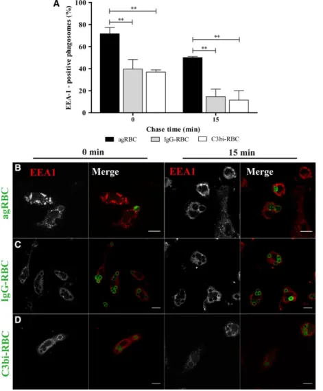
Fig. 1.Early endosome antigen 1 acquisition by phagosomal membranes containing different phagocytic particles in bone marrow-derived macrophages. The interaction of early endosomes with phagosomes containing different particles was assessed by the acquisition of EEA-1 (A–D). BMDM were exposed to different phagocytic particles for 15 min and then chased for the times indicated in the graph abscissa. After pulse-chase experiments, cells were fixed and stained with EEA-1 antibody and the positive phagosomes for the different chase time points were quantified in images acquired under a confocal microscope. (A) Quantification of the EEA-1-positive phagosomes. (B) EEA-1 staining of BMDM with agRBC-containing phagosomes. (C) EEA-1 staining of BMDM with IgG-RBC-containing phagosomes. (D) EEA-1 staining of BMDM with C3bi-RBC-containing phagosomes. In B–D, for each
immunofluorescence for all the phagocytic particles in BMDM. EEA-1 is responsible for tethering early endosomes to nascent phagosomes. As observed in Fig.1A, there was a decrease in EEA-1 association with all the RBC-containing phagosomes with time, suggesting that this Rab5 effector interacts transiently with the phagosomal membranes. The loss of EEA-1 in phagosomes containing opsonized RBC was faster than in phagosomes containing agRBC. At 15-min chase, a higher percentage of EEA-1-positive phago-somes was observed for phagosomes containing agRBC (50.070.92%) versus (14.67 6.70%) and (11.478.57%) for IgG-RBC and C3bi-RBC, respec-tively (Fig.1A and visualized in Fig.1B–D).
As the phagosomes mature, interaction with late components of the endocytic pathway such as late endosomes and lysosomes occurs, culminating in ery-throphagolysosome formation. RBC degradation takes place within this organelle, with subsequent trafficking
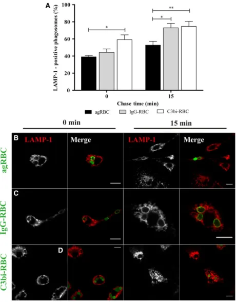
of heme to the cytosol. Interaction of phagosomes con-taining the different RBC models with the late endo-cytic compartment components was assessed by the acquisition of lysosomal-associated membrane protein (LAMP-1) in BMDM. At 15-min chase, LAMP-1 was present at higher levels in IgG-containing phagosomes (73.035.08%) and in C3bi-containing phagosomes (74.605.82%), compared to agRBC-containing phagosomes (52.704.45%), as shown graphically (Fig.2A) and illustrated in Fig.2B–D. Significant differences were also observed between agRBC- and C3bi-containing phagosomes (39.03 1.69% and 59.205.57%, respectively) for the same time point. These observations demonstrate that LAMP-1 acquisi-tion was faster in IgG- and C3bi-containing phago-somes and was well correlated with the fast loss of EEA-1 in these phagosomes (Fig.1A).
The kinetic profile for maturation of phagosomes containing different RBC in PM was followed as
Fig. 2.Lysosomal-associated membrane protein 1 acquisition by phagosomal membranes containing different phagocytic particles in bone marrow-derived macrophages. The interaction of the different phagosomes with late endocytic compartments (mainly late endosomes and lysosomes) was assessed by the acquisition of LAMP-1 (A–D).
BMDM were exposed to different phagocytic particles for 15 min and then chased for the times indicated in the graph abscissa. After pulse-chase experiments, cells were fixed and stained with LAMP-1 antibody and the positive phagosomes for the different chase time points were quantified in images acquired under a confocal microscope. (A) Quantification of the LAMP-1-positive phagosomes. (B) LAMP-1 staining of BMDM with agRBC-containing phagosomes. (C) LAMP-1 staining of BMDM with IgG-RBC-containing phagosomes. (D) LAMP-1 staining of BMDM with C3bi-RBC-containing phagosomes. In B–D, for each time point,
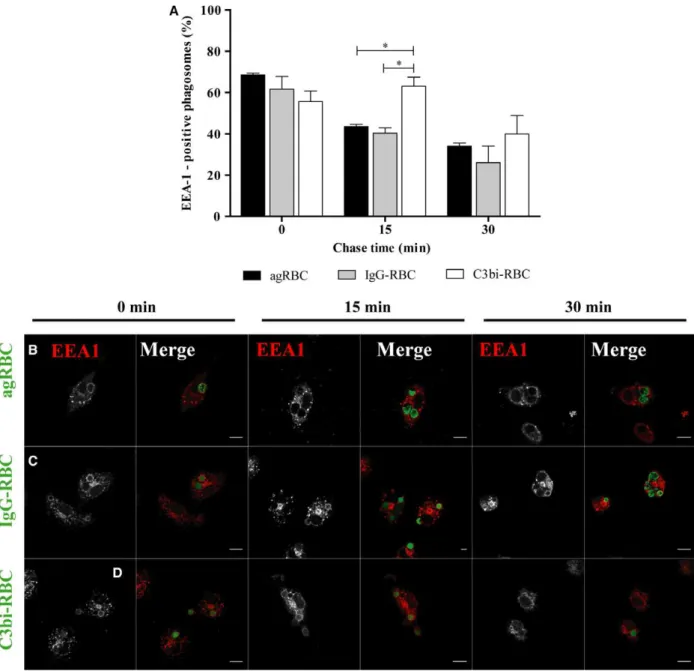
described above for BMDM. Again, the association of EEA-1 with phagosomal membranes was transient for the three different phagocytic particles (Fig.3A). However, EEA-1 remained associated with phago-somes containing C3bi-opsonized RBC for longer
periods of time compared with the other phagosomes and a significant statistical difference was observed for 15-min chase. Indeed, at 15-min chase time (63.10 4.33%), the phagosomes containing C3bi-opsonized RBC were still positive for EEA-1 in
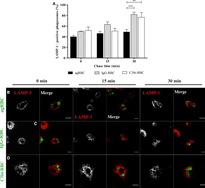
contrast with (43.51.03%) and (40.332.52%) for agRBC- and IgG-containing phagosomes, respectively (quantified in Fig.3A and visualized in Fig.3B–D). However, the delay in losing EEA-1 by the phago-somes containing C3bi-opsonized RBC did not have major consequences for the interaction of these phago-somes with late endocytic components (Fig.4A–D).
Similar to what was observed for BMDM, phagolyso-some formation was faster for opsonized RBC-con-taining phagosomes than for those conRBC-con-taining agRBC.
Comparing the results obtained in Figs2 and 4, phagolysosome biogenesis is slower in PM than in BMDM for all RBC particles. This outcome can explain, at least in part, the lower phagocytic capacity
of PM in comparison with BMDM as previously reported [42]. Indeed, during erythrophagolysosome biogenesis, besides membrane fusion with components of the endocytic pathway, membrane fission also occurs. This latter process is critical for recycling some phagosomal membrane components, such as phago-cytic receptors, back to the macrophage plasma mem-brane where they will participate in the engulfment of new RBC. Thus, a delay in phagolysosome biogenesis will affect phagocytosis.
In addition to their differing origins [38,43], BMDM and PM also have different gene expression profiles for the type and abundance of phagocytic receptors at their cell surfaces [44]. This might influence the homeo-static turnover of the different RBC models. For instance, BMDM express more high-affinity IgG bind-ing receptors than PM and the profile of the receptors that can be involved in the internalization of aged RBC is completely different [44].
Our results also showed that in both types of pri-mary macrophages, phagolysosome formation is faster for opsonized RBC-containing phagosomes than for those containing agRBC. These results are similar to those obtained by our group for smooth muscle cells [20], suggesting that RBC processing is conserved among professional and nonprofessional phagocytes. Although both types of macrophages have several PS receptors on their surface that could recognize agRBC, the PS receptor TIM-4 seems to be critical in the engulfment process for both type of macrophages. TIM-4 is highly expressed on PM [38] and is also the main receptor mediating phagocytosis of PS-bearing targets in BMDM [45]. This receptor is involved in phagosome stabilization via actin polymerization [46], which may explain why phagolysosome biogenesis of agRBC particles is slower than phagolysosome biogen-esis of opsonized RBC. Indeed, the interaction of the newly formed phagosomes with components of the endocytic pathway only occurs after actin depolymer-ization on the nascent phagosome. Therefore, inhibi-tion or a delay in actin depolymerizainhibi-tion around phagosomes will have a negative impact on phagolyso-some biogenesis. Furthermore, and in contrast with Fc- and complement-mediated phagocytosis, PS recep-tor-mediated phagocytosis is immunologically silent and induces an anti-inflammatory response [47]. This can also explain why phagolysosome biogenesis of agRBC is slower than opsonized RBC phagolysosome biogenesis. Indeed, as Fc and complement receptors are involved in the rapid removal and killing of bacte-ria from the circulation to avoid infection, it is to be expected that phagosomes whose formation depends on these receptors have to mature rapidly.
Additionally, phagocytosis through either of these receptors usually results in the formation of reactive oxygen species and pro-inflammatory cytokine secre-tion [48,49]. Finally, our data also demonstrate that different opsonins that interact with different phago-cytic receptors leading to different signaling responses do not result in major differences in terms of phagolysosome biogenesis in both types of primary macrophages.
In conclusion, this work identifies the differences involved in erythrophagocytosis of different in vitro RBC models in two different types of macrophages. Although the phagocytic cells and phagocytic particles of different origin could limit to some extent the con-clusions at the physiological level, our findings are a step forward in the identification of the molecular mechanisms of agRBC engulfment, which remain poorly understood.
Acknowledgements
We are grateful to ‘Matadouro da Beira Serra’ for providing the sheep blood for the experiments. We thank Dr. Sandra Gamboa and Dr. Fernando Delgado from ESAC for collecting sheep blood for the prelimi-nary experiments of this manuscript. We thank Win-chil Vaz and Mark Gibson for the critical reading of this manuscript. We also would like to thank Sara Silva for collecting human blood.
This work was supported by iNOVA4Health—UID/
Multi/04462/2013, a program financially supported by FCT through national funds and cofunded by FEDER under the PT2020 Partnership Agreement and FCT to OVV.
PhD fellowship reference: SFRH/BD/90258/2012.
Author contributions
OVV conceived and designed the project. IBS acquired the data. OVV and IBS analyzed and interpreted the results. OVV and IBS wrote the manuscript.
References
1 Bratosin D, Mazurier J, Tissier JP, Estaquier J, Huart JJ, Ameisen JC, Aminoff D and Montreuil J (1998) Cellular and molecular mechanisms of senescent erythrocyte phagocytosis by macrophages. A review. Biochimie80, 173
–195.
2 Soares MP and Hamza I (2016) Macrophages and iron metabolism.Immunity44, 492
–504.
Bosman GJCGM (2008) Erythrocyte vesiculation: A self-protective mechanism?Br J Haematol141, 549
–556. 4 Low PS and Kannan R (1989) Effect of hemoglobin
denaturation on membrane structure and IgG binding: role in red cell aging.Prog Clin Biol Res319, 525
–546. 5 Lang F, Gulbins E, Lang PA, Zappulla D and F€oller
M (2010) Ceramide in suicidal death of erythrocytes. Cell Physiol Biochem26, 21
–28.
6 Bosman GJCGM, Werre JM, Willekens FLA and Novotny VMJ (2008) Erythrocyte ageing in vivo and in vitro: structural aspects and implications for transfusion.Transfusion Med18, 335
–347.
7 Lang KS, Lang PA, Bauer C, Duranton C, Wieder T, Huber SM and Lang F (2005) Mechanisms of suicidal erythrocyte death.Cell Physiol Biochem15, 195–202. 8 Kay M (2005) Immunoregulation of cellular life span.
Ann N Y Acad Sci1057, 85
–111.
9 Lutz HU (2004) Innate immune and non-immune mediators of erythrocyte clearance.Cell Mol Biol (Noisy-le-Grand, France)50, 107–116.
10 de Back DZ, Kostova EB, van Kraaij M, van den Berg TK and van Bruggen R (2014) Of macrophages and red blood cells; A complex love story.Front Physiol5, 1
–11. 11 Lutz HU, Nater M and Stammler P (1993) Naturally
occurring anti-band 3 antibodies have a unique affinity for C3.Immunology80, 191–196.
12 Lang KS, Duranton C, Poehlmann H, Myssina S, Bauer C, Lang F, Wieder T and Huber SM (2003) Cation channels trigger apoptotic death of erythrocytes. Cell Death Differ10, 249
–256.
13 Bosman GJCGM (2013) Survival of red blood cells after transfusion: processes and consequences.Front Physiol4, 1
–8.
14 Franco RS, Puchulu-Campanella ME, Barber LA, Palascak MB, Joiner CH, Low PS and Cohen RM (2013) Changes in the properties of normal human red blood cells during in vivo aging.Am J Hematol88, 44
–51. 15 Ghashghaeinia M, Cluitmans JC, Akel A, Dreischer P,
Toulany M, Koberle M, Skabytska Y, Saki M,€
Biedermann T, Duszenko Met al.(2012) The impact of erythrocyte age on eryptosis.Br J Haematol157, 606–614.
16 Polak-Jonkisz D and Purzyc L (2013) Ca influx versus efflux during eryptosis in uremic erythrocytes.Blood Purif34, 209
–210.
17 Kempe DS, Akel A, Lang PA, Hermle T, Biswas R, Muresanu J, Friedrich B, Dreischer P, Wolz C, Schumacher Uet al.(2007) Suicidal erythrocyte death in sepsis.J Mol Med85, 269
–277.
18 Warren MK and Vogel SN (1985) Bone marrow-derived macrophages: development and regulation of differentiation markers by colony-stimulating factor and interferons.J Immunol134, 982–989.
19 Zhang X, Goncalves R, Mosser DM (2008) The isolation and characterization of murine macrophages.
Curr Protoc ImmunolUnit 14.1. doi: 10.1002/ 0471142735.im1401s83.
20 Viegas MS, Estronca LMBB and Vieira OV (2012) Comparison of the kinetics of maturation of phagosomes containing apoptotic cells and IgG-opsonized particles.PLoS One7, e48391.
21 Chow C-W, Downey GP and Grinstein S (2004) Measurements of phagocytosis and phagosomal maturation.Curr Protoc Cell Biol. Unit 15.7. doi: 10. 1002/0471143030.cb1507s22.
22 Linehan E, Dombrowski Y, Snoddy R, Fallon PG, Kissenpfennig A and Fitzgerald DC (2014) Aging impairs peritoneal but not bone marrow-derived macrophage phagocytosis.Aging Cell13, 699–708. 23 Lutz HU and Bogdanova A (2013) Mechanisms tagging
senescent red blood cells for clearance in healthy humans.Front Physiol4, 1
–15.
24 Lutz HU (2012) Naturally occurring anti-band 3 antibodies in clearance of senescent and oxidatively stressed human red blood cells.Transfus Med Hemotherapy39, 321
–327.
25 Kay MM (1981) Isolation of the phagocytosis-inducing IgG-binding antigen on senescent somatic cells.Nature
289, 491
–494.
26 Gottlieb Y, Topaz O, Cohen LA, Yakov LD, Haber T, Morgenstern A, Weiss A, Berman KC, Fibach E and Meyron-Holtz EG (2012) Physiologically aged red blood cells undergo erythrophagocytosis in vivo but not in vitro.Haematologica97, 994
–1002.
27 Lee SJ, Park SY, Jung MY, Bae SM and Kim IS (2011) Mechanism for phosphatidylserine-dependent erythrophagocytosis in mouse liver.Blood117,
5215–5223.
28 Fens MHAM, van Wijk R, Andringa G, van Rooijen KL, Dijstelbloem HM, Rasmussen JT, Vooght KMK, Schiffelers RM, Gaillard CAJM and Solinge WW (2012) A role for activated endothelial cells in red blood cell clearance: implications for vasopathology. Haematologica97, 500
–508.
29 Soares MP and Hamza I (2016) Macrophages and iron metabolism.Immunity44, 492–504.
30 Theurl I, Hilgendorf I, Nairz M, Tymoszuk P, Haschka D, Asshoff M, He S, Gerhardt LM, Holderried TA, Seifert Met al.(2017) On-demand erythrocyte disposal and iron recycling requires transient macrophages in the liver.Nat Med22, 945–951.
31 Bogdanova A, Mihov D, Lutz H, Saam B, Gassmann M and Vogel J (2007) Enhanced erythro-phagocytosis in polycythemic mice overexpressing erythropoietin. Blood110, 762
–769.
33 Shapiro S, Kohn D, Gershon H (1993) A role for complement as the major opsonin in the sequestration of erythrocytes from elderly and young donors.Br J Haematol83, 648–654.
34 Costa L, Moura E, Moura J and de Sousa M (1998) Iron compounds after erythrophagocytosis: chemical characterization and immunomodulatory effects. Biochem Biophys Res Commun247, 159
–165. 35 Bratosin D, Mazurier J, Tissier JP, Slomianny C,
Estaquier J, Russo-Marie F, Huart JJ, Freyssinet JM, Aminoff D, Ameisen JCet al.(1997) Molecular mechanisms of erythrophagocytosis. Characterization of the senescent erythrocytes that are phagocytized by macrophages.Comptes Rendus l’Academie des Sci–Ser III320, 811–818.
36 Packer BJ and Kreier JP (1986) Plasmodium berghei malaria: effects of acute-phase serum and erythrocyte-bound immunoglobulins on erythrophagocytosis by rat peritoneal macrophages. Infect Immun 51, 141
– 146.
37 Cassado AA, D’Imperio Lima MR and Bortoluci KR (2015) Revisiting mouse peritoneal macrophages: heterogeneity, development, and function.Front Immunol6, 1
–9.
38 Ghosn EEB, Cassado AA, Govoni GR, Fukuhara T, Yang Y, Monack DM, Bortoluci KR, Almeida SR, Leonard A, Herzenberg LAet al.(2010) Two physically, functionally, and developmentally distinct peritoneal macrophage subsets.Proc Natl Acad Sci USA107, 2568
–2573.
39 Vieira OV, Botelho RJ and Grinstein S (2002) Phagosome maturation: aging gracefully.Biochem J
366, 689
–704.
40 Flannagan RS, Jaumouille V and Grinstein S (2012) The cell biology of phagocytosis.Annu Rev Pathol7,
61–98.
41 Fairn GD and Grinstein S (2012) How nascent phagosomes mature to become phagolysosomes.Trends Immunol33, 397–405.
42 Wang C, Yu X, Cao Q, Wang Y, Zheng G, Tan TK, Zhao H, Zhao Y, Wang Y and Harris DCH (2013) Characterization of murine macrophages from bone marrow, spleen and peritoneum.BMC Immunol14, 6.
43 Yona S, Kim KW, Wolf Y, Mildner A, Varol D, Breker M, Strauss-Ayali D, Viukov S, Guilliams M, Misharin Aet al.(2013) Fate mapping reveals origins and dynamics of monocytes and tissue macrophages under homeostasis.Immunity38, 79
–91.
44 Ley K, Pramod AB, Croft M, Ravichandran KS and Ting JP (2016) How mouse macrophages sense what is going on.Front Immunol7, 1–15.
45 Flannagan RS, Canton J, Furuya W, Glogauer M and Grinstein S (2014) The phosphatidylserine receptor TIM4 utilizes integrins as coreceptors to effect phagocytosis.Mol Biol Cell25, 1511
–1522.
46 Mazaheri F, Breus O, Durdu S, Haas P, Wittbrodt J, Gilmour D and Peri F (2014) Distinct roles for BAI1 and TIM-4 in the engulfment of dying neurons by microglia.Nat Commun5, 1
–11.
47 Nairz M, Haschka D, Demetz E and Weiss G (2014) Iron at the interface of immunity and infection.Front Pharmacol5, 1–10.
48 Aderem A and Underhill DM (1999) Mechanisms of phagocytosis in macrophages.Annu Rev Immunol17,
593–623.
49 Ricklin D, Hajishengallis G, Yang K and Lambris JD (2010) Complement: a key system for immune surveillance and homeostasis.Nat Immunol11, 785