Universidade Nova de Lisboa
Instituto de Higiene e Medicina Tropical
Efflux pump activity in drug resistance of
Staphylococcus aureus
Sofia Maria Mourão Marques dos Santos Costa
DISSERTATION PRESENTED TO OBTAIN THE Ph.D. DEGREE IN BIOMEDICAL SCIENCES, SPECIALIZATION MICROBIOLOGY
Universidade Nova de Lisboa
Instituto de Higiene e Medicina Tropical
Efflux pump activity in drug resistance of
Staphylococcus aureus
Author: Sofia Maria Mourão Marques dos Santos Costa
Supervisor: Professor Isabel Couto
Tutorial Commission: Professor Isabel Couto Professor Miguel Viveiros Professor Filomena Pereira
Dissertation presented in fullfilment of the necessary requirements to obtain the Ph.D. degree in Biomedical Sciences, specialization Microbiology.
Thesis Publications
This Dissertation is based on original publications, listed here in chronological order:
Costa SS, Ntokou E, Martins A, Viveiros M, Pournaras S, Couto I, Amaral L. Identification of
the plasmid encoded qacA efflux pump gene in the methicillin-resistant Staphylococcus aureus
(MRSA) strain HPV107, a representative of the MRSA Iberian clone. Int J Antimicrob Agents.
2010;36:557-61.
Costa SS, Falcão C, Viveiros M, Machado D, Martins M, Melo-Cristino J, Amaral L, Couto I.
Exploring the contribution of efflux on the resistance to fluoroquinolones in clinical isolates of
Staphylococcus aureus. BMC Microbiol. 2011;11:e241.
Costa SS, Mourato C, Viveiros M, Melo-Cristino J, Amaral L, Couto I. Description of plasmid
pSM52, harboring the gene for Smr efflux pump, and its involvement in resistance to biocides
in a MRSA strain. Int J Antimicrob Agents. 2013;41:490-2.
Costa SS, Viveiros M, Amaral L, Couto I. Multidrug efflux pumps in Staphylococcus aureus:
an update. Open Microbiol J. 2013;7:59-71.
Costa SS, Junqueira E, Palma C, Viveiros M, Melo-Cristino J, Amaral L, Couto I. Resistance to
Acknowledgments
I would like to aknowledge all the people that made this Thesis possible.
To Professor Isabel Couto, for accepting me as her PhD student and providing the opportunity to work on such an interesting area of research. Most of all, I would also like to thank for all the guidance, teaching, support and patience during these last years of both the MSc and the PhD Thesis.
To Professor Miguel Viveiros, for all the great discussions that always pushed my work a step forward.
To Professor Leonard Amaral, for all the interesting talks on science. To Professor Filomena Pereira, for all the support during the work.
To Professor José Melo-Cristino. for providing the collection of Staphylococcus aureus
strains that allowed us to develop all the work presented in this Thesis.
To Professor Hermínia de Lencastre, for providing the Staphylococcus aureus strains HPV107 and RN4220.
To Professor Ricardo Parreira, for always being available to discuss with me RNA protocols and for all the help in the phylogenetic analysis.
To Professor João Piedade, for all the helpful discussions. To Professor Ilda Sanches, for access to PFGE facilities.
To Professor Stefan Scharwz for accepting me in his lab at the Institute of Farm Animal Genetics, Friedrich-Loeffler-Institut (FLI), and to Kristina Kadlec, for all the teaching and support during my visit.
To Professor Patrick Butaye, for the opportunity to work with Staphylococcus aureus of animal origin.
To my two Claudetes, the Master students Cláudia Mourato and Cláudia Palma as well to Elisabete Junqueira, for their contribution to the work developed in this Thesis. It was a pleasure working with the three of you.
To Celeste, who first characterized the clinical strains studied in this work.
To Clara Leandro and Francisco Dores, who carried out the adaptation and reversion of
the Staphylococcus aureus reference strain to ethidium bromide, the starting point of
this journey.
To Anna, for the English revision of this Thesis.
To all the colleagues, past and present, of the Micas team, Marta Martins, Liliana Rodrigues, Ana Martins, Diana Machado, Pedro Costa, Jorge Ramos, Ana Armada, Nádia Charepe, Susana Costa, Antónia Pinto, Carlos Serra, Vânia Silva, Lisa Machado, Pedro Cerca and Samuel Francisco, for all the friendship during these years at IHMT. To D. Fernanda for the precious help provided.
To all the colleagues of the Unit of Medical Microbiology, in particular Cristina Branco and Catarina Farinha, for all the friendship.
To all the special people I had the privilege to meet at IHMT, Ana Catarina, Gabriella, Joana, Daniela, Hanna, Zé João and Sofia, with whom I created a wonderful family of friends that is very dear to me.
To my dear friends Diana, Vera and Luís, Mónica, Denisa and João, and Américo, for all the support throughout this adventure and for all the encouraging afternoon talks.
To my “little” pearls, Susana, Sara, Sandra, Inês and Joana, for all the joy they bring to my life.
To my godparents, my oncles and my cousins for all the love and support that they gave me throughout these last years and for being so comprehensive about all my absences.
To my mother.
ix
Resumo
Efflux pump activity in drug resistance of Staphylococcus aureus
Sofia Maria Mourão Marques dos Santos Costa
Palavras-Chave: Staphylococcus aureus; bombas de efluxo; fluoroquinolonas; biocidas; multirresistência
Staphylococcus aureus é um importante agente patogénico, para o qual estão
descritos vários mecanismos de resistência a compostos antimicrobianos, sendo a resistência mediada por bombas de efluxo (MDR-EP) o menos bem caracterizado. Foi objectivo desta Dissertação avaliar a contribuição global destes sistemas de efluxo, em particular, de NorA, Smr e QacA, para a resistência a fluoroquinolonas e a outros compostos antimicrobianos em S. aureus.
Foi estudada uma colecção de isolados clínicos de S. aureus resistentes à ciprofloxacina e estirpes de referência. A actividade de efluxo foi avaliada por metodologias baseadas no transporte de brometo de etídeo (EtBr), substrato de EPs, e pela determinação da concentração mínima inibitória para substratos de MDR-EPs na presença/ausência de inibidores de efluxo. A informação obtida foi complementada pela análise por RT-qPCR da expressão de genes que codificam para as principais MDR-EPs de S. aureus e seus reguladores e pesquisa de mutações associadas aos principais fenótipos de resistência estudados.
x Em relação às bombas codificadas em plasmídeos, verificou-se que tanto QacA, codificada num plasmídeo associado à multirresistência, como Smr, codificada num plasmídeo sem genes de resistência adicionais, desempenham um papel importante na resistência a biocidas, podendo contribuir para a persistência e disseminação em ambiente hospitalar de estirpes resistentes a biocidas e potencialmente resistentes a antibióticos.
A caracterização global dos isolados de S. aureus de origem clínica revelou uma contribuição significativa do efluxo para a resistência aos compostos antimicrobianos, com a identificação de um grupo de isolados com actividade de efluxo aumentada e correlacionável com susceptibilidade reduzida a fluoroquinolones e biocidas. A incubação com inibidores de efluxo, em particular as fenotiazinas, promoveu uma redução dos níveis de resistência, sem contudo resultar na reversão do fenótipo de resistência. Os estudos de expressão génica não revelaram uma correlação directa entre maior actividade de efluxo e expressão génica, sugerindo que os isolados clínicos podem já estar adaptados para uma resposta por efluxo na presença de compostos nocivos. Observou-se ainda uma multiplicidade de respostas mediadas por efluxo aos diferentes substratos e suas concentrações, variáveis no padrão temporal de expressão génica, níveis de expressão e genes envolvidos.
Os resultados obtidos evidenciam que o efluxo constitui parte da resposta inicial da célula aos compostos antimicrobianos, a qual, no caso das fluoroquinolonas, é seguida pela aquisição de mutações nos genes alvo. Mostrou-se ainda que a pressão exercida por biocidas induz a resistência cruzada a fluoroquinolonas.
xi
Abstract
Efflux pump activity in drug resistance of Staphylococcus aureus
Sofia Maria Mourão Marques dos Santos Costa
Keywords: Staphylococcus aureus; efflux pumps; fluoroquinolones; biocides; multidrug resistance.
Staphylococcus aureus is a major human pathogen, for which several resistance
mechanisms have been described. Of these, resistance mediated by multidrug efflux pumps (MDR-EP) is characterized to a lesser extent. The aim of this Dissertation was to evaluate the overall contribution of MDR-EP, in particular, of NorA, Smr and QacA to
S. aureus resistance towards fluoroquinolones and other antimicrobials.
The collection studied comprised ciprofloxacin-resistant S. aureus clinical isolates plus reference strains was studied. Efflux activity was assessed by methodologies based on the MDR-EP substrate ethidium bromide (EtBr) and determination of minimum inhibitory concentrations of MDR-EP substrates in the presence/absence of efflux inhibitors. RT-qPCR was used to evaluate the expression level of MDR-EP genes and their regulators. Mutations associated with resistance were screened in genes of interest.
The relevance of NorA to antimicrobial resistance was analyzed in the S. aureus
stress response to EtBr. It was shown that the presence/withdrawal of this compound promoted an increase/decrease of norA expression level and subsequent reduction/increase of susceptibility towards fluoroquinolones, biocides and dyes. We also analyzed the norA alleles within the clinical isolates collection and found that norAI was the prevalent allele. The results obtained substantiated the intricate regulatory system of NorA, with the identification of multiple factors that contributed to the modulation of the norA gene and NorA activity.
xii and proliferation of biocide-resistant strains and potentially antibiotic-resistant strains in the hospital environment.
The characterization of the clinical isolates collection revealed efflux as an important component of resistance, with detection of a group of isolates with increased efflux activity, which could be correlated with increased resistance to fluoroquinolones, biocides and dyes. The efflux inhibitors tested, in particular phenothiazines, were efficient in reducing resistance levels, yet without promoting reversion of the resistance phenotype. Gene expression assays revealed no direct correlation between increased efflux activity and levels of gene expression, suggesting that clinical isolates may be primed to efflux noxious compounds. A multiplicity of efflux-mediated responses to different inducers and their concentrations was observed, varying in the temporal pattern of expression, genes overexpressed and levels of expression.
The results obtained support the role of efflux as a first-line response of S.
aureus to antimicrobial compounds, which in the case of fluoroquinolones, is followed
by occurrence of target-based mutations. Evidence was also obtained showing that the pressure exerted by biocides can lead to cross-resistance to fluoroquinolones.
xiii
Table of Contents
Thesis Publications………....iii
Acknowledgments………....vii
Resumo………..ix
Abstract……….xi
Index of Figures………xvii Index of Tables……….xx
List of Abbreviations………xxii
List of Units………..xxiv
CHAPTER 1. General Introduction ... 1
1.1. Staphylococcus aureus: the making of a major pathogen ... 3
1.2. Resistance to antimicrobial agents in S. aureus ... 8
1.2.1. Antibiotics: modes of action and mechanisms of resistance ... 8
1.2.2. Evolution of antibiotic resistance in S. aureus ... 10
1.2.3. Biocides: modes of action and mechanisms of resistance ... 12
1.2.4. Resistance to biocides in S. aureus ... 15
1.3. Multidrug efflux pumps ... 16
1.3.1. Major Facilitator Superfamily (MFS) ... 18
1.3.2. Small Multidrug Resistance (SMR) family ... 19
1.3.3. Multidrug and Toxic compound Extrusion (MATE) family ... 19
1.3.4. Resistance-Nodulation-Cell Division (RND) superfamily ... 20
1.3.5. ATP-Binding Cassette (ABC) superfamily ... 20
1.4. Multidrug efflux pumps in S. aureus ... 21
1.4.1. Chromosomally-encoded multidrug efflux pumps ... 21
1.4.2. Plasmid-encoded multidrug efflux pumps ... 27
1.4.3. Regulation of S. aureus multidrug efflux pumps ... 31
xiv 1.6. Efflux inhibitors: a pathway to circumvent the clinical impact of
efflux pumps ... 36
1.7. Dissertation Objectives and Outline ... 38
1.8. References ... 40
PART I. Characterization of the physiological role of
S. aureus
multidrug efflux pumps
CHAPTER 2. Revising the role of NorA ... 57
2.1. The role of NorA in the adaptative response of Staphylococcus aureus to stress stimuli ... 60
2.1.1. Introduction ... 60
2.1.2. Material and Methods ... 60
2.1.3. Results ... 66
2.1.4. Discussion ... 71
2.1.5. Conclusions ... 75
2.1.6. References ... 76
2.2. The genetic diversity of the norA gene ... 78
2.2.1. Introduction ... 78
2.2.2. Material and Methods ... 78
2.2.3. Results and discussion ... 80
2.2.4. Conclusions ... 90
2.2.5. References ... 91
CHAPTER 3. The role of QacA in efflux-mediated resistance ... 93
3.1. Identification of the plasmid encoded qacA efflux pump gene in the methicillin-resistant Staphylococcus aureus (MRSA) strain HPV107, a representative of the MRSA Iberian clone ... 96
3.1.1. Introduction ... 96
xv
3.1.3. Results and discussion ... 99
3.1.4. Conclusions ... 105
3.1.5. References ... 105
3.2. Role of pSM39, a multiresistance plasmid harboring the qacA gene for biocide resistance, on the resistance phenotype of a MRSA strain ... 107
3.2.1. Introduction ... 107
3.2.2. Material and Methods ... 108
3.2.3. Results... 110
3.2.4. Discussion and conclusions ... 117
3.2.5. Conclusions ... 120
3.2.6. References ... 121
CHAPTER 4. The role of Smr in efflux-mediated resistance ... 125
4.1. Introduction ... 127
4.2. Material and Methods ... 128
4.3. Results ... 132
4.4. Discussion ... 137
4.5. Conclusions ... 139
4.6. References ... 139
PART II. Efflux-mediated resistance to antimicrobial agents in
S.
aureus
CHAPTER 5. Resistance to fluoroquinolones mediated by efflux ... 143
5.1. Introduction ... 146
5.2. Material and Methods ... 147
5.3. Results ... 153
5.4. Discussion ... 161
5.5. Conclusions ... 165
xvi
CHAPTER 6. Resistance to antimicrobial compounds mediated by
efflux ... 169
6.1. Introduction ... 171
6.2. Material and Methods ... 173
6.3. Results and discussion ... 175
6.4. Conclusions ... 187
6.5 References ... 188
PART III. Efflux as an important player in the emergence of
antimicrobial resistance in
S. aureus
CHAPTER 7. Efflux as a first-line response to antimicrobial agents in
S. aureus
... 193
7.1. Introduction ... 195
7.2. Material and Methods ... 197
7.3. Results ... 197
7.4. Discussion ... 205
7.5. Conclusions ... 221
7.6. References ... 226
PART IV. General Discussion and Conclusions
CHAPTER 8. General Discussion ... 231
CHAPTER 9. Conclusions and Future Perspectives………...255
xvii
Index of Figures
Figure 1.1. Image of a S. aureus strain grown in blood-agar medium. ... 3
Figure 1.2. Map depicting the percentage of invasive MRSA isolates reported in 2011 by 28
of the European countries participating in the European Antimicrobial
Resistance Surveillance Network (EARS-Net). ... 7
Figure 1.3. Representation of the modes of action of antibiotics in the bacterial cell and the
main mechanisms of resistance to surpass their activity. ... 8
Figure 1.4. Representation of the five families of MDR efflux pumps and their distribution
among Gram-positive and Gram-negative bacteria. ... 17
Figure 1.5. Representation of the structural model of MFS transporters with 12 TMS and 14
TMS. ... 18
Figure 1.6. Representation of the structural model of a SMR transporter with four TMS. ... 19
Figure 1.7. Representation of the structural model of a MATE transporter with 12 TMS. ... 20
Figure 2.1. SmaI macrorestriction profiles of ATCC25923 and its derivatives,
ATCC25923EtBr and ATCC25923EtBr_rev for contamination control throughout the
processes of adaptation to EtBr and reversion. ... 66
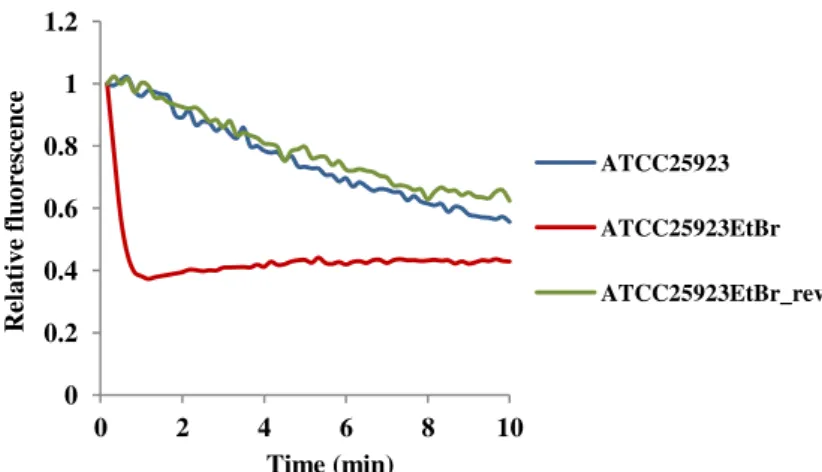
Figure 2.2. Assessment of efflux activity by real-time fluorometry for ATCC25923 and its
derivatives ATCC25923EtBr and ATCC25923EtBr_rev... 68
Figure 2.3. Quantification of the expression level of efflux pump genes and of the regulator
mgrA for the adapted ATCC25923EtBr and reverted ATCC25923EtBr_rev strains. ... 69
Figure 2.4. Alignment of the norA promoter region of ATCC25923 and its derivatives
ATCC25923EtBr and ATCC25923EtBr_rev strains. ... 70
Figure 2.5. Multiple alignment of norA sequences from S. aureus clinical strains,
ATCC25923 and norAI, norAII and norA1199. ... 81
Figure 2.6. Phylogenetic analysis of norA sequences… ... 83
Figure 3.1. Restriction products of the qacA/B internal fragment digested with AluI and
qacA/B amplification products from colonies selected after plasmid curing with
xviii
Figure 3.2. Analysis of ethidium bromide accumulation and efflux by a semi-automated
fluorometric method. ... 103
Figure 3.3. Assessment of EtBr efflux activity by real-time fluorometry of the strains
RN4220, SM39 and SM39cured. ... 113
Figure 3.4. Physical map and genetic organization of the S. aureus plasmid pSM39...115
Figure 3.5. Genetic organization of pSM39 and the location of the transposon Tn552 and of
the region with high homology with the S. epidermidis plasmid pSK105. ... 117
Figure 4.1. Physical map and genetic organization of the S. aureus plasmid pSM52. ... 133
Figure 4.2. Assessment of efflux activity by fluorometry of the strains SM52, RN4220 and
RN4220:pSM52. ... 136
Figure 5.1. Real-time EtBr accumulation/efflux for the representative strains ATCC25923
(reference), SM6 (EtBrCW-negative) and SM52 (EtBrCW- positive). ... 157
Figure 5.2. SmaI macrorestriction profiles of S. aureus clinical isolates. ... 159
Figure 6.1. Characterization of reference and clinical isolates according to their efflux
capacity. ... 177
Figure 6.2. Effect of the efflux inhibitors thioridazine and verapamil, at subinhibitory
concentrations, on the MIC values of ciprofloxacin and norfloxacin for
EtBrCW-positive, EtBrCW-intermediate and EtBrCW-negative groups. ... 180
Figure 6.3. Effect of the efflux inhibitors thioridazine and verapamil, at a subinhibitory
concentration, on the MIC values of ciprofloxacin and norfloxacin for the
EtBrCW-intermediate strain SM15. ... 181
Figure 6.4. Effect of the efflux inhibitors thioridazine and verapamil, at subinhibitory
concentrations, on the MIC values of several biocides for EtBrCW-positive,
EtBrCW-intermediate and EtBrCW-negative groups. ... 184
Figure 7.1. Diagram of the exposure processes subjected to the three strains in study and
discrimination of the phenotypic and genotypic characterization performed in
each step of the process. ... 201
Figure 7.2. SmaI macrorestriction profiles of the three S. aureus strains in study at the
xix
Figure 7.3. Evolution of the MIC values of ethidium bromide, ciprofloxacin and cetrimide
for the three strains in study throughout the 20-day exposure to ethidium
bromide, ciprofloxacin and cetrimide. ... 207
Figure 7.4. Assessment of EtBr efflux for the strains in study at the beginning and at the end
of the exposure process to ethidium bromide, ciprofloxacin and cetrimide. ... 215
Figure S1. Quantification of the expression of efflux pump genes and regulators by RT-qPCR
of strains ATCC25923, SM2 and SM50 at different time points of the exposure to
EtBr...264
Figure S2. Quantification of the expression of efflux pump genes and regulators by RT-qPCR
of strains ATCC25923, SM2 and SM50 at different time points of the exposure to
CIP...265
Figure S3. Quantification of the expression of efflux pump genes and regulators by RT-qPCR
of strains ATCC25923, SM2 and SM50 at different time points of the exposure to
xx
Index of Tables
Table 1.1. Examples of commonly used biocides, their modes of action and applications. ... 13
Table 1.2. Chromosomally-encoded MDR efflux pumps described so far in S. aureus. ... 22
Table 1.3. Plasmid-encoded MDR efflux pumps described so far in S. aureus. ... 28
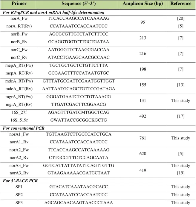
Table 2.1. Primers used in this study. ... 65
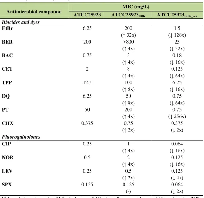
Table 2.2. MIC values of biocides, dyes and fluoroquinolones for ATCC25923 and its
derivatives ATCC25923EtBr and ATCC25923EtBr_rev... 67
Table 2.3. Characterization of the promoter and structural regions of the norA allelesof S.
aureus clinical isolates in study. ... 85
Table 2.4. Determination of the level of expression of the circulating norA alleles. ... 87
Table 2.5. Characterization of the S. aureus clinical isolates regarding their norA alleles,
QRDR mutations and MIC values for several known NorA substrates. ... 89
Table 3.1. Characterization of the S. aureus strain ATCC25923, MRSA strain HPV107, and
HPV107cured susceptibility profiles against antibiotics, biocides and dyes in the
absence and presence of efflux inhibitors. ... 100
Table 3.2. MIC values of biocides, dyes and antibiotics for the strains in study in the absence
and presence of efflux inhibitors. ... 111
Table 3.3. Description of the ORFs present in pSM39 and the best match obtained with
BlastP analysis. ... 114
Table 4.1. Description of the ORFs present in pSM52 and the best match obtained with the
BlastP analysis. ... 134
Table 4.2. MIC values of antibiotics, biocides and dyes for the strains in study in the absence
and presence of efflux inhibitors. ... 135
Table 5.1. Primers used in this study. ... 152
Table 5.2. Genotypic and phenotypic characterization of S. aureus clinical isolates... 153
Table 5.3. EP gene expression analysis by RT-qPCR of representative S. aureus exposed to
xxi
Table 7.2. List of primers used in this study. ... 204
Table 7.3. MIC values of antibiotics, biocides and dyes for the strain ATCC25923 at the
beginning and at the end of the exposure to ethidium bromide, ciprofloxacin and
cetrimide. ... 209
Table 7.4. MIC values of antibiotics, biocides and dyes for the strain SM2 at the beginning
and at the end of the exposure to ethidium bromide, ciprofloxacin and cetrimide.210
Table 7.5. MIC values of antibiotics, biocides and dyes for the strain SM50 at the beginning
and at the end of the exposure to ethidium bromide, ciprofloxacin and cetrimide.211
Table 7.6. Effect of the efflux inhibitor thioridazine on the MIC values of the three inducing
agents at the beginning and at the end of exposure for the three strains in study. . 213
Table 7.7. Gene overexpression of efflux pump genes and regulators by RT-qPCR of strains
ATCC25923, SM2 and SM50 at different time points of the exposure to EtBr. ... 217
Table 7.8. Gene overexpression of efflux pump genes and regulators by RT-qPCR of strains
ATCC25923, SM2 and SM50 at different time points of the exposure to CIP. .... 218
Table 7.9. Gene overexpression of efflux pump genes and regulators by RT-qPCR of strains
ATCC25923, SM2 and SM50 at different time points of the exposure to CET. ... 219
Table S1. MIC values of ciprofloxacin and norfloxacin for strains representative of the
EtBrCW-positive, EtBrCW-intermediate and EtBrCW-negative groups, in the
absence and presence of efflux inhibitors………262
Table S2. MIC values of ethidium bromide for strains representative of the EtBrCW-positive,
EtBrCW-intermediate and EtBrCW-negative groups, in the absence and presence of
efflux inhibitors………262
Table S3. MIC values of biocides for strains representative of the positive,
EtBrCW-intermediate and EtBrCW-negative groups, in the absence and presence of efflux
xxii
List of abbreviations
A – adenine
ABC –adenosine 5’-triphosphate (ATP)-binding cassette
ACR – acriflavine
Ala – alanine
BAC – benzalkonium chloride
BER – berberine
C – citosine
CA-MRSA – community-associated methicillin-resistant Staphylococcus aureus
CCCP – carbonyl cyanide m-chlorophenylhydrazone
cDNA – complementary DNA
CET - cetrimide
CHL – chloramphenicol
CHX – chlorhexidine diacetate
CHXg – chlorhexidine digluconate
CIP – ciprofloxacin
CoNS – coagulase-negative staphylococci
CPZ – chlorpromazine
CT– threshold cycle
CTAB – hexadecyltrimethylammonium bromide
CV- crystal violet
D/Asp – aspartic acid
Del – deletion
DMSO – dimetilsulphoxide
DNA – deoxyribonucleic acid
dNTP – deoxyribonucleoside triphosphate
DQ – dequalinium chloride
E/Glu – glutamic acid
ECOFF – epidemiological cut-off value
EI - efflux inhibitor
ERY – erythromycin
EtBr – ethidium bromide
F/Phe – phenylalanine
G – guanine
xxiii
HA-MRSA – hospital-associated methicillin-resistant Staphylococcus aureus
Ins – insertion
IS – insertion sequence
K/Lys – lysine
K - guanine / timine.
Lac – lactate
LA-MRSA – livestock-associated methicillin-resistant Staphylococcus aureus
LEV – levofloxacin
M - adenine / citosine
MATE – multidrug and toxic compound extrusion
MBC – minimum bactericidal concentration
MDR – multidrug resistance
MFS – major facilitator superfamily
MGE – mobile genetic element
MHB – Mueller-Hinton broth
MIC – minimum inhibitory concentration
MLST – multilocus sequence typing
MRSA – methicillin-resistant Staphylococcus aureus
MSSA – methicillin-susceptible Staphylococcus aureus
N/Asn – asparagine
NAL – nalidixic acid
NOR – norfloxacin
OD – optical density
ORF – open reading frame
OXA – oxacillin
P/Pro - proline
PBP – penicillin-binding protein
PBS – phosphate buffered saline
PCR – polymerase chain reaction
PEN – penicillin
PFGE – pulsed-field gel electrophoresis
PMF – proton motive force
PT – pentamidine isothionate salt
Q/Gln – glutamine
QAC – quaternary ammonium compounds
xxiv
RES – reserpine
RIF – rifampicin
RNA – ribonucleic acid
RND – resistance-nodulation-cell division
RT – room temperature
RT-qPCR – quantitative reverse transcription polymerase chain reaction
S/Ser – serine
SCCmec– staphylococcal cassette chromosome mec
SMR – small multidrug resistance
SPX – sparfloxacin
ST – sequence type
T – timine
TET – tetracycline
TMS – transmembrane segment
Tn – transposon
TPP – tetraphenylphosphonium bromide
TSA – tryptone soy agar
TSB – tryptone soy broth
TZ – thioridazine
UTR – untranslated region
VER – verapamil
VISA – vancomycin-intermediate resistant Staphylococcus aureus
VRSA - vancomycin-resistant resistant Staphylococcus aureus
W - adenine / timine
Y/Tyr – tyrosine
List of Units
bp – base pair; kb – kilobase; Mb – megabase
ºC – degrees Celsius
rpm – rotation per minute
msec – millisecond; sec – second;
min – minute; h – hour
nm – nanometer; µm – micrometer;
cm – centimeter
mL – milliliter; L – liter
µg – micrograma; mg – milligram
pmol – picomole
mM – millimolar; M - molar
cfu – colony forming unit
µF – microfaraday
kV – kilovolt
1
CHAPTER 1
2 1. General Introduction
1.1. Staphylococcus aureus: the making of a major pathogen………..3
1.2. Resistance to antimicrobial agents in S. aureus………...8
1.2.1. Antibiotics: mode of action and mechanisms of resistance………8
1.2.2. Evolution of antibiotic resistance in S. aureus………..10
1.2.3. Biocides: modes of action and mechanisms of resistance……….12
1.2.4. Resistance to biocides in S. aureus………...15
1.3. Multidrug efflux pumps………...………...16
1.3.1. Major Facilitator Superfamily (MFS)………...18
1.3.2. Small Multidrug Resistance (SMR) family………...19
1.3.3. Multidrug and Toxic compound Extrusion (MATE) family………...19
1.3.4. Resistance-Nodulation-Cell division (RND) superfamily………...20
1.3.5. ATP-Binding Cassette (ABC) superfamily………...20
1.4. Multidrug efflux pumps in S. aureus……….21
1.4.1. Chromosomally-encoded multidrug efflux pumps………21
1.4.2. Plasmid-encoded multidrug efflux pumps………...27
1.4.3. Regulation of S. aureus multidrug efflux pumps………...31
1.5. Additional efflux pumps in S. aureus……….35
1.6. Efflux inhibitors: a pathway to circumvent the clinical impact of efflux
pumps………......36
1.7. Dissertation Objectives and Outline………..38
3
1.1.
Staphylococcus aureus
: the making of a major pathogen
The species Staphylococcus aureus is one of the 48 species comprised in the
Staphylococcus genus, which was established in 1883 to describe a group of bacteria
responsible for inflammation and suppuration [48, 100]. During the 20th century, the classification criteria of this genus underwent several revisions [100]. Currently, it includes Gram-positive cocci with a diameter varying from 0.5 to 1.5 μm that can occur as single cells, in pairs, tetrads, short chains or as irregular grape-like clusters. These cocci are non-motile, non-sporogenic, and in some cases possessing a microcapsule. They are facultative anaerobes and producers of the enzyme catalase, with the exception
of Staphylococcus aureus subspecies anaerobius, Staphylococcus saccharolyticus and
some strains of Staphylococcus aureus subsp. aureus. S. aureus (Figure 1.1) is a producer of the enzyme coagulase, a trait traditionally used to differentiate it from other species, generally designated as coagulase-negative staphylococci (CoNS). Nonetheless, other species are also coagulase-positive, including Staphylococcus intermedius,
Staphylococcus pseudintermedius, Staphylococcus delphini and some strains of
Staphylococcus hyicus, among others [32, 100].
Figure 1.1. Image of a S. aureus strain
grown in blood-agar medium.
Staphylococci are ubiquitous in nature, although their presence is more common in the skin, skin glands and mucous membranes of mammals, in a permanent or transient status [32, 100]. Primates are the natural hosts
of S. aureus, although they can also be found
in domestic animals, poultry, hares and rodents [32].
4 individuals [101, 223]. It is recognized as a key factor for the development of S. aureus
infections, with the anterior nares being proposed as the potential starting point of propagation of S. aureus strains to other sites of the body like the skin. It has been also shown that persistent carriers have higher S. aureus loads and an elevated risk for acquisition of S. aureus infections [101, 223]. Although the majority of S. aureus
infections are attributable to colonizing strains, transmission can also occur, either directly, by skin-to-skin contact, or indirectly by contact with contaminated surfaces and objects [123].
Infections caused by this bacterium can take the form of mild superficial lesions, such as the skin and soft-tissue infections cellulitis and impetigo, more common in community-associated infections; life-threatening systemic infections like bacteremia, endocarditis, pneumonia and osteomyelitis, more common in nosocomial-associated infections; and toxin-mediated syndromes, such as food poisoning, scalded skin syndrome and toxic shock syndrome [32, 123]. S. aureus is a frequent primary pathogen [139]. In general, for an infection to take place, a first step of local colonization of the tissue surface occurs, followed by an invasion of the local tissue, an evasion from the host defense mechanisms and a final dissemination to other body sites [123]. S. aureus
elaborates a plethora of virulence factors that play an important role in the pathogenesis of the infection [32]. These virulence factors are involved in attachment (e.g., collagenase and clumping factors), in tissue invasion (e.g., proteases, lipases and hyaluronidase) and in evasion of the host defense mechanisms (e.g., leukocidins, like Panton-valentine leukocidin and γ-toxin, and protein A) [32, 43, 62]. Some strains can also secret additional virulence factors involved in toxin-mediated syndromes, such as the toxic shock syndrome toxin-1 (TSST-1), several enterotoxins (SEA, SEB, SECn, SED, SEE, SEG, SHE, and SEI) responsible for food poisoning and exfoliative toxins (ETA and ETB), responsible for the scalded skin syndrome [32, 43, 62]. Other virulence factors, such as coagulase, the arginine catabolic mobile element and bacteriocin have a poorly defined role in virulence and pathogenesis [62]. The S. aureus capacity to produce this array of virulence factors contrasts with the less virulent CoNS, some of which can cause opportunistic infections in humans.
5 Mb, have been made available (http://www.ncbi.nlm.nih.gov/genome). Comparative analysis of the earlier published genomes revealed that 75% of the genome, named
“core” genome, is conserved and mainly comprised of genes with housekeeping
functions, whereas the remaining 25%, the “accessory” genome, is variable and
constituted by mobile genetic elements (MGEs) [117]. These MGEs include integrated prophages, genomic islands, pathogenecity islands, staphylococcal cassette chromosomes (SCC), plasmids, transposons (Tn) and insertion sequences (IS), playing an important role in the pathogenesis, virulence, resistance and host-adaptation of S.
aureus given that they often carry virulence, toxin and resistance determinants [60, 130,
134].
Besides its pathogenic potential and virulence, S. aureus also shows a remarkable ability to develop and/or acquire a diversity of resistance mechanisms towards antibiotics and other antimicrobial compounds. Of major concern are the methicillin-resistant S. aureus (MRSA) strains, resistant to all β-lactam antibiotics. Since the description of the first MRSA strain, more than five decades ago, they have become a major cause of nosocomial infections worldwide [23, 40, 145]. These MRSA strains with onset in hospitals are denominated hospital-associated MRSA (HA-MRSA). MRSA infections are associated with an increased burden, adding to infections caused by methicillin-susceptible S. aureus (MSSA) [63, 102]. The association between MRSA bacteremia and higher mortality rates is still debatable [63, 102, 217]. However, there is strong evidence that MRSA infections are implicated in a higher morbidity [63] and higher hospital costs [63, 119]. More recently, MRSA strains with onset in the community have emerged, being designated community-associated MRSA (CA-MRSA). These strains were shown to be distinct from the traditional HA-MRSA strains; they belong to different clonal lineages, show diverse genetic backgrounds, and albeit
more susceptible to antibiotics other than β-lactams, they are generally more virulent
6 recent studies have reported that these CA-MRSA strains have acquired additional resistance determinants [138, 200]. More recently, new MRSA clones that are not related to contemporary CA-MRSA or HA-MRSA clones have emerged among livestock, being designated livestock-associated MRSA (LA-MRSA) [51]. Among these, a particular clone (ST398) was first detected in pig farmers and later found to be prevalent among pigs and in other food-producing animals like cattle and poultry [51]. Reports of the occurrence of this LA-MRSA clone in livestock have arisen from several countries, including cases of animal-to-person transmission [49]. This transmission was found to occur mainly in people in close contact with animals, like pig farmers and slaughterhouse workers, but these strains were found to be poor colonizers of humans and it was proposed that their carriage results from continued exposure rather than stable colonization [65]. Furthermore, they appear not to be associated with a high infectious risk for humans [51]. A recent study revealed a likely human ancestral origin for this clonal lineage as MSSA that experienced a human-to-livestock jump followed by host adaptation to livestock and acquisition of additional resistance determinants [172]. Of particular interest is the identification of new resistance determinants in these LA-MRSA strains [98, 221]. These findings highlight the potential role of S. aureus as a zoonotic pathogen and of animals as reservoirs for new MRSA strains [72, 172]. These LA-MRSA strains may also act as a reservoir for the emergence and dissemination of new resistance genes. Similarly, reports on the occurrence of MRSA strains in a variety of food samples like meat products, milk, fish and other food products have been published, but information is still scarce to support the role of MRSA as a potential food-borne pathogen [5, 222].
7
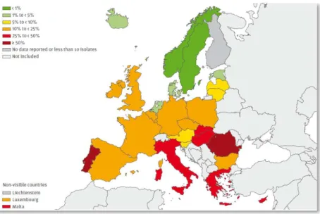
Figure 1.2. Map depicting the percentage (%) of invasive MRSA isolates reported in 2011 by 28 of the European countries participating in the European Antimicrobial Resistance Surveillance
Network (EARS-Net). Reproduced from [46].
Some European countries have been indicating a decreasing trend of invasive MRSA infections [46], probably supported by application of improved measures of control and prevention of MRSA in healthcare settings [102]. Yet, at least one third of the European countries remain with proportion rates above 25% [46, 102]. In addition, some evidence indicates that this decrease is not followed by the overall rates of S.
aureus infections, thus implying a rise in the number of MSSA infections [63].
8
1.2.
Resistance to antimicrobial agents in
S. aureus
1.2.1. Antibiotics: modes of action and mechanisms of resistance
Antibiotics are natural, semi-synthetic or synthetic compounds with selective toxicity towards bacteria, causing death or growth inhibition, thus allowing the host defense mechanisms to cope with the infection [28]. Antibiotics can exert their antibacterial action by (Figure 1.3-A); (i) the inhibition of nucleotide biosynthesis (e.g., sulfonamides); (ii) the inhibition of DNA synthesis (e.g., quinolones); (iii) the inhibition of RNA synthesis (e.g., rifamycin); (iv) the inhibition of protein synthesis (e.g., macrolides); (v) the inhibition of cell wall biosynthesis (e.g., β-lactams); and (vi) the disorganization of the cell membrane (e.g., peptides) [28].
Figure 1.3. Representation of the modes of action of antibiotics in the bacterial cell (A) and the
main mechanisms of resistance to surpass their activity (B).
Sulfonamides Trimethoprim DNA Replication Protein Synthesis Quinolones Nitrofurans Nitroimidazoles RNA Transcription mRNA Rifamycins Aminoglycosides Chloramphenicol Macrolides Tetracyclines Fusidic Acid Oxazolidinones Streptogramins Cell Wall Biosynthesis β-lactams Bacitracin Cycloserine Ethambutol Ethionamide Fosfomycin Glycopeptides Isoniazid Membrane integrity Peptides Nucleotide Biosynthesis Topoisomerase Efflux pump Drug-inactivating enzyme
Modified drug target
Modified cell wall protein
Alteration of drug target
Inhibition of drug uptake
Activation of drug efflux pumps Drug enzymatic inactivation
9 The introduction of antibiotics to treat infectious diseases was one of the hallmarks of 20th century medicine. Shortly after, the first antibiotic-resistant bacteria were described. Since then, the development of new antibiotics has been accompanied by an increase in antibiotic-resistant bacterial strains and diversity of the mechanisms used to surpass their effect. These mechanisms include (Figure 1.3-B); (i) enzymatic
degradation or modification of the antibiotic, exemplified by the action of β-lactamases
that cleave the β-lactam ring of β-lactam antibiotics and acetylation of
aminoglycosides by acetyltransferases, respectively [228]; (ii) modification of the target, such as the occurrence of mutations in the DNA gyrase and topoisomerase IV that diminishes their affinity to quinolones [112]; (iii) reduction of the intracellular concentration of the antibiotic, via its extrusion by efflux pumps and/or reduction of its entry by altered cellular permeability [108], (iv) protection of the target site, exemplified by the overproduction of peptidoglycan in S. aureus increasing the number of vancomycin target sites, thus trapping the antibiotic in the cell wall and preventing its action [122]. Nowadays, at least one mechanism of resistance is described for each class of commonly used antibiotics [34]. Specific physiological states can also influence the susceptibility of bacteria to antibiotics, such as biofilm formation, growth conditions that induce slow growth rates, as well as phenotypic variation within a bacterial population [78].
Some bacteria are intrinsically resistant to one or more classes of antibiotics, such as Pseudomonas aeruginosa, while others are naturally susceptible to almost all antibiotics, as in the case of S. aureus. Yet, bacteria that are initially susceptible to an antibiotic can develop and/or acquire resistance through spontaneous mutations in target genes or by the acquisition of exogenous genes [34]. These exogeneous genes are usually localized in MGEs, such as plasmids and transposons, and can be acquired by horizontal gene transfer, through transformation, conjugation or transduction processes [34]. Many bacterial species may show multidrug resistance (MDR) phenotypes, such
as S. aureus, vancomycin-resistant enterococci, Mycobacterium tuberculosis,
Escherichia coli, Acinetobacter baumanii, Klebsiella spp., Enterobacter spp., and P.
aeruginosa, which may be responsible for major outbreaks in the hospital and/or in the
10 1.2.2. Evolution of antibiotic resistance in S. aureus
In the pre-antibiotic era, the mortality rate associated with invasive infections of
S. aureus surpassed 80% [198]. In the early 1940s, the β-lactam antibiotic penicillin was
introduced in the clinical practice, registering a drastic reduction in the mortality rate associated with these infections. Nevertheless, penicillin-resistant strains emerged shortly after, first in hospitals and then in the community. Underlying this resistance was the acquisition of a plasmid-located gene encoding a penicillinase (β-lactamase), an
enzyme that degrades the β-lactam ring rendering the antibiotic inactive [122]. By 1948,
more than 50% of the S. aureusclinical isolates were β-lactamase producers, a number that increased significantly in the following years, currently reaching 80 to 90% of the total clinical isolates worldwide [40].
Following penicillin, new antibiotics were introduced in the clinical practice,
including aminoglycosides, new β-lactams, chloramphenicol, tetracycline and
macrolides. However, strains resistant to these agents emerged rapidly, as well as the first strains showing resistance to multiple antibiotics. Many of the resistance mechanisms to these antibiotics were encoded in MGEs, including large multiresistance plasmids [126].
To overcome resistance to penicillin, semi-synthetic β-lactam antibiotics were developed that were not susceptible to the action of β-lactamases, such as methicillin and oxacillin. However, resistance once again emerged swiftly and in the year following the introduction of methicillin in clinical practice were published the first reports of resistance in clinical strains [6, 87]. These MRSA strains emerged in the UK, but soon spread, first, to healthcare institutions in other European countries and, later on, around the world. The underlying mechanism of resistance to methicillin is the synthesis of an additional altered penicillin-binding-protein (PBP), PBP2a or PBP2’, that shows a
diminished affinity for β-lactam antibiotics [73, 216]. This additional PBP is coded by
the mecA gene, comprised in the mobile element staphylococcal cassette chromosome
mec (SCCmec) that integrates into the S. aureus chromosome at a specific site within
orfX gene that encodes a ribosomal methyltransferase [15, 35]. To date, eleven SCCmec
11 resistance determinants to other antibiotics, such as aminoglycosides, macrolides, lincosamides and streptogramins as well as to heavy-metals like mercury [40, 194]. Recently, a new SCCmec element was described carrying a divergent mec gene [194].
Fluoroquinolones were developed to treat Gram-negative infections, showing a narrow spectrum of activity against Gram-positive bacteria [80]. Although not generally used to treat S. aureus infections, fluoroquinolone resistance emerged rapidly in this bacterium, probably due to an extensive exposure in the hospital environment. Resistance originates through spontaneous mutations in the two targets, DNA gyrase and topoisomerase IV and by efflux [79]. An interesting aspect of fluoroquinolone resistance in S. aureus is the build up of evidence as a selective factor for MRSA strains [80].
12 other therapeutical alternatives are the glycylcycline tigecycline, the lipoglycopeptide telavancin and the cephalosporin ceftaroline. However, resistance to these agents has already been reported [56, 64], including cross-resistance between vancomycin and daptomycin [33]. The use of older antibiotics, alone or in combination, is also being pondered as an option for standard therapy, including quinupristin-dalfopristin, trimethoprim-sulphamethoxazole, chloramphenicol and tetracycline [56, 64].
1.2.3. Biocides: modes of action and mechanisms of resistance
Biocides are compounds that possess a broad spectrum of action and inhibit cell growth or promote cell death [135]. They are classified according to their applicability; as antiseptics, when for application on living tissues, such as the skin and mucuous membranes; as disinfectants, for the decontamination of inanimate surfaces; as preservatives, for use in consumer products [59, 135]. Their use is widespread in healthcare settings, playing an important role in the prevention and control of nosocomial infections. Historically, biocides have been in use for several centuries, as exemplified by vinegar for the cleansing of wounds. However, it was in the late 19th century and early 20th century that other agents were introduced, including chlorine-releasing agents, phenols, organomercurials, cationic compounds, such as the quaternary ammonium compounds (QACs), and chlorhexidine [179]. A large assortment of molecules is currently registered as biocides in the USA and Europe [191]. The last decades have also witnessed a massive increase in the use of these compounds in consumer products, animal husbandry and in several industries, intensifying the exposure of the general population to these agents [59].
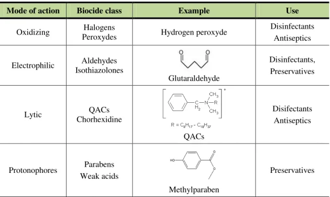
13 binding to cellular nucleophiles; as lytic (e.g., chlorhexidine and QACs), when they promote the destabilization of the cell membrane; or as protonophores (e.g., weak acids, parabens), which disturb the membrane pH gradient [24, 61, 146]. The activity of a biocide can be influenced by several factors, including its concentration, pH, temperature, the time of contact, formulation, presence of interfering material like organic matter, as well as the nature, load, localization and condition of the microorganisms upon which the biocide acts [178-181]. Biocides usually show low selectivity, acting against different types of microorganisms, with varying degree of activity against bacteria, spores, fungi, viruses and protozoa [181].
Table 1.1. Examples of commonly used biocides, their modes of action and applications.
Mode of action Biocide class Example Use
Oxidizing Halogens
Peroxydes Hydrogen peroxyde
Disinfectants Antiseptics
Electrophilic Aldehydes
Isothiazolones
Glutaraldehyde
Disinfectants, Preservatives
Lytic QACs
Chorhexidine
QACs
Disifectants Antiseptics
Protonophores Parabens
Weak acids
Methylparaben
Preservatives
QAC: quaternary ammonium compound.
14 resistance [191, 192]. These questions are accompanied by a difficulty in establishing cut-off values to distinguish between biocide-resistant and biocide-susceptible bacteria, with many authors preferring to apply the expressions reduced susceptibility or tolerance instead of resistance. In the context of an antibiotic, when measuring minimum inhibitory concentration (MIC) values to establish the susceptibility of a bacterium to a given antibiotic, this cut-off value is expressed as the breakpoint, specific for each combination of antibiotic and bacteria and above which bacterial strains are deemed resistant to that antibiotic, consequently treatment failure is anticipated [121]. In the context of biocides, not used therapeutically, many of the parameters establishing breakpoints are not applicable. Furthermore, biocides are usually used in formulations, at high-concentrations, with one or more active ingredients and excipients, which enhance their activity. They have an unspecific effect on the cell and are required to achieve a rapid killing of bacteria. Thus, the use of MIC values to reflect changes in the susceptibility of bacteria to biocides is considered not the most appropriate methodology and no guidelines have been established to differentiate resistance from susceptibility. However, many studies use MICs since these may be useful for a first assessment of the expected effect of a biocide on a given microorganism [18, 129, 180]. In the current year, tentative biocide epidemiological cut-off (ECOFF) values, based on MIC and minimum bactericidal concentrations (MBC), have been proposed for some bacterial species, including S. aureus, and a restricted number of biocides, namely benzalkonium chloride, chlorhexidine, triclosan and sodium hypochlorite [9].
15 been associated with the occurrence of mutations in the gene fabI encoding an enzyme involved in fatty acid biosynthesis in several bacteria, including S. aureus [16], E. coli
and M. tuberculosis (homolog inhA) [224].
In general, reduced susceptibility to biocides has been associated with chromosomally or plasmid-encoded efflux pumps that are capable of extruding a broad range of substances [168, 170]. For example, it has been shown that exposure to triclosan and household antimicrobial cleaning agents promoted an efflux-mediated reduced susceptibility to those biocides and several antibiotics, such as tetracycline, ciprofloxacin and trimethoprim in Gram-negative bacteria [29, 205]. Resistance to biocides may also emerge by acquisition of MGEs. A wide variety of plasmids have been identified carrying resistance determinants to cationic compounds and heavy-metals [168, 170, 177]. In the last two decades, plasmid-borne resistance to several biocides, including chlorhexidine, cetrimide, triclosan and benzalkonium chloride has been reported in both Gram-negative and Gram-positive bacteria [59, 168, 170].
1.2.4. Resistance to biocides in S. aureus
As a prevalent nosocomial pathogen, S. aureus is under constant exposure to antiseptics and disinfectants. Among standard actions for prevention of nosocomial infections, hand hygiene is of paramount importance. Hand formulations are mainly alcohol-based (ethanol and/or isopropanol), many times supplemented with other biocides, such as chlorhexidine, QACs, triclosan and povidone-iodine [227]. Adding up to these measures, guidelines for MRSA infection control generally rely on the use of biocides for the skin decolonization of patients [30].
16 isolates [14], and more recently in human clinical isolates [232], have the capacity to expel several biocides, like QACs, chlorhexidine, diamidines and dyes and have been correlated with reduced susceptibility towards those compounds [14, 170]. Of particular interest is the occurrence of these genes on multiresistance plasmids of S. aureus and other staphylococci that already convey resistance to several antibiotics and heavy-metals. In addition, all chromosomally multidrug resistance (MDR) efflux pumps described so far in S. aureus, including NorA, NorB, NorC, MepA and MdeA have the potential to extrude biocides. However, the assessment of their involvement in biocide resistance is more difficult to ascertain, since these chromosomal efflux pumps occur naturally and the evaluation of the role played by each individual pump is a complex task. Nevertheless, some studies have associated a biocide reduced susceptibility phenotype to the overexpression of the genes coding for these pumps [31, 83, 105].
1.3.
Multidrug efflux pumps
Efflux pumps are present in both eukaryotic and prokaryotic cells. The analysis of several published genomes reveals that drug efflux pumps may constitute 6% to 18% of all the transporters found in any bacterial cell [108]. The physiological role of these systems in bacteria has been related to the elimination of endogenous metabolites that are noxious to the cell, the secretion of virulence determinants, and in cell stress responses, suggesting that antimicrobial compounds are fortuitous substrates of these transporters [167, 171].
The first efflux systems to be associated with antibiotic resistance were identified in 1980 in tetracycline-resistant isolates of E. coli and corresponded to Tet proteins [137]. Since then, numerous efflux systems have been identified either in the chromosome or in plasmids of bacteria and associated with resistance to antibiotics and other antimicrobial agents [168, 170].
17 classes of antimicrobial compounds, being designated MDR efflux pumps. Concerning the energy source, efflux systems are divided in primary active transporters, which use the free energy of ATP hydrolysis to extrude compounds against a concentration gradient, exemplified by ABC transporters [103] and secondary active transporters that use the electrochemical potential gradient of the cytoplasmic membrane, the proton motive force (PMF), to drive the extrusion of their substrates by an antiport drug:H+ mechanism. This type of transporter includes pumps of the major facilitator superfamily (MFS) [52, 159, 182], the small multidrug resistance (SMR) family [7, 8], the multidrug and toxic compound extrusion (MATE) family [110] and the resistance-nodulation-cell division (RND) superfamily [154] (Figure 1.4). Some MATE transporters can also use the membrane gradient of sodium ions as energy source [110].
Gram-negative and Gram-positive bacteria differ in the organization and composition of their cell wall. Whilst Gram-negative bacteria possess two membrane layers spaced by the periplasm, Gram-positive bacteria have a single cell membrane. Thus, the majority of the efflux systems present in Gram-negative bacteria is a tripartite complex that may belong to the RND, ABC or MFS families (Figure 1.4) [173]. In Gram-positive bacteria, the efflux systems are composed of a single component that traverses the entire cell membrane and belong to the MFS, MATE, SMR or ABC families (Figure 1.4) [173].
Figure 1.4. Representation of the five families of MDR efflux pumps and their distribution among Gram-positive and Gram-negative bacteria.
Outer membrane
Periplasm
Inner membrane
Gram–negative bacteria
Cytoplasm A cr A A cr A M ac A M ac A Em rA Emr A Several antimicrobials H+ Macrolides H+ Nalidixic acid Novobiocin AcrB TolC TolC MacB TolC EmrB
RND ABC MFS
Several antimicrobials
ATP ADP + Pi ABC
H+/Na+ Aminoglycosides Fluoroquinolones Cationic compounds
H+
MATE
LmrA NorM NorA
MFS Fluoroquinolones Cationic compounds H+ Smr SMR Cationic compounds Gram–positive bacteria
18 1.3.1. Major Facilitator Superfamily (MFS)
The Major Facilitator Superfamily comprehends more than 1,000 transporters grouped in, at least, 59 families among prokaryotic and eukaryotic cells [52, 182]. They are associated with the transport via antiport or sinport of several molecules, including antimicrobials, sugars, intermediates of the Krebs cycle and oligosaccharides [159, 182]. Hydropathy analysis of the MFS members supported a further division of the transporters according to their number of transmembrane segments (TMS), either 12 or 14 (Figure 1.5) [163]. Representatives of this superfamily are the S. aureus efflux pumps QacA/B and NorA, E. coli YdiY and EmrB and Mycobacterium smegmatis LfrA [182].
Figure 1.5. Representation of the structural model of MFS transporters with 12 TMS (A) and 14
TMS (B). The conserved motifs and respective consensus sequence are represented by full circles. An
upper-letter case indicates an amino acid that occurs at a high frequency (> 70%) at a defined position, whereas a low-letter case indicates an amino acid that occurs at a low frequency (40-70%) at a defined position. The letter x stands for any amino acid [160, 173].
-NH2 lg x x x x x Pv x P G x L a x D r k G r x x x l x x l x G R q x x x g a a x g G x x G x x P Gx l G x x x P G L -COOH Out In Motif D2 Motif A
Motif B Motif C Motif G
-NH2 lD n x x v T vA l P G x L a x D r k G r x x x l x x l x G R q x x x g a a x g G x x G x x PG x l
wxW
lF x
lPvNgi xG x Dx L x lg xx x G a x v g x x l -COOH Out In Motif D1 Motif A
Motif B Motif C
Motif F
Motif H
Motif E
A
19 1.3.2. Small Multidrug Resistance (SMR) family
Transporters that belong to this family are small proteins (100-140 amino acids) that possess four TMS (Figure 1.6). They are responsible for the extrusion of lipophilic compounds, such as QACs, DNA intercalating dyes and some toxic metabolites, by an antiport mechanism [7, 8]. It has been suggested that these transporters may function as oligomeric complexes, inasmuch as some studies indicate that the E. coli pump EmrE functions as a tetramer [7, 127]. The efflux pumps Smr of S. aureus, Mmr of M.
tuberculosis, EmrE of E. coli and EbrA of Bacillus subtilis are representatives of this
family [7, 8].
Figure 1.6. Representation of the structural model of a SMR transporter with four TMS. The
conserved motifs and respective consensus sequence are represented by full circles An upper-letter case indicates an amino acid that occurs at a high frequency (> 70%) at a defined position, whereas a low-letter case indicates an amino acid that occurs at a low frequency (40-70%) at a defined position. The letter x stands for any amino acid [160, 173].
1.3.3. Multidrug and Toxic compound Extrusion (MATE) family
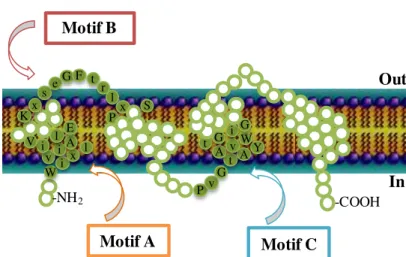
This family of transporters has been identified only recently hence it is not fully characterized. Sequence analysis demonstrates that bacterial MATE transporters possess a membrane topology similar to MFS transporters, with 12 TMS (Figure 1.7) [110]. They perform the extrusion of several antimicrobials by an antiport mechanism using either the proton or the sodium membrane gradient [110]. Members of this family are
-NH2 Wi x l v i A i ll E V Kx se G F t
20 the efflux pumps MepA of S. aureus, NorM of Vibrio parahaemolyticus, YdhE of E. coli, PmpM of P. aeruginosa and NorM of Neisseria meningitidis [110, 168].
Figure 1.7. Representation of the structural model of a MATE transporter with 12 TMS. There are
no conserved motifs identified to date for MATE transporters, but highly conserved sequences are represented by full circles [156].
1.3.4. Resistance-Nodulation-Cell division (RND) superfamily
RND transporters are usually found in Gram-negative bacteria, where they function as a tripartite complex [154]. The RND transporter is located in the inner membrane, presenting 12 TMS, and its periplasm domain associates with an outer membrane protein with aid of an adapter, a membrane fusion protein [154]. Some members of this superfamily are the AcrAB-TolC of E. coli, MexAB-OprM of P.
aeruginosa and MtrCDE of Neisseria gonorrhoeae [168].
1.3.5. ATP-Binding Cassette (ABC) superfamily
The ABC superfamily comprises a large group of transporters found in both prokaryotic and eukaryotic cells that explore the free energy of the ATP hydrolysis to energize the transport of multiple substrates [103]. Examples of the bacterial representatives of the ABC superfamily are the efflux pumps Msr(A) and AbcA of S.
aureus, LmrA of Lactococcus lactis, VcaM of Vibrio cholerae and EfrAB of E. faecalis
[168].
-NH2
-COOH
Out
21
1.4.
Multidrug efflux pumps in
S. aureus
Efflux-mediated resistance in S. aureus has been mainly associated with resistance towards fluoroquinolones and cationic compounds usually used as biocides [166, 170]. MDR efflux pumps have been identified in the chromosome and on plasmids of S. aureus. They all have the capacity to extrude a diverse array of antimicrobials, but while the majority of the chromosomally-encoded MDR efflux pumps extrude both fluoroquinolones and cationic compounds, plasmid-encoded MDR efflux pumps expel only cationic compounds.
Multidrug resistance is usually defined as resistance to at least one agent of three or more classes of antibiotics [128]. Though, due to the wide array of antimicrobial agents other than antibiotics that bacterial MDR efflux pumps can extrude, in this Thesis, we will use a broader definition for multidrug resistance, considering it as resistance to three or more classes of antimicrobial agents.
1.4.1. Chromosomally-encoded multidrug efflux pumps
To date, nine chromosomal MDR efflux pumps have been identified for S.
aureus (Table 1.2). In general, their substrate specificity is somewhat superimposed, all
Table 1.2. Chromosomally-encoded MDR efflux pumps described so far in S. aureus.
Efflux pump Familya Regulator(s)b Substrate specificity c References
NorA MFS MgrA, NorG(?) Fluoroquinolones (hydrophilic)
QACs (e.g. TPP, benzalkonium chloride); Dyes (EtBr, rhodamine) [88, 152, 233]
NorB MFS MgrA, NorG Fluoroquinolones (hydrophilic and hydrophobic); Tetracycline
QACs (e.g. TPP, cetrimide); Dyes (e.g. EtBr) [207]
NorC MFS MgrA(?), NorG Fluoroquinolones (hydrophilic and hydrophobic)
Dyes (rhodamine) [208, 213]
MepA MATE MepR Fluoroquinolones (hydrophilic and hydrophobic); Glycylcyclines (tigecycline)
QACs (e.g. TPP, cetrimide, benzalkonium chloride); Dyes (EtBr) [96, 133]
MdeA MFS n.i. Fluoroquinolones (hydrophilic); virginiamycin, novobiocin, mupirocin, fusidic acid
QACs (e.g. TPP, benzalkonium chloride, dequalinium); Dyes (EtBr) [82, 231]
SepA n.d. n.i. QACs (e.g. benzalkonium chloride); Biguanidines (chlorhexidine); Dyes (acriflavine) [149]
SdrM MFS n.i. Fluoroquinolones (hydrophilic)
Dyes (EtBr, acriflavine) [230]
LmrS MFS n.i.
Linezolide; Phenicols (chloramphenicol, florfenicol); trimethoprim, erythromycin, kanamycin, fusidic acid
QACs (e.g. TPP); Detergents (sodium docecyl sulphate); Dyes (EtBr)
[50]
NorD MFS n.i. Not identified to date, but presumed to be similar to other Nor efflux pumps [42]
a


![Figure 1.7. Representation of the structural model of a MATE transporter with 12 TMS. There are no conserved motifs identified to date for MATE transporters, but highly conserved sequences are represented by full circles [156]](https://thumb-eu.123doks.com/thumbv2/123dok_br/15770231.641211/46.892.163.765.228.445/representation-structural-transporter-conserved-identified-transporters-conserved-represented.webp)