online | memorias.ioc.fiocruz.br
Outbreaks of acute Chagas disease related to the acci-dental ingestion of food contaminated with Trypanosoma cruzi have been registered in several Brazilian regions, such as in Teutônia, Rio Grande do Sul, (Nery-Guimarães et al. 1968, Neves da Silva 1968), Catolé do Rocha, Paraí-ba (Shikanai-Yasuda et al. 1991) and the Brazilian Ama-zon (Valente et al. 1999, 2009, Pinto et al. 2001).
In 2005, in the Brazilian state of Santa Catarina (SC), an outbreak of acute Chagas disease was associated with the ingestion of sugar cane juice, which was likely con-taminated with T. cruzi by a naturally infected insect vec-tor during its preparation (Steindel et al. 2008). All the infected people had acquired the infection at the same municipality of Navegantes and the commonality between
individuals was the ingestion of sugar cane juice at the same time and place. Twenty-four people were infected and developed acute Chagas disease, three cases of which were fatal. A total of nine T. cruzi strains were isolated through haemoculture from the infected individuals by Dr Mario Steindel [Microbiology and Parasitology Division, Federal University of Santa Catarina (UFSC), Florianó- polis, SC] who had also isolated T. cruzi from one Triatoma tibiamaculata collected in the neighbourhood and from marsupials (Didelphis aurita and Didelphis marsupialis) captured in the same area. A molecular and isoenzymic study of the isolated strains was initially performed by Steindel et al. (2008). These authors detected the presence of T. cruzi II (Anonymous 1999) in the human isolates, double infection with T. cruzi I and T. cruzi II in the tri-atomine and T. cruzi I in the marsupials.
Our laboratory (Gonçalo Moniz Research Centre-Fiocruz, state of Bahia) received the isolated strains in liver infusion tryptose (LIT) culture media from Dr Ma-rio Steindel in 2006. All the isolates were submitted to biological characterisation by inoculation into mice to evaluate their behaviour in an experimental vertebrate host and to determine their isoenzymic patterns (Miles et al. 1980, Andrade et al. 1983, Andrade & Magalhães Financial support: FAPESB (to SGA), FAPEMIG, CNPq, CAPES
(to AMM)
† In memoriam
+ Corresponding author: [email protected] Received 25 April 2011
Accepted 22 September 2011
Biological, biochemical and molecular features of
Trypanosoma cruzi
strains isolated from patients infected through oral transmission
during a 2005 outbreak in the state of Santa Catarina, Brazil:
its correspondence with the new
T. cruzi
Taxonomy Consensus (2009)
Sonia Gumes Andrade1/+, Rozália Figueira Campos1,2, Mario Steindel3,
Marcos Lázaro Guerreiro1, Juracy Barbosa Magalhães1,†, Marcio Cerqueira de Almeida1,
Joice Neves Reis4, Viviane Corrêa Santos5, Helder Magno Silva Valadares5,7,
Mitermayer Galvão dos Reis6, Andréa Mara Macedo5
1Laboratório de Chagas Experimental, Autoimunidade e Imunologia Celular 4Laboratório de Epidemiologia Molecular 6Laboratório de Patologia Celular e Molecular, Centro de Pesquisas Gonçalo Moniz-Fiocruz, Salvador, BA, Brasil 2Universidade Estadual de Feira de Santana, Feira de Santana, BA, Brasil 3Departamento de Microbiologia e Parasitologia,
Universidade Federal de Santa Catarina, Florianópolis, SC, Brasil 5Departamento de Bioquímica e Imunologia,
Universidade Federal de Minas Gerais, Belo Horizonte, MG, Brasil 7Universidade Federal de São João Del-Rey, Divinópolis, MG, Brasil
We examined strains of Trypanosoma cruzi isolated from patients with acute Chagas disease that had been acquired by oral transmission in the state of Santa Catarina, Brazil (2005) and two isolates that had been obtained from a marsupial (Didelphis aurita) and a vector (Triatoma tibiamaculata). These strains were characterised through their biological behaviour and isoenzymic profiles and genotyped according to the new Taxonomy Consensus (2009) based on the discrete typing unities, that is, T. cruzi genotypes I-VI. All strains exhibited the biological behaviour of biodeme type II. In six isolates, late peaks of parasitaemia, beyond the 20th day, suggested a double infection with biodemes II + III. Isoenzymes revealed Z2 or mixed Z1 and Z2 profiles. Genotyping was performed using three poly-morphic genes (cytochrome oxidase II, spliced leader intergenic region and 24Sα rRNA) and the restriction fragment length polymorphism of the kDNA minicircles. Based on these markers, all but four isolates were characterised as T. cruzi II genotypes. Four mixed populations were identified: SC90, SC93 and SC97 (T. cruzi I + T. cruzi II) and SC95 (T. cruzi I + T. cruzi VI). Comparison of the results obtained by different methods was essential for the correct iden-tification of the mixed populations and major lineages involved indicating that characterisation by different methods can provide new insights into the relationship between phenotypic and genotypic aspects of parasite behaviour.
1997). The studies revealed that all the samples exhibit-ed the biological patterns of biodeme type II (Andrade et al. 2006b) presumably corresponding to T. cruzi II major lineages (Anonymous 1999).
In 2009, during a commemorative event for the cen-tenary of the discovery of Chagas disease, a new consen-sus for T. cruzi subdivision into major groups was estab-lished. Six discrete typing units (DTUs) (Tibayrenc 1998, 2003, Brisse et al. 2000) were formally recognised and characterised as the genotypes T. cruzi I through T. cruzi VI (Zingales et al. 2009). The epidemiological relevance of the six DTUs is not completely established, but at least four of the genotypes (T. cruzi I, II, V and VI) appear to be primarily involved with human disease. T. cruzi III and IV are rarely isolated from humans, but these genotypes, along with T. cruzi I, have been implicated in outbreaks of orally transmitted, acute cases of Chagas disease, es-pecially in the Amazon Basin (Valente et al. 2009).
The present report is an attempt to compare the results obtained from the former characterisation of the strains isolated from the outbreak of Chagas disease due to oral infection in 2005 (Andrade et al. 2006b) to a new set of recently obtained data, using biological (biodemes) (An-drade 1974, 1985, An(An-drade & Magalhães 1997), biochemi-cal (zymodemes) (Miles et al. 1980, Andrade et al. 1983) and molecular approaches. The molecular approaches were targeted to both the kinetoplast [maxicircle and minicircle restriction fragment length polymorphism(RFLP) analy-ses] and nuclear [internal transcribed spacer (ITS)
splice-leader and 24Sα rRNA gene analyses] genomes. The main
goal of this broad study was not only to determine the ma-jor genotypes of the strains in accordance with the new classification (Zingales et al. 2009) but also to characterise the molecular variability of the samples, identifying poly-clonal or mixed populations and correlating the findings to the biological behaviour of the strains.
SUBJECTS, MATERIALS AND METHODS
Biodemes characterisation - T. cruzi strains - A total of 11 strains were studied as follows: nine strains (SC94, SC95, SC96, SC97, SC98, SC99, SC100, SC101, SC102) isolated by haemoculture from acute human cases of Chagas disease acquired by oral transmission after the ingestion of a contaminated sugar cane juice in Nave-gantes (Steindel et al. 2008), one strain (SC90) isolated from an oposum (Didelphis aurita) and one strain (SC93) from a triatomine (T. tibiamaculata), both from the same area as the disease outbreak. The cultures were received from the Protozoology Laboratory of UFSC and kindly provided by Dr Mario Steindel.
Inoculation in mice - The culture forms in LIT medium were submitted to three successive washing in phosphate buffered saline (PBS), pH 7.2, under centrifugation at 750 g. After resuspension in PBS, pH 7.2, the pellet was exam-ined in a Neubauer chamber and the number of metacyclic forms in 1 µL was evaluated. For mice in the first passage group, 104-204 metacyclic forms were used as the inocula.
Experimental groups - For each strain, blood forms obtained from the first passage group were intraperito-neally inoculated into 60 Swiss mice weighing 15-20 g.
The behaviour of parasites was evaluated in the second and fifth passages in mice. Inoculum: 5 x 10⁴ blood trypomastigotes.
Parasitaemia - Parasitaemia was evaluated daily in a sample of five mice per experimental group by count-ing the number of trypomastigotes in 5 µL of peripheral blood in 50 microscopic fields (400X).
Cumulative mortality - The cumulative mortality was registered and expressed as the percentage of dead ani-mals for each group until 30 days after the initial infection, excluding those killed for the histopathological study.
Histopathology - Three mice were killed at the fol-lowing time points: the seventh, 10th, 14th, 20th and 30th days after infection. Tissue sections of the heart, skeletal muscles, liver and spleen were fixed in 10% for-malin and embedded in paraffin and 5-µm sections were stained with haematoxylin-eosin.
Isoenzymic characterisation - The strains were culti-vated into Warren medium and the isoenzymic extracts were obtained according to Miles et al. (1980). The en-zymes that distinguish biodemes types I, II and III (An-drade et al. 1983) were used as follows: aspartate amino transferase (ASAT), alanino aminotransferase (ALAT), phosphoglucomutase (PGM) and glucosephosphate isomerase (GPI). Thin-layer starch-gel electrophoresis was performed by application of 30 V/cm for 90 min for ALAT and for 60 min for ASAT and 20 V/cm for 150 min for GPI and for 120 min for PGM. The enzymes ALAT
and ASAT were developed with 0.1 M PBS and βNAD
and examined by ultraviolet light. For the enzymes GPI and PGM, 0.3 M TRIS/HCl buffer and nicotinamide ade- nine dinucleotide phosphate were used in addition to
0.36 mM MTT (dimethilthiazole 2-γ l 2 -5 diphenil tetra -zolium bromide), 10.06% agar gel and 0.03 mM phenazine metasulfate. As a control for isoenzyme characterisation, the prototypes of each of the three biodeme types were included in each electrophoretic run: Peruvian (type I), 12SF (type II) and Colombian (type III).
Molecular characterisation of the kinetoplast ge-nome - RFLP of kDNA minicircle variable regions: schizodemes were performed according to Avila et al. (1990), using the previously described oligonucleotide primers S35 and S36 to amplify fragments of 330 bp. Each polymerase chain reaction (PCR) round had two negative controls (ultra-pure water and normal serum from a human patient). Following the PCR, the minicir-cle amplicons were digested with RsaI, HinfI and EcoRI restriction endonucleases, electrophoresed in a 7% poly-acrylamide gel and visualised using the system Eagle Eye I after ethidium bromide staining. Well-defined and reproducible bands identified in the electrophoretic pro-files were scored for the presence or absence of charac-teristics in each tested T. cruzi isolate.
designed to amplifya fragment of 375 bp from T. cruzi maxicircle DNA, as described by Freitas et al. (2006). Fol-lowing PCR, the COII amplicons were digested with the AluI restriction enzyme and the fragments were analysed on a silver-stained 6% polyacrylamide gel, which enabled the differentiation of T. cruzi I, II and III-VI genotypes. For RFLP-COII standards, we used DNA obtained from strains or clones that exhibited mitochondrial haplotype A, corresponding to T. cruzi I (Col 17G2: fragments of 30, 81 and 264 bp), haplotype C, corresponding to T. cruzi II (JG: fragments of 81, 82 and 212 bp) and haplotype B (CL Brener: fragments of 81 and 294 bp). The B haplotype is associated with T. cruzi III, IV, V and VI genotypes.
Molecular characterisation of the nuclear genome - Molecular typing of the intergenic regions of the T. cruzi mini-exon gene - The molecular typing of the ITS of the splice-leader was performed in accordance to Burgos et al. (2007) using the primers TcIII
(5’-CTC-CCCAGTGTGGCCTGGG-3’)and UTCC
(5’-CGTAC-CAATATAGTACAGAAACTG-3’). This PCR strategy, which targets the spliced leader intergenic region (SL-IR) gene, was devised to distinguish populations be-longingto T. cruzi III and IV (amplicons of ~200 bp) from T. cruzi I, II, V and VI, which present fragments of ~150 bp (Burgos et al. 2007). The PCR products were analysed by electrophoresisin 2% agarose gels stained with ethidium bromide. As reference amplicons for the SL-IRgenes, we used DNA obtained from strains and clones that present fragments of ~200 bp (231, T. cruzi III) and ~150 bp (CL Brener, T. cruzi VI).
Amplification of the D7 domain of the 24Sα rRNA -
The 24Sα rRNA analysis was conducted according to
Souto et al. (1996) using the primers D71 (5’-fluores-cein-AAGGTGCGTCGACAGTGTGG-3’) and D72
(5’-TTTTCAGAATGGCCGAACAGT-3’). As PCR amplifi-cation controls for this gene, we used DNA from JG (a fragment of 125 bp, rDNA type 1), Col 17G2 (a fragment of 110 bp, rDNA type 2) and SO3 cl5 (fragments of 110 and 125 bp, rDNA type 1/2). The PCR products were analysed by electrophoresis in silver-stained 6% polyacrylamide gels or, to better determinate the allele sizes, fluorescent products were analysed on a 6% denaturing polyacrylam-ide gel of an automatic laser fluorescent sequencer (GE Healthcare, Milwaukee, Wisconsin, USA) and compared with the fluorescent DNA fragments of 50-500 bp using the Allelelocator software (GE Healthcare).
RESULTS
Biological characterisation (biodemes) - Inocula-tion of each strain into Swiss outbreed mice determined the patent infection of the experimental animals. Strain characterisation was based on the following parameters: evolution of parasitaemia, virulence, mortality rate and pathogenicity for mice, histopathological lesions and tis-sue tropism. The evaluation of these parameters identi-fied a predominant pattern of biodeme type II (Table I).
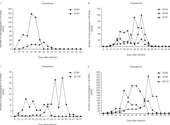
Parasitaemia - Parasitaemia was evaluated in the ex-perimental groups in the second or fifth passages into mice. Parasitemic curves are shown in Fig. 1A-D. Strains isolated either from a marsupial or a triatomine (SC90 and SC93), as well as the strains SC94, SC95, SC100, which were isolated from human patients, showed irregu-lar peaks from the 12th to the 20th day, characteristic of biodeme type II. Strain SC94 was the most virulent, with the earliest parasitemic peak. Strains SC96, SC97, SC98, SC99, SC101 and SC102, isolated from six of the infected humans, exhibited an early and low parasitemic peak from the 12th to the 20th day and a late peak after the 20th day, which are characteristic of biodeme type III.
TABLE I
Biodemes and zymodemes: strains from oral transmission outbreak in the state of Santa Catarina, Brazil, 2005
Identification
Biodemes (passages into mice)
Zymodemes (culture forms)
Strains Origin Biodeme types PGM GPI ASAT ALAT
SC90 Didelphis aurita II Z1 Z1/Z2 Z1 Z1
SC93 Triatoma tibiamaculata IIa Z1 Z2 Z1 Z2
SC94 Human II Z2 Z2 ND Z2
SC95 Human II Z1 Z2 ND ND
SC96 Human IIa Z2 Z2 Z2 Z1
SC97 Human IIa Z1 Z2 Z1 Z1
SC98 Human IIa Z2 Z2 Z1 Z1
SC99 Human IIa Z1 Z2 Z2 Z1
SC100 Human II Z2 Z2 ND Z1
SC101 Human IIa Z2 Z2 Z2 Z1
SC102 Human II Z2 Z2 Z2 Z1
a:strains that showed an early and a late peak of parasitaemia, suggesting double infection with biodemes II and III; ALAT:
Mortality - Table II shows the cumulative mortal-ity rates until the 30th day post-infection for the mice infected with each strain. The percentage mortality for the mice infected with the strains SC90, SC93, SC95 and SC98 was zero. Strains SC96, SC97, SC100, SC101 and SC102 caused 50% mortality of the infected mice; strains SC94 and SC99 caused 100% mortality.
Virulence - Based on the mortality rate at 30 days, as shown in Table II, and the parasitaemia and the severity of tissue lesions, the strains were classified as “low lence”, corresponding to 0% mortality, “medium lence”, corresponding to 50% mortality and “high viru-lence”, corresponding to 100% mortality. The strains SC94 and SC99 exhibited high virulence and produced intense cardiac lesions in mice.
Isoenzymes patterns - Table I shows the results of the isozymic profiles for PGM, GPI, ASAT and ALAT analysis of the strains. The strain SC90, from D. aurita, exhibited the profile of zymodeme Z1 in PGM, ASAT and ALAT analyses and a mixed profile Z1/Z2 in GPI
analysis; strain SC93, from T. tibiamaculata, exhibited a zymodeme Z2 profile when analysed by the enzymes GPI and ALAT and a Z1 profile when analysed by PGM and ASAT. The nine strains isolated from humans (SC94-SC102) exhibited a Z2 profile in eight of the re-ferred strains when analysed by the GPI isoenzyme (SC100 was not performed) and in six of these strains when analysed by PGM.
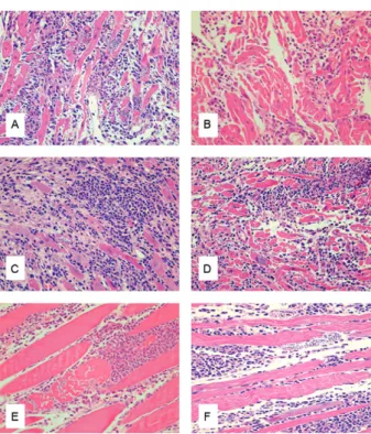
Histopathological evaluation - Pathogenicity in mice was evaluated by the intensity of the necrotic and inflam-matory lesions in the heart and skeletal muscles (Figs 2, 3). Strains with parasitemic peaks between 12-20 days post-infection (SC90, SC94 and SC100) exhibited histo-pathological lesions characteristics of biodeme type II, showing mild myocarditis from the seventh to 10th day of infection with diffuse mononuclear infiltrations and presence of T. cruzi amastigotes into the cardiac myo-cells. The inflammatory lesions and cardiac parasitism increased until the 20th day post-infection. The strain SC94, which exhibited high virulence (Table II),
pre-Fig. 1: parasitemic curves of the several strains, disclosed the profiles of the biodemes types (I, II, III) and in some cases the combination of the types II and III. A: strain SC90 showed the parasitemic pattern of the biodeme type II of low virulence. SC94 showed an early high peak cor-responding to biodeme type II of high virulence; B: strain SC93 shows the pattern of biodeme type II and the strains SC96 and SC97 a double parasitemic pattern of biodemes types II and III; C: strains SC95 and SC100 show an early parasitemic peak corresponding to biodeme type II and a late peak of the biodeme type III strains; D: strains SC98, SC99 and SC101 show the early peak characteristic of biodeme type II strains and a late peak characteristic of biodeme type III.
120 100 80 60 40 20 0
8 10 12 14 16 17 18 20 21 22232425 22 28 30 31 34 36 40 Days after infection
Parasitemia B
Number of trypomastigoes in 50 fields
(400X)
SC93 SC96 SC97
40 35 30 25 20 15 10 5 0
8 10 12 14 16 18 20 22 25 27 29 31 34 36 40 45 47 49 51 Days after infection
Parasitemia C
Number of trypomastigoes in 50 fields
(400X)
SC95 SC100
45 180
160 140 120 100 80 60 40 20 0
8 10 12 14 16 18 20 22 25 Days after infection
Parasitemia A
Number of trypomastigoes in 50 fields
(400X)
27 31 33 35 37 40 42 SC90 SC94
47 340
320 300 280 260 240 220 200 180 140 120 100 80 60 40 20 0
8 10 12 14 16 18 20 22 25
Days after infection Parasitemia D
Number of trypomastigoes in 50 fields
(400X)
27 29 31 34 36 40 45 SC98
SC99
sented with early inflammatory infiltrations in the myo-cardium and skeletal muscle from the seventh to the 10th day post-infection, with variable evolution to intense car-diac lesions and intense parasitism of carcar-diac cells from the 14th to the 20th day (Fig. 2A, B). The strains that presented double peaks of parasitaemia (SC96, SC97, SC98, SC99, SC101 and SC102) presented histopatholog-ical lesions and intracellular parasitism that were more prominent from the 20th to 30th days post-infection, with intense inflammatory lesions in the myocardium (Fig. 3A-D) and skeletal muscles (Fig. 3E, F).
Molecular analysis - Typical results from the molecu-lar characterisation of the 11 isolates can be found in Figs 4-7. Compiled molecular data are presented in Table III.
RFLP-COII profiles - Analysis of RFLP-COII pro-files for T. cruzi populations showed the presence of all three possible haplotypes (Fig. 4). The isolates SC94, SC96, SC98, SC99, SC100, SC101 and SC102 presented the haplotype C (fragments of 212, 81 and 82 bp) charac-teristic of T. cruzi II. The isolates SC90, SC93 and SC97 showed both A and C haplotypes (fragments of 264, 212, 81/82 and 30 bp), corresponding to a mixture of popula-tions belonging to T. cruzi I and II. The SC95 isolate presented both C and B haplotypes (fragments of 294, 212 and 81/82 bp), corresponding to a mixture of popula-tions belonging to T. cruzi II and one of T. cruzi III, IV, V or VI (Table III).
Molecular typing of the intergenic regions of T. cruzi spliced leader genes - The analysis of the polymorphism of the ITS region of the spliced leader genes was per-formed according to Burgos et al. (2007). Using this strategy, all T. cruzi populations presented PCR amplifi-cationproducts of ~150 bp, characteristic of T. cruzi I, II, V or VI (Fig. 5) (Table III).
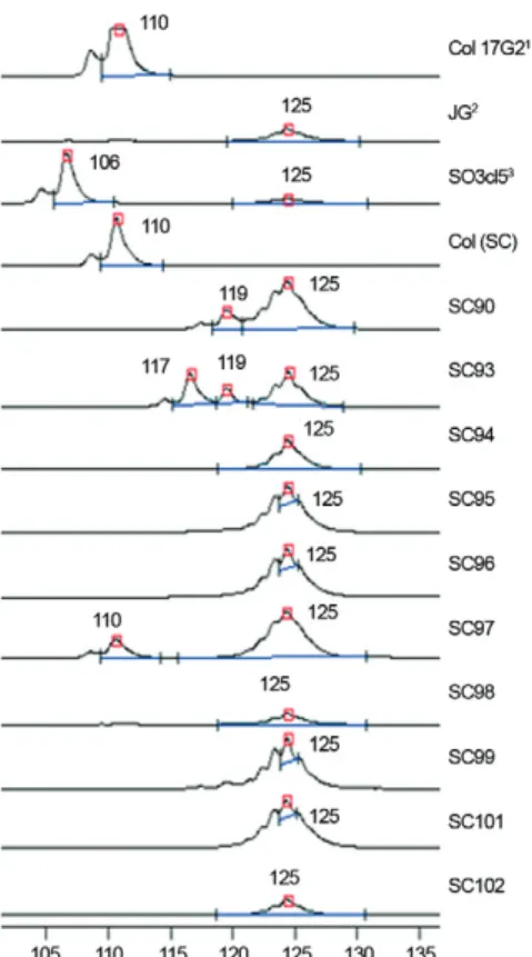
Analysis of the 24Sα rRNA gene - The 24Sα DNA
analysis revealed that the isolates SC94, SC95, SC96, SC98, SC99, SC100, SC101 and SC102 displayed am-plicons of 125 bp, characteristic of T. cruzi II, IV or VI (Figs 6, 7). Therefore, based on all the molecular data, we concluded that the isolate SC-95 consisted of a mix-ture of populations belonging to T. cruzi II and VI geno-types, as the ITS analysis rules out the possibility of a
mixture including T. cruzi IV. The SC97 isolate showed fragments of both 110 and 125 bp and the SC90 and SC93 isolates exhibited fragments of ~117 and 125 bp, all of which are compatible with a probable mixture of T. cruzi I and II (Table III).
RFLP of kDNA minicircle variable regions - Isolates SC96, SC97, SC98, SC99 and SC-101 were character-ised by the schizodeme analysis with RsaI, EcoRI and HinfI endonucleases. All three endonucleases generated profiles with genetic distances indicating a high degree of genetic differentiation. The restriction with EcoRI showed that SC90 and SC93 shared 86% similarity with one another and 70% similarity with isolates SC94, SC96 and SC97. The other three isolates (SC98, SC99 and SC101) formed a new profile with 78% similarity among themselves. The restriction with HinfI clustered five isolates (SC96, SC97, SC98, SC99 and SC101) with 75% similarity. Again, the marsupial and triatomine iso-lates grouped with 80% similarity. These strains showed greater heterogeneity when restricted with RsaI.
TABLE II
Cumulative mortality rates of mice/virulence of strains
Low virulence (0% mortality)
Medium virulence (until 50% mortality)
High virulence (100% mortality)
SC90a SC96c SC94c
SC93b SC97c SC99c
SC95c SC100c
-SC98c SC101c
-SC102c
-a: isolated from Didelphis aurita; b: isolated from Triatoma
tibiamaculata; c: isolated from humans.
DISCUSSION
The 2005 outbreak of oral T. cruzi infection that oc-curred in SC resulted in the infection of several people at the same locality and under the same circumstances, leading to the conclusion that all individuals were infect-ed with the same T. cruzi strain. The present study was a unique opportunity to investigate the biological and bio-chemical behaviour of these strains after human infection and passage through an experimental animal and to corre-late these characteristics of the parasite with their molecu-lar patterns according to the New Consensus on T. cruzi Nomenclature, proposed in 2009 (Zingales et al. 2009).
In the present investigation, several methods were used for biological characterisation: parasitaemia, mor-tality, virulence, histopathological lesions, tissue tropism and pathogenicity in mice. Based on these parameters, all the isolated strains exhibited the patterns described for biodeme type II, as described previously (Andrade et al. 2006b). Interestingly, six of the nine human strains classified as biodeme type II presented double peaks of parasitaemia (SC96, SC97, SC98, SC99, SC101 and
SC102) in experimental mice, with the early low peak characteristic of this biodeme and a late peak after the 20th day that corresponds to the parasitaemia pattern of biodeme type III. These findings suggest the presence of a mixed infection. In the early phase of the infection by these strains, the histopathological patterns were characteristic of biodemes type II; however, lesions and intracellular parasitism were more prominent from the 20th to 30th days post-infection, with intense inflam-matory lesions in the myocardium and skeletal muscles, which is characteristic of infection with strains belong-ing to biodeme type III. Additionally, the isoenzymic characterisation of the nine strains isolated from humans presented Z1 or Z2 profiles by PGM, ALAT and ASAT
Fig. 4: polymerase chain reaction (PCR)-restriction fragment length polymorphism profiles of the cytochrome oxidaseII gene after AluI digestion on silver stained 6% polyacrylamide gel. 1-4: Trypanosoma cruzi reference strains; 5-14: T. cruzi isolates from the oral outbreak; MW: 1 kb Plus DNA Ladder (Invitrogen®); NC: negative PCR control;
NC Dig: negative AluI digestion.
Fig. 5: spliced leader intergenic region profiles on bromide ethidium stained 2% agarose gel. 1-3: Trypanosoma cruzi reference strains; 4-13:
T. cruzi isolates from the oral outbreak; MW: 1 kb Plus DNA Ladder (Invitrogen®); NC: negative polymerase chain reaction control.
Fig. 6: 24Sα rDNA profiles on silver stained 6% polyacrylamide gel
electrophoresis. 1-4: Trypanosoma cruzi reference strains; 5-14: T. cruzi isolates; MW: 25 bp DNA Ladder (Invitrogen®); NC: negative
polymerase chain reaction control. Fig. 3: histopathological lesions of the heart and skeletal muscles of
analyses and Z2 profiles by GPI analysis. The presence of a variant pattern of PGM analysis for the strains iso-lated from humans and a mixed Z1/Z2 profile for the strains SC92 and SC93 isolated from T. tibiamaculata has been previously observed by Steindel et al. (2008), who demonstrated for the first time the transmission of a mixed infection by this vector in SC. According to Miles and Cibulskis (1986), some strains isolated from either single mammals or vectors may consist of heterogeneous mixtures; combinations of different isoenzymes eletro-phoretic profiles could help elucidate epidemiological questions, define taxa and detect genetic exchanges. The strain SC90 (isolated from D. aurita), initially described as T. cruzi I (Steindel et al. 2008) by multiloci isoen-zymes, was revealed in the present investigation via mo-lecular characterisation to be a mixture of T. cruzi I + T. cruzi II genotypes. Steindel et al. (1995) previously reported mixed T. cruzi I + T. cruzi II infections in adult Panstrogylus megistus in SC. The occurrence of mixed populations in many of the isolates of SC may explain the apparent incongruity of the zymodeme profiles ob-served for these populations, which present as either Z1 or Z2, depending on the isoenzyme used for analysis.
Some characteristics emerge from these studies, such as the high virulence of two strains isolated from hu-mans (SC94 and SC99), both characterised as the T. cru-zi II genotype, and exhibited 100% mortality in the early
phase of acute experimental infection. It was shown that the strains isolated either from the marsupial (SC90) or from the insect vector (SC93) exhibited low virulence, al-though they contained mixed strains (T. cruzi I + T. cruzi II). Pena et al. (2011), utilising vector-derived mixed TcI/ TcII infective trypomastigotes (SC90-SC102), showed that T. cruzi II, but not T. cruzi I strains, were selected by both human and murine macrophages in vitro and by peritoneal cavity cells of Balb/c mice in vivo.
Molecular characterisation revealed the presence of T. cruzi II parasites in all isolates, which may reflect the fact that this is the major T. cruzi lineage associated with human cases in southern Brazil (Freitas et al. 2005). However, a combination of T. cruzi II with other types was observed in four samples (SC90, SC93, SC95 and SC97). To identify these mixtures and the correct ma-jor lineages, simultaneous analyses using three different molecular approaches were necessary.
The initial RFLP analysis of the COII gene revealed the presence of different combinations of the three pos-sible haplotypes, corresponding to three cases of mixed populations of T. cruzi I + T. cruzi II (SC90, SC93, SC97) and a single case (SC95) of a mixed population T. cruzi II and either T. cruzi III, IV, V or VI. To discriminate among these possibilities, molecular typing of the SL-IR was applied. Identification of mini-exon amplicons of 150 bp characteristic of T. cruzi I, II, V or VI ruled out
TABLE III
Identification of phylogenetic discrete typing units (DTU)
for the Trypanosoma cruzi isolates employing
the cytochrome oxidase(CO)II haplotypes spliced
leader intergenic region (SL-IR) and 24Sα rDNA markers
T. cruzi
isolates
COII haplotypes
rDNA 24Sα (bp)
SL-IR (~bp)
T. cruzi
DTUsb
Col 17G2a A 110 150 I
JGa C 125 150 II
231a B 110 200 III
Can IIIa B 125 200 IV
SO3 cl5a B 110 + 125 150 V
CL Brenera B 125 150 VI
SC90 A + C 119 + 125 150 I + II
SC93 A + C 117 + 119 + 125 150 I + II
SC94 C 125 150 II
SC95 B + C 125 150 II + VI
SC96 C 125 150 I
SC97 A + C 110 + 125 150 I + II
SC98 C 125 150 II
SC99 C 125 150 II
SC101 C 125 150 II
SC102 C 125 150 II
a: T. cruzi strains or clones used as reference to COII
haplo-types, 24α rDNA and SL-IR marker; b: according to Zingales
et al. (2009).
Fig. 7: automatic laser fluorescent DNA sequencer electrofluoro-grams presenting the Trypanosoma cruzi amplicons obtained by the
analysis of 24Sα rDNA gene. Numbers at the peaks refer to the size of
the amplicons in base pairs. Control patterns: 1rDNA group 1; 2rDNA
the possibility of SC95 existing as a mixture of T. cruzi II with T. cruzi III or IV. Finally, analysis of the 24Sα rRNA gene demonstrated that SC-95 exhibited only am-plicons of 125 bp, characteristic of T. cruzi II, III or VI, resulting in the conclusion that SC95 constitutes a mix-ture of T. cruzi II and T. cruzi VI.
The presence of mixtures of different genotypes in isolates of the SC strains from infected humans, the mammalian reservoir and the insect vector could have in-fluenced the clinical presentation of the disease, as the le-thality in the acute phase was unusually high (12.5%). The unusually high mortality index in this outbreak contrasts with clinical surveys that have demonstrated a mortality rate varying from 5-9% in the acute phase of infections. We cannot exclude the possibility that the inoculum and the route of infection affect the parasite burden and con-sequently the severity of the disease in infected individu-als. However, different studies have demonstrated that the presence of mixed populations can interfere in the course and severity of experimental infections and in the host immune response (Rodrigues et al. 2010).
Experimentally, multiple infections performed by Andrade et al. (2006a) presented worsening of the infec-tion and tissue lesions in mice successively inoculated with strains of the three different T. cruzi biodemes (Y, 21SF and Colombian strains). In the triple-infected mice, it was possible to re-isolate the three inoculated strains, taking into account their biological behaviour, after suc-cessive passages in young mice (Andrade et al. 2006a). In the present study, the different genotypes detected by a combination of molecular techniques in several sam-ples isolated from human cases reflect the complexity of the tentative correlations between the parasite isolates and the clinical manifestations of Chagas disease.
Considering the different approaches in the present in-vestigation, we conclude that oral transmission represents an important factor for the emergence of acute Chagas disease outbreaks, even for non-endemic areas. Biologi-cal, biochemical and molecular analyses detected the pres-ence of mixed infections with different genotypes, possi-bly related to the different clinical courses of the acute human cases, as observed during the SC oral outbreak. Interestingly, a concordance between the biological and isoenzymic characters confirmed the presence of mixed infections, not only in the vector but also in the vertebrate reservoir and in human isolates. The heterogeneity of the isolated strains was confirmed by genotyping according to the new T. cruzi taxonomy (Zingales et al. 2009).
REFERENCES
Andrade SG 1974. Caracterização de cepas do Trypanosoma cruzi
isoladas no Recôncavo Baiano. Rev Patol Trop 3: 65-121. Andrade SG 1985. Morphological and behavioural
characteriza-tion of Trypanosoma cruzi strains. Rev Soc Bras Med Trop 18 (Suppl.): 39-46.
Andrade SG, Campos RF, Sobral KSC, Magalhães JB, Guedes RSP, Guerreiro ML 2006a. Reinfections with strains of Trypanosoma cruzi of different biodemes as a factor of aggravation of myocar-ditis and myositis in mice. Rev Soc Bras Med Trop 39: 1-8. Andrade SG, Magalhães JB 1997. Biodemes and zymodemes of
Trypanosoma cruzi strains: correlations with clinical data and experimental pathology. Rev Soc Bras Med Trop 30: 27-35.
Andrade SG, Magalhães JB, Sobral KS, Rosado AP, Oliveira PLS 2006b. Caracterização de cepas do Trypanosoma cruzi isoladas durante surto agudo de doença de Chagas por transmissão oral em Santa Catarina, Brasil. Rev Soc Bras Med Trop 39 (Suppl. 1): 7. Andrade V, Brodskyn C, Andrade SG 1983. Correlation between
isoenzyme patterns and biological behaviour of different strains of Trypanosoma cruzi. Trans R Soc Trop Med Hyg 77: 796-799.
Anonymous 1999. Recommendations from a Satellite Meeting. Mem
Inst Oswaldo Cruz 94 (Suppl. I): 429-432.
Avila H, Gonçalves AM, Neheme NS, Morel CM, Simpson L 1990. Schizodeme analysis of Trypanosoma cruzi stocks from South and Central America by analysis of PCR-amplified minicircle variable region sequences. Mol Biochem Parasitol 42: 175-188.
Brisse S, Dujardin JC, Tibayrenc M 2000. Identification of six
Trypanosoma cruzi lineages by sequence-characterised ampli-fied region markers. Mol Biochem Parasitol 111: 95-105.
Burgos JM, Atcheh J, Bisio M, Duffy T, Valadares HM, Seidenstein ME, Piccinali R, Freitas JM, Levin MJ, Macchi L, Macedo AM, Freilij H, Schijman AG 2007. Direct molecular profiling of minicircle signatures and lineages of Trypanosoma cruzi blood-stream populations causing congenital Chagas disease. Int J Par-asitol 37: 1319-1327.
Freitas JM, Augusto-Pinto L, Pimenta JR, Bastos-Rodrigues L, Gon-çalves VF, Teixeira SM, Chiari E, Junqueira AC, Fernandes O, Macedo AM, Machado CR, Pena SD 2006. Ancestral genomes, sex and the population structure of Trypanosoma cruzi. PLos
Pathogens 2: 226-235.
Freitas JM, Lages-Silva E, Crema E, Pena SD, Macedo AM 2005. Real time PCR strategy for the identification of major lineages of Trypanosoma cruzi directly in chronically infected human tis-sues. Int J Parasitol 35: 411-417.
Miles MA, Cibulskis RE 1986. Zymodeme characterization of
Trypanosoma cruzi. Parasitology Today 2: 94-101.
Miles MA, Laham SM, Souza AA, Povoa M 1980. Further enzymic characters of Trypanosoma cruzi and their evaluation for strain identification. Trans R Soc Trop Med Hyg 74: 221-237.
Nery-Guimarães F, Silva NN, Clausell DT, de Mello AL, Rapone T, Rodrigues N 1968. Um surto epidêmico de doença de Chagas de provável transmissão digestiva ocorrida em Teutônia (Estrela-Rio Grande do Sul). Hospital (RJ) 73: 1767-1804.
Neves da Silva N, Clausell DT, Nólibos H, de Mello AL, Ossanai J, Rapone T, Snell T 1968. Surto epidêmico de doença de Chagas com provável contaminação oral. Rev Inst Med Trop Sao Paulo
10: 265-276.
Pena DA, Eger I, Nogueira L, Heck N, Menin A, Bafica A, Steindel M 2011. Selection of TcII Trypanosoma cruzi population following macrophage infection. J Infect Dis 204:478-486.
Pinto AY, Harada GS, Valente VC, Araújo JE, Gomes FS, Souza GC, Valente SA 2001. Acometimento cardíaco em pacientes com doença de Chagas aguda em microepidemia familiar, em Abaetetuba, na Amazônia Brasileira. Rev Soc Bras Med Trop 34: 413-419.
Rodrigues CM, Valadares HMS, Francisco AF, Arantes JM, Campos CF, Carvalho AT, Filho OAM, Araújo MSS, Arantes RME, Chi-ari E, Franco GR, Machado CR, Pena SDJ, FChi-aria AMC, Macedo AM 2010. Coinfection with different Trypanosoma cruzi strains interferes with the host immune response to infection. PLoS Negl
Trop Dis 4: e846.
Shikanai-Yasuda MA, Marcondes CB, Guedes LA, Siqueira GS, Ba- rone AA, Dias JCP, Amato Neto V, Tolezano JE, Peres BA, Ar-ruda Jr. ER, Lopes MH, Shiroma M, Chapadeiro E 1991. Possi-ble oral transmission of acute Chagas’ disease in Brazil. Rev Inst
Souto RP, Fernandes O, Macedo AM, Campbell DA, Zingales B 1996. DNA markers define two major phylogenetic lineages of
Trypanosoma cruzi. Mol Biochem Parasitol 83: 141-152.
Steindel M, Pacheco LK, Scholl D, Soares M, Moraes MH, Eger I, Ko-smann C, Sincero TCM, Stoco PH, Murta SMF, Carvalho-Pinto CJ, Grisard EC 2008. Characterization of Trypanosoma cruzi iso-lated from humans, vectors and animal reservoirs following an outbreak of acute human Chagas disease in Santa Catarina state, Brazil. Diag Microbiol Infect Dis 60: 25-32.
Steindel M, Toma HK, Ishida MM, Murta SM, Carvalho Pinto CJ, Grisard EC, Schlemper Jr. BR, Ribeiro-Rodrigues R, Roma- nha AJ 1995. Biological and isoenzymatic characterization of
Trypanosoma cruzi strains isolated from sylvatic reservoirs and vectors from the state of Santa Catarina, southern Brazil. Acta Trop 60: 167-177.
Tibayrenc M 1998. Genetic epidemiology of parasitic protozoa and other infectious agents: the need for an integrated approach. Int
Parasitol 28: 85-104.
Tibayrenc M 2003. Genetic subdivisions within Trypanosoma cruzi
(discrete typing units) and their relevance for molecular epide-miology and experimental evolution. Kinetoplastid Biology and Disease 2: 12.
Valente SA, Valente VC, Fraiha Neto H 1999. Considerações sobre a epidemiologia e transmissão da doença de Chagas na Amazônia Brasileira. Rev Soc Bras Med Trop 32 (Suppl. 2): 51-55.
Valente SAS, Valente VC, Pinto AYN, César MJB, Santos MP, Mi-randa CO, Cuervo P, Fernandes O 2009. Analysis of an acute Chagas disease outbreak in the Brazilian Amazon: human cases, triatomines, reservoir mammals and parasites. Trans R Soc Trop
Med Hyg 103: 291-297.