INTRODUCTION
Address to: Dra. Vanete Thomaz-Soccol. Rua Francisco H. dos Santos 100, Centro Politécnico, Jardim das Américas, 81531-980 Curitiba, PR, Brasil.
Phone: 55 41 3361-3272
e-mail: [email protected]
Received 5 April 2013
Accepted 3 June 2013
Comparison of conventional serology and PCR
methods for the routine diagnosis of
Trypanosoma cruzi
infection
Soraia Reda Gilber
[1],[2],
Silvana Maria Alban
[1],
Luiza Gobor
[3],
Jessica de Oliveira Bescrovaine
[3],
Márcia Iurico Myiazaki
[4]and Vanete Thomaz-Soccol
[1][1]. Programa de Pós-Graduação em Processos Biotecnológicos, Universidade Federal do Paraná, Curitiba, PR. [2]. Setor de Imunologia, Laboratório Central do Estado do Paraná, Curitiba, PR. [3]. Programa de Pós-Graduação em Biotecnologia, Universidade Positivo, Curitiba, PR. [4]. Serviço de Doença de Chagas, Hospital de Clínicas, Universidade Federal do Paraná, Curitiba, PR.
ABSTRACT
Introduction: Trypanosoma cruzi, a fl agellated protozoan, is the etiologic agent of Chagas disease, and it is estimated that
approximately 5 million people in Brazil are infected with this parasite. This work aimed to compare the current diagnostic methods for Chagas disease, including conventional serological (IFAT and ELISA) and molecular techniques (PCR), to introduce PCR as an auxiliary technique. Methods: A total of 106 chagasic patients were evaluated: 88 from endemic areas of Parana, 6 from São Paulo, 3 from Minas Gerais, 3 from Rio Grande do Sul, 1 from Bahia and 5 from the Santa Catarina T. cruzi outbreak.
The samples were analyzed by conventional serological methods (IFAT, ELISA), hemoculture and PCR to confi rm Chagas
disease. Results: When IFAT was used to determine antibody levels, the sensitivity was 81.7% for patients with the cardiac form of the disease and 100% for the other clinical forms. In contrast, ELISA showed 84% sensitivity and 100% specifi city. The use of serological and molecular techniques and their implications for the diagnosis of Chagas disease in non-endemics area are
discussed. Conclusions: PCR constitutes an excellent support methodology for the laboratory diagnosis of Chagas disease due to its high sensitivity and specifi city.
Keywords: Chagas disease. Inconclusive diagnosis. Routine diagnosis.
Chagas disease is an anthropozoonosis caused by
Trypanosoma (Schizotrypanum) cruzi (Chagas, 1909). It is
estimated that 16 to 18 million people are infected with the
parasite in 19 Latin American countries, and that approximately
50,000 individuals will die annually due to Chagas disease1.
In Brazil, approximately 5 million people are infected with
T. cruzi, with 60% residing in urban areas, similar to other
countries2,3. Despite a signifi cant decrease in the incidence of
T. cruzi infection in humans, numerous chronic chagasic patients
still receive medical attention and are treated in such reference centers as the University Hospital of the Universidade Federal do Paraná4. Asymptomatic T. cruzi carriers with positive
serology and blood donors with inconclusive serology are often
identifi ed in blood banks. Considering that these individuals do not present clinical signs and receive no defi nitive diagnosis, it
is possible that such individuals carry antibodies against another Trypanosomatidae, including T. rangeli.
The World Health Organization recommends two methods that use different principles and different antigens to determine a serological diagnosis of T. cruzi infection5. The indirect
immunofl uorescence antibody test (IFAT) and the enzyme-linked immunosorbent assay (ELISA) are widely used due to the diffi culty of directly visualizing the parasite in chronic
patients6. However, serological cross-reactivity may reduce the
sensitivity of these tests in regions where other Kinetoplastida (Leishmania spp. and T. rangeli) parasites are present7.
Molecular diagnostic methods have enabled the more precise identifi cation of pathogenic agents; indeed, such molecular techniques as the polymerase chain reaction (PCR) have improved diagnostic specifi city and the ability to identify the type of parasite circulating in a specifi c area6,8-10. With regard
to Chagas disease, however, PCR has presented highly variable levels of sensitivity. The reasons for this include variations in the sample volume and sample preservation, the methods used for DNA extraction, the primers selected, the quality of the reagents,
the thermocycling conditions and the intermittent presence and
METHODS
Regardless, PCR is more sensitive than parasitological methods12 and can be introduced as an auxiliary technique
for diagnosing Chagas disease in patients with inconclusive serology. In this work, serology, hemoculture and PCR techniques were simultaneously performed in an attempt to verify the presence of T. cruzi in patients with the chronic, acute
and indeterminate forms of disease. Trypanosoma rangeli was
also investigated in patients with positive serology who were negative by PCR for T. cruzi.
Study population
All the individuals enrolled in the study signed a term
of informed consent and filled out an epidemiological questionnaire.
One hundred and six patients from 10 to 89 (average 55.32) years of age were enrolled in this study. Eighty eight out of these came from different endemic areas of Chagas disease of the State of Paraná, Brazil and also fi ve patients from an outbreak in Santa Catarina, six from São Paulo, three from Minas Gerais, three from Rio Grande do Sul and one from Bahia. We included 15 non-infected individuals as negative controls.
Venous blood (30mL) was collected using a vacutainer system (Becton Dickinson) for the following assays: a) 5mL for serology; b) 5mL in ethylenediaminetetraacetic acid (EDTA) for DNA extraction and PCR; c) 20mL inEDTA for hemoculture. All the patients were surveyed in different medical specialties
at the UPFR University Hospital.
Serological assays
The IFAT assays were performed using IFI-Imunocruzi®, Fluoline M® and Fluoline G® kits (bioMérieux) in accordance with the manufacturer’s instructions. Each serum sample was diluted (1/20-1/2560) in phosphate-buffered saline (PBS), pH 7.2 and incubated for 30min at 37°C with T. cruzi epimastigotes
pre-adsorbed onto immunofluorescence glass slides. The unbound immunoglobulins were removed by washing the slides twice with PBS, followed by incubation with fl uorescein-labeled anti-human IgG or IgM conjugates at 37°C for 30min; the unbound conjugates were removed by washing with PBS. The slides were mounted with buffered glycerin (pH 9.5) and were observed under a fl uorescence microscope at 400x magnifi cation (Olympus BH2). Positive and negative sera were used as controls. The cutoff for IFI was 1/40.
ELISA was performed using the commercial kit Chagatek® (bioMérieux) in accordance with the manufacturer’s recommendations. Briefl y, sera were diluted 1/20 in phosphate-buffered saline-Tween 20 (0.05%) containing 1% bovine serum albumin. A 200µL aliquot of diluted serum was added to the wells of a microtiter plate and incubated for 30min at 37°C. After 6 washes in 0.1M PBS to remove the unbound immunoglobulin, the samples were incubated at 37°C with a 1:10 dilution of peroxidase-labeled an anti-human IgG conjugate. Following another washing step, the microplates
were detected with the addition H2O2 in 50mM citrate buffer
(pH 3.2) and 0.01mM TMB in 0.1N HCl. After a 20min incubation at room temperature, the reaction was stopped by the addition of 2N sulfuric acid. The absorbance of the samples was measured at 450nm using a microplate reader. The mean absorbance of the negative controls plus 0.100 was used for determining the cutoff, in accordance with the manufacturer’s instructions. A borderline ELISA result was defi ned as reactivity
within 2 standard deviations of the control. Hemoculture
Hemoculture was performed as proposed by Kopp4. Briefl y,
20mL of heparinized venous blood was collected from each individual in a vacutainer system and centrifuged at 600 x g for
30min at 4°C. The plasma was collected and stored at -20°C. The buffy coat was resuspended in lmL of physiological serum at 0.9% and distributed into 10 tubes containing CCS medium. The cultures were maintained at 28°C and examined weekly over a three-month period. The positive cultures were divided into two aliquots: the fi rst was grown in LIT medium supplemented with 10% fetal bovine serum for DNA extraction of the parasite, and the remaining aliquot was cryopreserved in liquid nitrogen (-196°C).
DNA extraction
DNA was extracted from two sources: from the epimastigotes of positive cultures and from the patients’ blood. The parasites obtained from cultures were washed twice with PBS, and the resulting pellet was stored at -20°C until use. DNA extraction was performed using the protocol proposed by Sambrook
et al.13, and the resulting the DNA was stored at -70°C until use.
DNA extractions from the patients’ blood and artifi cially contaminated blood were achieved by the BOOM method using a Nuclisens® kit (bioMérieux, France). Briefl y, 10µL of buffy coat was resuspended in 900µL of lysis buffer (guanidine thiocyanate buffer solution, 5mol/L) and maintained at 4°C overnight. Then,
50µL of silica gel was added; following homogenization, the
mixture was centrifuged for 3min at 10,000 x g. The supernatant
was discarded, and the silica containing the immobilized DNA was subjected to successive washing steps to remove any contaminants. The DNA was eluted from the silica and stored at -20°C until use.
PCR technique
To perform the specifi city and sensitivity tests, authenticated negative samples were contaminated with parasites from
cultures in LIT medium. The T. cruzi, T. rangeli, Leishmania (Viannia) braziliensis and L. (Leishmania) amazonensis strains
were used to determine specificity. The positive controls consisted of 20ng of T. cruzi or T. rangeli DNA, and the negative
controls consisted of a reaction mixture without DNA.
PCR amplifi cation of the repetitive nuclear sequence
using primers TCZ1 and TCZ2
Amplifi cation reactions were performed in a fi nal volume of 25µL containing 10mM Tris-HCl (pH 8.3), 1.5mM MgCl2,
50mM KCl, 0.01% gelatin, 200µM each dNTP, 25pmol each
RESULTS
TABLE 1 -Samples and results of different laboratory diagnoses.
Positive samples
Group Number of samples hemoculture IFI ELISA PCR
Cardiac 60 6 49 46 44
Digestive 29 3 29 29 28
Indeterminate 12 1 12 12 4
Acute 5 1 4 2 5
Negative control 10 0 0 0 0
Total 106 11 94 89 81
IFI: indirect immunofl uorescence; ELISA: enzyme-linked immunosorbent assay; PCR: polymerase chain reaction. USA). Different amounts of DNA extracted from the
patients’ blood (2, 4, and 8µL) or 20ng of culture DNA was then added. The cycle amplifi cation was performed using a Thermo Hybaid thermal cycler with the following conditions: 5min at 94°C (initial denaturation), 35 cycles of 1min at 95°C, 1min at 65°C and 1min at 72°C and a fi nal extension of 5min at 72°C. The primer sequences used were TCZ1, 5’CGAGCTCTTGCCCACACGGGTGCT3’, and TCZ2, 5’CCTCCAAGCAGC GGATAGTTCAGG3’.
PCR amplifi cation of the P542 repetitive sequence
of T. rangeli using primers R1 and R2
Amplifi cation reactions were performed in a fi nal volume of 25µL containing 10mM Tris-HCl (pH 8.3), 1.0mM MgCl2,
50mM KCl, 0.01% gelatin, 100µM each dNTP, 12.5 pmol
each primer, 1U Taq DNA polymerase and 4µL of DNA
extracted from the patients’ blood or 20ng of culture DNA. The cycling conditions used were as follows: 5min at 94°C (initial denaturation); 2 cycles of 1min at 94°C, 1min at 65°C and 1min at 72°C; 10 cycles of 1min at 94°C, 1min from 65°C to 55°C, with a decrease of 1°C in each cycle, and 1min at 72°C; 18 cycles of 1min at 94°C, 1min at 55°C and 1min at 72°C and a fi nal extension of 5min at 72°C.
The primer sequences used were R1 (F), 5’CGC GGCTCGCACTGCACCTC3’, and R2, (R) 5’GGC GCATCCACCGAGCACTG3’.
For all the reactions, the amplifi ed products were subjected to 1.6% agarose gel electrophoresis, stained with ethidium
bromide, visualized under UV light and digitally recorded.
Statistical analysis
The degree of agreement between the serology and PCR results was determined by calculating the Kappa (k) values with
95% confi dence intervals. The analysis of agreement test was analyzed using web-based software developed by Laboratório de Epidemiologia e Estatítisca (FAFESP), which is available
online at http://lee.dante.br/pesquisa/kappa/ kappaupload.html. Kappa values of 0.01 indicated no concordance, values between 0.1 and 0.4 indicated weak concordance, values between 0.41 and 0.60 indicated clear concordance, values between 0.61 and 0.80 indicated strong concordance, and values between 0.81 and
1.00 indicated nearly complete concordance.
Ethical considerations
The study was approved by the Research Ethics Committees of the Federal University of Paraná (1161.009/2006-01), by the
Universidade Positivo and by CONEP, MS (Register in CONEP:
11163 and Register in: Doc: CAAE: 0105.0.094.000-10 Protocol
no. 114/2010).
Serological diagnosis
The comparative results of the two serological methods and PCR for the diagnosis of Chagas disease are shown in Table 1.
For the IFAT technique, 94 of 106 patients presented positive antibodies, with titers varying from 1/40 to 1/2,560. Two patients (1.60%) presented inconclusive serology (titer 1/20). Four patients from the 2005 outbreak of acute Chagas disease in Santa Catarina State also presented positive IgM serology, with titers of 1/40, 1/80 and 1/640. All the negative controls
were below the cutoff level.
Using the ELISA approach, 89 of 106 patients presented positive IgG antibodies. Ten (9.43%) patients presented borderline absorbance and were considered inconclusive cases; the samples with inconclusive serology were all blood bank donors. All the patients that were positive by IFAT were also positive by ELISA, except for the patients with acute disease. Two patients who presented IgM titers of 1/40 and 1/80 had no detectable antibodies
by ELISA. All the negative controls were below the cutoff.
Hemoculture
Of the 106 hemocultures conducted using the buffy coat from patients presenting positive serology, eleven (10.4%) were positive. These cases permitted the isolation of fi ve T. cruzi
strains. Ten cultures were isolated from chronic patients and one from an acute case.
Validation and sensitivity of PCR for Trypanosoma cruzi and Trypanosoma rangeli
Therefore, the technique was sensitive enough to detect a single parasite in 10mL of blood. The specifi city analysis showed that
TCZ1/TCZ2 amplifi ed DNA from T. cruzi (strain 152 Y and
isolate HC45), T. rangeli, L. braziliensis and L. amazonensis.
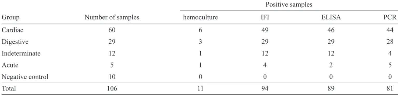
Therefore, we conclude that this primer pair permits the amplifi cation of PCR fragments from parasites belonging to the order Kinetoplastida (Figures 1A and 1B).
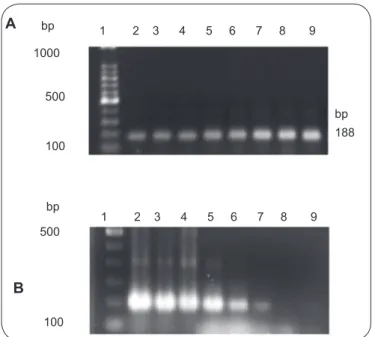
Evaluation of the sensitivity of primer pair R1/R2 showed that it was able to detect DNA of a single parasite. Amplifi cations were conducted with cultures of various trypanosomatids (T. rangeli, T. cruzi and Leishmania) to determine specifi city.
As expected, the T. rangeli cultures were positive when
using primer pair R1/R2, whereas the T. cruzi cultures were all
negative. Only one culture of L. (L.) amazonensis was positive
using these primers, but the amplifi cation signal was weak, showing 90% specifi city (Figures 2A and 2B).
Patient evaluation using the PCR technique
When using the primer pair TCZ1/TCZ2, the expected
188-bp band (Figure 3) was detected in 81 of the 106 Chagas patients. Sixty-nine of these patients presented positive PCR and serology, and nine showed inconclusive or positive serology in only one of the serological assays. Two of the seronegative patients were positive by PCR.
None of the individuals with inconclusive serology presented
the expected T. rangeli band when their blood was subjected to
1 2 3 4 5 6 7 8 9
1 2 3 4 5 6 7 8 9 bp
1000
500
100
bp 188
bp
500
100
A
B
FIGURE 1 -A: Evaluation of the sensitivity of the primer pair TCZ1/TCZ2 for
the PCR amplifi cation of Trypanosoma cruzi DNA. Lane 1: 100-bp molecular
weight standard; Lanes 2 through 9: PCR amplifi cation of T. cruzi DNA isolated
from 1, 5, 10, 50, 100, 150, 300 and 600 parasites. Electrophoresis was performed using a 1.6% agarose gel. B: Specifi city test of the primer pair TCZ1/TCZ2 for the PCR amplifi cation of T. cruzi DNA. Lane 1: 100-bp molecular weight standard;
Lane 2: reference strain of T. cruzi (152 Y); Lane 3: T. cruzi isolated from humans
(HC 45); Lane 4: T. rangeli (strain CHOACHI); Lane 5: T. rangeli (SC 58);
Lane 6, L. braziliensis (CUR 22); Lane 7: L. amazonensis (LEM 690); Lane 8:
negative control (water); Lane 9: negative control (human blood negative for
T. cruzi). Electrophoresis was performed using a 1.6% agarose gel. bp: base pairs.
1 2 3 4 5 6 7 8 9 10 11 12
1 2 3 4 5 6 7 8 9 bp
500
bp 500
FIGURE 2 - A: Sensitivity test of the primer pair R1/R2. Lane 1: 100-bp
molecular weight standard; Lanes 2 through 11: Trypanosoma rangeli DNA
from 1, 5, 10, 50, 100, 150, 200, 250, 300 and 670 parasites; Lane 12: negative control (water). B: Specifi city test of the primer pair R1/R2. Lane 1: 100-bp molecular weight standard; Lane 2: T. cruzi (HC 43); Lane 3: T. cruzi (HC 10);
Lane 4, T. cruzi (reference strain 152 Y); Lane 5: Leishmania braziliensis (CUR
22); Lane 6: L. amazonensis (LEM 690); Lane 7: T. rangeli (SC 58 strain);
Lane 8: T. rangeli (CHOACHI strain); Lane 9: negative control. The expected
fragment was 450 bp. bp: base pairs.
PCR using primer pair R1/R2. None of the negative controls
showedamplifi cationfragmentsspecifi c forT. cruzi. Comparison of molecular and
serological methods
Data from the 106 patients were considered to determine the sensitivity and specifi city of the laboratory methods in question (Table 1).
The sensitivity of IFAT was 81.7% for the cardiac form of the disease and 100% for patients with the digestive and indeterminate clinical forms. For patients from epidemic areas presenting acute disease, 80% were diagnosed using IFAT IgM
measurement. The ELISA technique showed 100% sensitivity for patients with the digestive and indeterminate forms of the disease; the sensitivity was 76.7% for patients with the cardiac form. PCR using primer pair TCZ1/TCZ2 showed 76.4% (81/106) positivity for the cardiac patients. A specifi city of 100% was observed for all the tests.
The calculated Kappa value with a 95% confi dence interval was k = 0.27 (0.093; 0.439) for IFAT and PCR, k = 0.23 (0.05; 0.42) for ELISA and PCR and k = 0.80 (0.615; 0.988) for IFAT
and ELISA.
1 2 3 4 5 6 7 8 9 10 11 12 13 14
188bp
500bp
200
FIGURE 3 -PCR using the primer pair TCZ1/TCZ2 and blood from patients
DISCUSSION
The World Health Organization has recently recognized Parana State in southern Brazil as an intradomiciliary
transmission-free zone for T. cruzi. However, the parasite has
been circulating among humans in chronic cases14, and a sylvatic
cycle with risks of occasional transmission has been registered15.
Routine diagnostic techniques in reference laboratories, such as the Central Laboratories (LACENs), are necessary to assist chronic patients or those with an undetermined diagnosis. The techniques used in this work aimed to determine safer means to confi rm or invalidate a diagnosis of Chagas disease.
The IFAT technique was sensitive and specifi c, and ELISA produced inconclusive results more frequently than IFAT. This fi nding can be explained by the fact ELISA is a qualitative rather than a quantitative test. Nonetheless, serological tests are the gold standard, even for the diagnosis of chronic
T. cruzi infection, due to their high sensitivity and specifi city
and because antibody levels remain elevated even decades after infection16-19.
In this study, hemoculture presented only 10.4% positivity, and the positivity rate is known to vary by geographical region and the stage of illness. While, Luz et al.20 observed 90%
positivity in untreated chronic chagasic patients and Fernandes et al.21 observed 76% positivity among children infected with
T. cruzi from the state of Rio Grande do Sul. Indeed,
parasitological methods usually present low sensitivity during the chronic phase of Chagas disease12,22,23. Trypanosoma cruzi
parasitemia is highly variable, and this biological feature can affect the results of parasitological methods.
The positivity rate was 76.4% using primer pair TCZ1/ TCZ2 to detect T. cruzi DNA by PCR (81/106). According to
the clinical classifi cation, the sensitivity of PCR varied from 33.3% to 96.5% (Table 1). Diagnosis was confi rmed by PCR in fi ve patients presenting inconclusive serology by ELISA but compatible epidemiology (living in an endemic area), the result was negative by PCR in three cases with inconclusive serology and with no accompanying epidemiology. Different rates of PCR
sensitivity have been reported in the literature. Steindel et al.24
demonstrated 100% positivity by PCR for 19 patients from an outbreak of acute Chagas disease in Navegantes, Santa Catarina
State. Fernandes et al.23 showed PCR positivity of 86.7% in
a study performed with 240 asymptomatic chronic chagasic patients from the state of Rio Grande do Sul.
The primer pair TCZ1/TCZ2 amplifi es a contiguous 188-bp segment of tandem, repetitive DNA sequences, i.e., on the same chromosome. This characteristic allows for an ‘all or nothing’ phenomenon when only a fraction of the extracted
DNA is analyzed25. For this reason, all the samples presenting
a negative PCR result were repeated twice using a higher DNA concentration, which resulted in a 50% increase in positivity. Picka et al.26 reported that PCR was the technique most likely
to resolve inconclusive serology for chagasic infections. PCR also constitutes an excellent tool for T. cruzi detection among transplant or immunosuppressed patients, as antibodies are
often absent in these cases. Because T. cruzi takes advantage of
suppressed immunity, PCR is the laboratory method of choice to detect circulating parasites, without exacerbating Chagas
disease in organ transplant patients.
Recently, an international survey11 evaluated PCR methods
for the detection of T. cruzi, showing large variations in accuracy
and a lack of quality controls worldwide among the 48 reviewed PCR studies. This international collaborative study was launched by expert PCR laboratories from 16 countries from which the four best performance tests were evaluated. The results indicated the limitations of PCR for the diagnosis of patients with chronic
disease, and the authors11 recommended PCR only for
post-treatment follow-up, the diagnosis of congenital disease in newborns, post-organ transplantation control, AIDS patients
and oral transmission patients.
Importantly, chronic patients with negative serology were
positive by conventional PCR in our study. Nevertheless, our
analysis used fresh blood samples instead of samples that had been stored at 4°C for two years, as was performed by Schijman
et al11.
Public health service reference laboratories (LACENs) are responsible for the diagnosis of infectious diseases, and, as recommended by the Ministry of Health27, two or more
methodologies must be available to confi rm a diagnosis of
Chagas disease. As these laboratories regularly receive many
samples awaiting diagnosis confi rmations, a standardized PCR method would be very useful.
Our data show that PCR constitutes an auxiliary methodology for the laboratory diagnosis of individuals rejected by blood banks due either to inconclusive serology for T. cruzi or when
one of the gold standard serology techniques is negative. None of the patients who were positive by serology and negative by PCR for T. cruzi (TCZ1/2) were positive for T. rangeli.
REFERENCES
The authors declare that there is no confl ict of interest.
1. World Health Organization. Chagas Disease, Brazil: interruption of
transmission. Weekly Epidemiol Rec 2000; 75:153-160.
2. World Health Organization. Chagas Disease: control and elimination.
Executive Board 2008; 124:1-4.
3. Dias JP, Macedo VO. Doença de Chagas. In: Coura JR, editor. Dinâmica
das Doenças Infecciosas e Parasitárias. Rio de Janeiro: Guanabara Koogan; 2005. p. 557-593.
4. Kopp RL. Variabilidade genética de Panstrongylus megistus BURMEISTER,
1835 (Hemiptera: Reduviidae: Triatominae) e sua implicação na
epidemiologia de Trypanosoma cruzi CHAGAS, 1909 em dois focos no
Estado do Paraná [Thesis]. [Curitiba (PR)]: Universidade Federal do Paraná;
2002. 204 p.
5. Médecins Sans Frontières. International meeting: new diagnostic tests are
urgently needed to treat patients with Chagas disease. Rev Soc Bras Med
Trop 2008;41:315-319.
6. Junqueira AV, Chiari E, Wincker P. Comparison of the polymerase chain
reaction with two classical parasitological methods for the diagnosis of Chagas’disease in an endemic region of north-eastern Brazil. Trans R Soc Trop Med Hyg 1996; 90:129-132.
7. Amato Neto V, Amato VS, Tuon FF, Gakiya E, Marchi CR, Souza
RM. False-positive results of a rapid K39-based strip test and Chagas disease. Int J Infect Dis 2009; 13:182-185.
8. Kirchhoff LV, Votava JR, Ochs DE, Moser DR. Comparison of PCR and
microscopic methods for detecting Trypanosoma cruzi. J Clin Microbiol
1996; 34:1171-1175.
9. Marcon GE, Andrade PD, Albuquerque DM, Souza W, Almeida
EA, Guariento ME, et al. Use for a nested-polymerase chain reaction
(N-PCR) to detect Trypanosoma cruzi in blood samples from chronic
chagasic patients and patients with doubtful serologies. Diagn Micr Infec Dis 2002; 43:39-43.
10. Sambrook J, Russel DW. Molecular Cloning: a laboratory manual. 3rd ed.
New York: Cold Spring Harbor Laboratory Press; 2001.
11. Schijman AG, Bisio M, Orellana L, Sued M, Duffy T, Mejia-Jaramillo
AM, et al. International Study to Evaluate PCR Methods for Detection of Trypanosoma cruzi DNA in Blood Samples from Chagas Disease
Patients. PLoS Negl Trop Dis 2011; 5:e931.
12. Freitas VLT, Silva SCV, Sartori AM, Bezerra RC, Westphalen EVN,
Molina TD, et al. Real-Time PCR in HIV/Trypanosoma cruziCoinfection
with and without Chagas Disease Reactivation: Association with HIV
Viral Load and CD4+Level. PLoS Negl Trop Dis 2011; 5:e1277.
13. Kopp RL, Miyazaki M, Thomaz Soccol V. Trypanosoma cruzi Chagas,
1909: genetic variability of isolates from chronic chagasic patients in the
Paraná state, Brazil.Braz J Biol and Technol 2005; 48:389-395.
14. Thomaz Soccol V, Barnabe C, Castro E, Luz E, Tibayrenc M.
Trypanosoma cruzi: isoenzyme analysis suggests the presence of an
active Chagas sylvatic cycle of recent origin in Paraná State, Brazil. Exp Parasitol 2002; 100:81-86.
15. Kirchhoff LV. American trypanosomiasis (Chagas’disease): a tropical
disease now in the Unites States. N Engl J Med 1993; 329:639-644.
16. Luz ZMP, Coutinho MG, Cançado, JR. Alta positividade de hemoculturas
repetidas em pacientes chagásicos. Rev Soc Bras Med Trop 1993;
26:66-67.
17. Krieger MA, Almeida E, Oelemann W, Lafaille JJ, Pereira JB, Krieger H,
et al. Use of recombinant antigens for the accurate immunodiagnosis of
Chagas disease. Am J Trop Med Hyg 1992; 46:427-434.
18. Godsel LM, Tibbetts RS, Olson CL, Chaudoir BM, Engman DM. Utility
of recombinant fl agellar calcium-binding protein for serdiagnosis of
Trypanosoma cruzi infection. J Clin Microbiol 1995; 33:971-974.
19. Umezawa ES, Shikanai-Yasuda MA, Gruber A, Pereira-Chioccola
V, ZingalesB. Trypanosoma cruzi defi ned antigens in the serological
evaluation of an outbreak of acute Chagas disease in Brazil (Catolé da Rocha, Paraíba). Mem Inst Oswaldo Cruz 1996; 91:87-93.
20. Fernandes CD, Tiecher FM, Fernandes DD, Henriques NMP, Steindel M.
High rates of positive hemocultures in children and teenagers infected by
Trypanosoma cruzi in the state of Rio Grande do Sul, Brazil. Mem Inst
Oswaldo Cruz 1999; 94:7-8.
21. Chiari E, Dias JCP, Lana M, Chiari CA. Hemocultures for the
parasitological diagnosis of human chronic Chagas’ disease. Rev Soc
Bras Med Trop 1989;22:19-23.
22. Gomes ML, Galvao LM, Macedo AM, Pena SD, Chiari E. Chagas’
disease diagnosis: comparative analysis of parasitological, molecular and serologic methods. Am J Trop Med Hyg 1999; 60:205-210.
23. Fernandes CD, Tiecher FM,Balbinot MM, Liarte DB, Scholl D, Steindel
M,et al. Effi cacy of benznidazol treatment for asymptomatic chagasic patients from state of Rio Grande do Sul evaluated during a three years
follow-up.Mem Inst Oswaldo Cruz 2009; 104:27-32.
24. Steindel M, Pacheco LK, Scholl D, Soares M,Moraes MH, Eger I, et al.
Characterization of Trypanosomacruzi isolated from humans, vectors and
animal reservoirs following an outbreak of acute human Chagas disease in Santa Catarina state, Brazil. Diag Microbiol Infect Dis 2008; 60:25-32.
25. Braz LMA, Raiz R, Amato-Neto V, Alárcon RS, Gakiya E, Okay TS.
The detection of Trypanossoma cruzi in Triatoma infestans: comparison
of a PCR- based assay with microscopical examination. Ann Trop Med Parasitol 2007; 101:461-465.
26. Picka MC, Meira DA, Carvalho TB, Peresi E, Marcondes-Machado
J. Defi nition of a diagnostic routine in individuals with inconclusive serology for Chagas disease. Braz J Infec Dis 2007; 11:226-233.
27. Ministério da Saúde. Secretaria de Vigilância em Saúde. Consenso