Ago nistic-like re spo nse s fro m the
to rus se m icircularis do rsalis e licite d by
GABA
A
blo ckade in the we akly e le ctric
fish
Gym notus carap o
1Departamento de Fisiologia, Faculdade de Medicina de Ribeirão Preto, Universidade de São Paulo, Ribeirão Preto, SP, Brasil
2MRC Centre for Synaptic Plasticity, Department of Anatomy,
School of Medical Sciences, University of Bristol, University Walk, Bristol, UK 3Departamento de Física, Universidade Católica de Goiás, Goiânia, GO , Brasil T.T. Duarte1, S.A.L. Corrêa2,
U.J. Santana3, A.S.F. Pereira1 and A. Hoffmann1
Abstract
Findings by our group have shown that the dorsolateral telencephalon of Gymnotus carapo sends efferents to the mesencephalic torus
semicircularis dorsalis (TSd) and that presumably this connection is involved in the changes in electric organ discharge (EOD) and in skeletomotor responses observed following microinjections of GABAA
antagonist bicuculline into this telencephalic region. Other studies have implicated the TSd or its mammalian homologue, the inferior colliculus, in defensive responses. In the present study, we explore the possible involvement of the TSd and of the GABA-ergic system in the modulation of the electric and skeletomotor displays. For this pur-pose, different doses of bicuculline (0.98, 0.49, 0.245, and 0.015 mM) and muscimol (15.35 mM) were microinjected (0.1 µL) in the TSd of the awake G. carapo. Microinjection of bicuculline induced
dose-dependent interruptions of EOD and increased skeletomotor activity resembling defense displays. The effects of the two highest doses showed maximum values at 5 min (4.3 ± 2.7 and 3.8 ± 2.0 Hz, P < 0.05) and persisted until 10 min (11 ± 5.7 and 8.7 ± 5.2 Hz, P < 0.05). Microinjections of muscimol were ineffective. During the interrup-tions of EOD, the novelty response (increased frequency in response to sensory novelties) induced by an electric stimulus delivered by a pair of electrodes placed in the water of the experimental cuvette was reduced or abolished. These data suggest that the GABA-ergic mechan-isms of the TSd inhibit the neural substrate of the defense reaction at this midbrain level.
Co rre spo nde nce
T.T. Duarte
Departamento de Fisiologia FMRP, USP
Av. Bandeirantes, 3900 14049-900 Ribeirão Preto, SP Brasil
Fax: + 55-16-633-0017 E-mail: [email protected] Research supported by FAPESP (Pre-master fellowship to T.T. Duarte, No. 00/08527-8).
Received August 31, 2005 Accepted March 30, 2006
Ke y words
•Electric fish
•Torus semicircularis dorsalis •GABA-ergic system •Electromotor modulation •Agonistic behavior
Intro ductio n
Gymnotiform weakly electric fishes modu-late the frequency of their electric organ discharge (EOD) in different behavioral con-texts, most robustly during courtship and aggressive encounters (1-3). The frequency
output cells of the PM and their axonic ter-minals innervate spinal motoneurons that drive the electric organ (4-6). Parvocells receive electrotonic inputs from and pro-duce chemical synapses back onto pace-maker and relay cells (7). The firing of PM can be modified by diencephalic inputs from the central-posterior/prepacemaker complex (CP/PPn), which contains separate cell clus-ters (8,9) or by mesencephalic inputs from the sublemniscal prepacemaker nucleus (SPPn) (3,10,11). Both CP/PPn and SPPn receive projections from the diencephalic nucleus electrosensorius (nE), which pro-vides an interface between the electrosensory processing performed by the deep layers, mainly VIIIC and VIIID, of the torus semicircularis dorsalis (TSd) and the pre-motor control of PM (12-14).
The TSd, a large and laminated midbrain structure, is the terminal station of the lateral lemniscus and is homologous to the inferior colliculus of mammals (15). In the wave-type electric fish Eigenmannia, the torus has
been related in particular to the jamming avoidance response, a frequency shift of the EOD that avoids jamming with neighboring conspecific EOD (16,17). The toral neurons are also involved in other types of behavior such as the pulse-types of Gymnotus and Hypopomus, in which jamming avoidance
response is absent and the TSd is involved in novelty detection (18). Gymnotus carapo
responds to sensory novelties with a tran-sient frequency increase (novelty response, NR) (19), a behavior which shows a partial habituation when the stimulus is repeated, and is influenced by seasonal variation (19). At the level of PM cells, the different patterns of EOD are mediated by gamma-aminobutyric acid (GABA) or glutamate re-ceptors (2,3,6,20-22). GABA is a major neu-rotransmitter in the central nervous system, acting essentially as a hyperpolarizing or inhibitory transmitter. GABA neurons are widespread within the nervous system of vertebrates including teleosts. Kennedy and
Heiligenberg (22) demonstrated GABA im-munoreactivity in the PM of the weakly electric fish Hypopomus. A study from our
laboratory showed that microinjection of the GABAA antagonist bicuculline into the dor-solateral telencephalon of G. carapo leads to
changes in EOD frequency and in skeleto-motor activity, supposedly by connections with the TSd (23).
In addition to the well-known involve-ment of the TSd and the optic tectum in sensory processing, some studies have im-plicated these structures or their mammalian homologues, the inferior and superior col-liculus, respectively, in integrative functions. In all vertebrates studied, they are concerned with orienting (24) and defensive responses (25). Defense-like reactions can be obtained from these regions by a variety of stimulat-ing agents, includstimulat-ing GABA blockers and excitatory amino acids (26,27). Glutamate microinjected into the regions of the mesen-cephalic tegmentum near the TS of the toad
Bufo paracnemis induces a flight response
(28).
reaction could be prevented by pretreatment with the NMDA receptor antagonist amino-7-phosphonoheptanoic acid.
To our knowledge, no such studies have been undertaken in teleosts, specifically in weakly electric fishes, in which the role of TSd in electrosensory processing and its connections with other brain regions have been the subject of several studies (12-14,30). The objective of the present study was to identify the local regulatory role of GABA receptors in the expression of the electro-and skeletomotor components of behavior to further explore the role of the TSd and to contribute to a better understanding of evo-lutionary processes. To achieve this, differ-ent amounts of bicuculline and an agonist (muscimol) of GABAA receptors were mi-croinjected into the TSd of awake, unre-strained G. carapo fish and the effects on the
EOD and on behavior were recorded.
Mate rial and Me thods
Thirty-five specimens of G. carapo
with-out sex distinction, originating from the Tietê River in the State of São Paulo, were pur-chased on the local market and kept in indi-vidual aquaria (40 x 22 x 20 cm) containing continuously aerated water with resistivity ranging from 20 to 30 kΩ/cm, pH 6-7, at 25º to 29ºC. The animals were 22.5 ± 0.44 cm long and weighed 44.5 ± 2.5 g. They were fed small live fish every 2 days and main-tained on a natural photoperiod. The experi-ments were carried out in compliance with the guidelines set up by Colégio Brasileiro de Experimentação Animal (Princípios Éticos na Experimentação Animal (1991), http:// www.cobea.org.br/etica.htm#3) and ap-proved by the Ethics Committee of our Insti-tution (No. 047/2003).
Surgical pro ce dure
The animals were submerged in a solu-tion of MS222 (0.2 g/L tricaine
methanesul-fonate; Sigma, St. Louis, MO, USA) until postural loss and disappearance of opercular movements were observed before surgery. During surgery, anesthesia was maintained by perfusion of a 0.18 g/L MS222 aerated solution into the animal’s mouth and across the gills. The skin was removed from the skull and a small hole was drilled above the midbrain to expose this region. A guide cannula prepared from a hypodermic needle (OD 0.7 mm, length 7 mm) was attached to the holder of the stereotaxic apparatus and placed over the exposed surface of the mid-brain at the level of the TSd, according to references of the atlas of the telencephalon of G. carapo (31) and the brain atlas of Apteronotus leptorhynchus (32). Coordinates
were adjusted as necessary according to his-tology. The orifice around the cannula was filled with a paste consisting of a mixture of equal parts of paraffin and glycerin, and the cannula was fixed to the skull with acrylic resin.
Expe rime ntal proce dure
con-nected to a 2-µL Hamilton microsyringe by PE10 polyethylene tubing. The injection needle was provided with a cursor made of a piece of polyethylene, placed with the aid of a caliper (Mitutoyo Industrial Ltd., Itatiba, SP, Brazil) 2.4 ± 0.24 mm above the end of the needle, and lowered through the guide cannula to reach the TSd. Microinjection of a constant volume of 0.1 µL took approxi-mately 10 s. This microinjection of 0.1 µl of solution spread to a diameter of 240 µm (34) and since the TSd of G. carapo is 1.7 mm
wide, 5 mm in its rostrocaudal extent and 1.4 mm deep, we considered that there was no diffusion away from this structure.
EO D acquisition and ale rting stimulus
The EOD signal was recorded with a pair of electrodes coupled to the internal wall of the experimental cuvette, perpendicular to the rostrocaudal axis of the fish and moni-tored by a Tektronix model 5111 storage oscilloscope (Beaverton, OR, USA). For EOD acquisition a 10/26-type 10-bit digital analogue data acquisition board (Lynx Ltd., São Paulo, SP, Brazil) was used, installed in a PC-AT 3.86 DX 40 computer and having the capacity to record signals of up to 10 kHz. The EOD frequencies, reported as Hz, were recorded over a period of 3 s 1.5 min following microinjection of the drugs and subsequently every 5 min for 30 min.
The alerting stimulus used to evoke a novelty response was released into the water by a Nikon-Kohden (Long Branch, NJ, USA). voltage/current generator model SEN 1101 through electrodes coupled to the internal wall of the experimental cuvette. In view of the characteristics of the EOD in this spe-cies, the stimulus consisted of square wave pulses of 1-ms duration, 70-Hz frequency and a 100-µA current, yielding a 150 mV/cm field in the median region of the electrodes (35). The alerting stimulus was applied for 2 s before and 5 min after the microinjection of the drugs.
Be havio ral re spo nse s
Skeletomotor activity was recorded us-ing a written shorthand description. This method was found to be most efficient for simultaneously recording in an easy manner a wide variety of patterns of the skeletomotor repertoire. The skeletomotor patterns ob-served in the present study following intra-cerebral drug microinjections were classi-fied according to the earlier study on G. carapo by Black-Cleworth (1). Patterns not
described in the previous paper were also noted and included. The following responses were recognized:
Rolling. The fish rolls over on its side. Tail curling. Pronounced lateral
undula-tions of the body with little or no forward movement.
Retreat. Indicated by forward or
back-ward rapid movements.
Postural change. The fish assumes
dor-sal or lateral decubitus and displays one of the above-described skeletomotor patterns.
Evaluation of agonistic ske le tomotor patte rns
The appearance and intensity of the skeletomotor patterns, recognized as ago-nistic responses (1), were evaluated. Each fish was scored according to an intensity scale (0-3) for each behavioral unit, where 0 = absence of the agonistic skeletomotor pat-terns; 1 = occasional occurrence; 2 = con-stant presence during the experiment, and 3 = constant presence with high intensity. The agonistic scores described were those ob-served for a period of 2 min, 4 min after the microinjections.
Histo lo gical pro ce dure s
inserted through the guide cannula. This microelectrode was equipped with a cursor that permitted it to be inserted to the same depth as the injection needle. A reference electrode was placed on the animal’s tail and a continuous current of 1 mA was passed for 5 s. The resulting electrolytic lesion permit-ted subsequent localization of the microin-jection sites. After removal of the acrylic resin, the head of the fish was detached and immersed in 10% formalin for 4 days, after which the brain was removed from the skull and embedded in paraffin. The midbrain was cut serially with a microtome into 15-µm frontal sections and stained by the Nissl method for subsequent analysis under the light microscope. The location of the micro-injection sites was recognized and plotted on a series of drawings of frontal sections of the mesencephalon.
D ata and statistical analysis
Electric organ discharge frequencies (Hz) prior to and following microinjection of drugs or saline were counted. For the construction of the curves showing the effects of the drugs, individual EOD frequencies were cal-culated and plotted as a function of the ex-perimental time. Mean value (± SEM) curves for each dose of bicuculline were constructed and compared with the curve for saline. Graphic presentations were performed us-ing Origin 6 and Corel Draw 9.
Specific parametric statistical tests were used to assess the significance of each ex-perimental treatment. The level of signifi-cance was set at P < 0.05 and the analyses were performed using the Sigma Stat soft-ware (version 2.03).
Expe rime ntal groups
Group 1 animals received a microinjec-tion of bicuculline (0.98 mM, N = 6; 0.49 mM, N = 6; 0.245 mM, N = 6; 0.015 mM, N = 6) into the TSd; group 2 animals received
a microinjection of muscimol (15.35 mM, N = 6) into the TSd, and group 3 animals received a microinjection of saline (N = 5) into the TSd. The animals were used only once.
Re sults
EO D fre que ncy
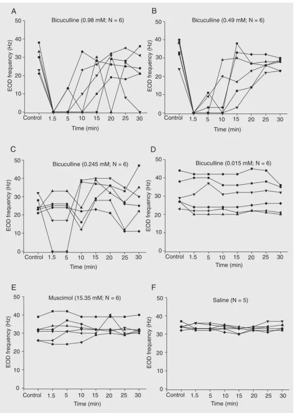
Figure 1. Time course of individual values of the electric organ discharge (EOD) frequencies (Hz) of awake
(11 ± 5.7 and 8.7 ± 5.2 Hz, respectively) were significantly different from the value for saline (31.8 ± 1.51, 31.8 ± 1.7 and 32.1 ± 1.4 Hz, respectively), at the same time inter-vals (P < 0.05, one-way ANOVA on ranks followed by the Dunnett multiple compari-sons test).
Nove lty re sponse
Animals of all groups responded with an increase in EOD frequency (novelty re-sponse) to the artificial electric stimulus pre-sented prior to the microinjections of saline (Figure 3A) or drug. During the EOD inter-ruptions elicited by the microinjection of the highest dose of bicuculline (0.98 mM), the NR did not occur (Figure 3B). After the other doses of 0.49 mM (Figure 3C) and 0.245 mM, the EOD interruptions were over-come by the NR.
Ske le tomotor re sponse
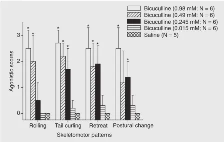
Microinjections of bicuculline into the TSd induced patterns of motor activity that resembled the components of agonistic be-havior (Figure 4). The motor responses were established at the same time as the EOD interruptions. The highest dose of bicucul-line (0.98 mM) elicited a strong behavioral activation expressed by a combination of different patterns: rolling, postural change, forward or backward (retreat) movements, and lateral undulations of the body (tail curl-ing). The agonistic scores of all skeletomotor patterns were different from the values ob-tained with saline (P < 0.05, unpaired t-test).
After lower doses (0.49 and 0.245 mM) a reduction of the number of patterns expressed by the animals, as well as of the intensity of the movements, was observed. With the dose of 0.49 mM, except for postural change, the agonistic scores of the skeletomotor patterns were different from saline (P < 0.05). Roll-ing was less expressed with the dose of 0.245 mM, but the agonistic scores for the
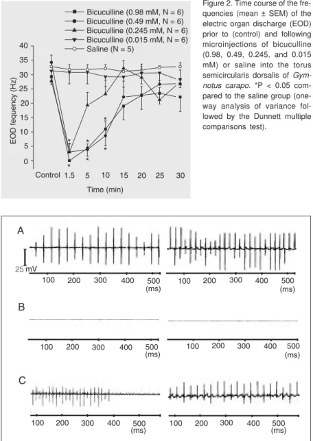
Figure 2. Time course of the fre-quencies (mean ± SEM) of the electric organ discharge (EOD) prior to (control) and following microinjections of bicuculline (0.98, 0.49, 0.245, and 0.015 mM) or saline into the torus semicircularis dorsalis of Gym-notus carapo. *P < 0.05 com-pared to the saline group (one-way analysis of variance fol-lowed by the Dunnett multiple comparisons test).
Figure 3. Examples of electric organ discharge in Gymnotus carapo before (left side) and during (right side) application of an alerting stimulus (square wave pulses of 5-ms duration, 70-Hz repetition frequency and a 100-µA current applied to the water of the experimental cuvette) in animals submitted to microinjections of saline (A), 0.98 mM bicuculline (B), and 0.49 mM bicuculline (C) into the torus semicircularis dorsalis. Vertical bar = 25 mV for A, B and C.
Histo lo gy
Only fish showing histologically con-firmed placement of the microinjections into the TSd were employed for data analysis. Figure 5A is a photomicrograph of a frontal section of the mesencephalon of G. carapo
showing a typical microinjection site in the most medial aspect of the deep layers of the TSd. Figure 5B is a series of drawings of frontal sections of the mesencephalon of several fish showing the location of sites from which EOD interruptions were elicited by bicuculline as well as sites outside the torus (N = 4) which were unresponsive to the drug.
D iscussio n
Our data demonstrate for the first time that microinjection of the GABA-ergic an-tagonist bicuculline into the TSd of the awake unrestrained weakly electric fish G. carapo
induces EOD alterations (variable duration of beat interruptions) that are dose-depend-ent, together with a strong skeletomotor re-sponse, by interfering with tonic toral GABA-ergic inhibition. The highest dose of bicu-culline (0.98 mM) not only caused the most pronounced EOD effect (long-lasting com-plete interruption), but also abolished the NR when the evoking stimulus was pre-sented during the EOD interruption.
Previous studies have shown that the de-crease in EOD frequency involves different brain regions such as the TSd, ventral por-tion of the nE, SPPn and PM (10,11). In
Eigenmannia, the pathway that controls EOD
deceleration starts with neurons in the TSd that are selective for positive frequency dif-ferences, subsequently involves the ventral nE, which in turn lowers the tonic activity of the SPPn via its GABA-ergic input. The diminished activity of the SPPn finally re-duces the glutamatergic NMDA-mediated input to the PM (10). Iontophoretic applica-tion of L-glutamate to the ventral nE or of Figure 4. Intensity scores of skeletomotor patterns evaluated over a period of 2, 4 min after
microinjection of different concentrations of bicuculline or saline into the torus semicircularis dorsalis of Gymnotus carapo. *P < 0.05 compared to the saline group (unpaired t-test)
Figure 5. A, Photomicrograph of a frontal section of the mesencephalon of a representative fish showing the electrolytic lesion placed at the injection site into the torus semicircularis dorsalis (TSd) (arrow). Scale bar = 500 µm. B, Schematic drawings of frontal sections of the mesencephalon of Gymnotus carapo, showing the TSd and associated brain structures. Circles indicate the location of sites inside the TSd in which microinjections of bicuculline elicited motor and electromotor responses. The number of points in the figure is less than the total number of fish used because of several overlaps. Squares indicate the location of sites outside the TSd that were unresponsive to the drug. Scale bar for drawing = 650 µm. CCb = corpus cerebelli; CP/PPn = central-posterior/prepacemaker complex; DFl = nucleus diffusus lateralis of inferior lobe; eTS = torus semicircularis efferents; flm = fasciculus longitudinalis medialis; LL = lateral lemniscus; nE = nucleus electrosensorius; MRF = mesencephalic reticular formation; PE = periglomerular nucleus; PGl = preglomerular nucleus, lateral subdivision; RF = reticular formation (rhombencephalon); TA = nucleus tuberis anterior; TeO = optic tectum; TSd = torus semicircularis dorsalis; tST = subtectal tract; TSv = torus semicircularis ventralis; VCb = valvula cerebelli; VCbm = valvula cerebelli pars medialis.
GABA into the SPPn results in a prompt reduction of the fish’s EOD frequency (11). In Hypopomus pinnicaudatus the activation
of the ventral nE provides glutamatergic activation of the SPPn, which in turn causes a long depolarization of relay cells due to selective activation of NMDA receptors (6). The sustained depolarized relay cells fail to transmit the pacemaker cell rhythm to the motoneurons that drive the electric organ, interrupting the regular EOD, while pace-maker cells keep firing regularly. So, when the relay cells repolarize, the EOD rhythm restarts at a frequency very close to that observed prior to the interruption (6).
The same pathway could be involved in the interruptions of the EOD described in this study in G. carapo since in this species
tract tracing experiments showed the exist-ence of a projection from the TSd to the nE (Duarte TT, Hoffmann A, Pereira ASF and Correa SAL, unpublished results). GABAA receptors of the TSd may play a role in modulating this pathway by controlling the intensity of the tonic inhibition of the pro-jecting neurons. Since the inhibitory effect is a tonic one, muscimol microinjections had no additional effect.
We also observed in the present study that the artificial electric stimulus did not trigger the NR during the EOD interruption elicited by the microinjection of the highest dose of bicuculline into the TSd. Other doses of bicuculline, muscimol or saline did not cause this effect. In Hypopomus, the
in-crease in EOD frequency that occurs in re-sponse to novelties can be elicited by excita-tory inputs from a subnucleus of the CP/PPn complex, the CP/PPn-G, onto the NMDA receptors of the pacemaker cells of the PM (2). In contrast, the EOD interruption is elic-ited through excitatory inputs from the SPPn to the somata of the relay cells of the PM (2). The liberated glutamate binding to NMDA receptors triggers a sustained depolarization in relay cells and, while depolarized, the electric organ no longer fires coherently, so
that the regular EOD is interrupted (6). We postulate that in our experiments, after mi-croinjection of the highest dose of bicucul-line into the TSd, the relay cells submitted to a sustained depolarization fail to transmit the rise of the pacemaker cell rhythm elic-ited by the alerting stimulus. With the lower doses of bicuculline this sustained depolar-ization may be overcome by inputs coming from a transient activation of the CP/PPn-G caused by the novelty stimulus.
The role of the electroreception system in the agonistic interaction of G. carapo is
considered to be of great importance and the discharge frequencies of the animals are posi-tively correlated with social status in domi-nance hierarchies (36). Two main signals are observed in this species during agonistic encounters: short-lived frequency increases or bursts that are used as threatening signals by dominant individuals and complete inter-ruption of discharge which may be of vari-able duration and indicates submission. Ac-cording to Black-Cleworth (1), the EOD interruptions result from a conflict between fleeing tendencies induced by an enemy pro-ducing electrical pulses and other tenden-cies, which vary in different situations, ren-dering the intensity of the fleeing responses proportional to the duration of the EOD interruption. Furthermore, the interruption of a discharge is a natural choice for an appeasement display.
In our experiments, microinjections of different doses of bicuculline into the deep layers of the TSd elicited different types of skeletomotor patterns. In nature, when two
G. carapo individuals interact, their
opponent, maximizing the effect of the di-pole moment. Rolling may be a method of cushioning the shock of a butt from the side, causing the fish to rotate away from the attack. A fish in the rolled posture also pre-sents less surface area to the opponent, so that butts may be less likely to occur (1). Responses like approach, parallel orienta-tion of two fish, and nipping and thrusting, that are part of the aggressive behavior (36), were not observed.
Since GABAA receptor blockade of the TSd in Gymnotus resulted in interruptions of
the EOD, together with strong skeletomotor activation expressed by a combination of different patterns of defensive behavior, we suggest that the GABA-ergic mechanisms of the TSd appear to inhibit the neural
sub-strate of the defense reaction at this midbrain level of the fish. In mammals, it has been demonstrated that sensory collicular input from a given spatial location is not used only for orientation but also to generate a motor output critical for survival. In toads of the genus Bufo, chemical or electrical
stimula-tion of the torus semicircularis induces spe-cies-specific defensive responses (28,37). We therefore postulate that collicular con-trol of defense systems is a phylogenetically old, retained mechanism since it is common to representatives of all vertebrate groups studied. We conclude that the electrosensory information arriving at the TSd is converted into electromotor and skeletomotor com-mands leading to orientation and defense responses.
Re fe re nce s
1. Black-Cleworth P. The role of electrical discharges in the nonrepro-ductive social behavior of Gymnotus carapo (Gymnotidae, Pisces).
Anim Behav Monogr 1970; 31: 1-17.
2. Kawasaki M, Heiligenberg W. Distinct mechanisms of modulation in a neuronal oscillator generate different social signals in the electric fish Hypopomus. J Comp Physiol [A] 1989; 165: 731-741. 3. Keller CH, Kawasaki M, Heiligenberg W. The control of pacemaker
modulations for social communication in the weakly electric fish
Sternopygus. J Comp Physiol [A] 1991; 169: 441-450.
4. Bennett MV. Electrolocation in fish. Ann N Y Acad Sci 1971; 188: 242-269.
5. Dye JC, Meyer JH. Electroreception. In: Bullock TH, Heiligenberg W (Editors), Electroreception. New York: Wiley; 1986. p 71-102. 6. Spiro JE. Differential activation of glutamate receptor subtypes on a
single class of cells enables a neural oscillator to produce distinct behaviors. J Neurophysiol 1997; 78: 835-847.
7. Smith GT, Lu Y, Zakon HH. Parvocells: a novel interneuron type in the pacemaker nucleus of a weakly electric fish. J Comp Neurol
2000; 423: 427-439.
8. Heiligenberg W, Finger T, Matsubara J, Carr C. Input to the medul-lary pacemaker nucleus in the weakly electric fish, Eigenmannia
(Sternopygidae, Gymnotiformes). Brain Res 1981; 211: 418-423. 9. Kawasaki M, Heiligenberg W. Individual prepacemaker neurons can
modulate the pacemaker cycle of the gymnotiform electric fish,
Eigenmannia. J Comp Physiol [A] 1988; 162: 13-21.
10. Metzner W. The jamming avoidance response in Eigenmannia is controlled by two separate motor pathways. J Neurosci 1993; 13: 1862-1878.
11. Heiligenberg W, Metzner W, Wong CJ, Keller CH. Motor control of the jamming avoidance response of Apteronotus leptorhynchus: evolutionary changes of a behavior and its neuronal substrates. J Comp Physiol [A] 1996; 179: 653-674.
12. Carr CE, Maler L, Heiligenberg W, Sas E. Laminar organization of the afferent and efferent systems of the torus semicircularis of gymnotiform fish: morphological substrates for parallel processing in the electrosensory system. J Comp Neurol 1981; 203: 649-670. 13. Bastian J, Yuthas J. The jamming avoidance response of
Eigen-mannia: Properties of a diencephalic link between sensory process-ing and motor output. J Comp Physiol [A] 1984; 154: 895-908. 14. Keller CH. Stimulus discrimination in the diencephalon of
Eigen-mannia: the emergence and sharpening of a sensory filter. J Comp Physiol [A] 1988; 162: 747-757.
15. Meek JR, Nieuwenhuys R. Holosteans and teleosts. In: Nieuwen-huys R, ten Donkelaar HJ, Nicholson C (Editors), The central ner-vous system of vertebrates. Berlin, Heidelberg, New York: Springer-Verlag; 1998. p 855.
16. Heiligenberg W, Rose G. Phase and amplitude computations in the midbrain of an electric fish: intracellular studies of neurons partici-pating in the jamming avoidance response of Eigenmannia. J Neurosci 1985; 5: 515-531.
17. Grau HJ, Bastian J. Neural correlates of novelty detection in pulse-type weakly electric fish. J Comp Physiol [A] 1986; 159: 191-200. 18. Lissmann HW. On the function and evolution of electric organs in
fish. J Exp Biol 1958; 35: 156-191.
19. Corrêa SAL, Hoffmann A. Novelty response in the weakly electric fish Gymnotus carapo: seasonal differences and the participation of the telencephalon in its modulation. Comp Biochem Physiol 1998; 119: 255-262.
20. Kawasaki M, Heiligenberg W. Different classes of glutamate recep-tors and GABA mediate distinct modulations of a neuronal oscillator, the medullary pacemaker of a gymnotiform electric fish. J Neurosci
1990; 10: 3896-3904.
brain-stem nucleus. Proc Natl Acad Sci U S A 1989; 86: 8993-8997. 22. Kennedy G, Heiligenberg W. Ultrastructural evidence of
GABA-ergic inhibition and glutamatGABA-ergic excitation in the pacemaker nucleus of the gymnotiform electric fish, Hypopomus. J Comp Physiol [A] 1994; 174: 267-280.
23. Santana UJ, Roque-da-Silva AC, Duarte TT, Correa SA. Interfer-ence with the GABAergic system in the dorsolateral telInterfer-encephalon and modulation of the electric organ discharge frequency in the weakly electric fish Gymnotus carapo. J Comp Physiol A Neuroethol Sens Neural Behav Physiol 2001; 187: 925-933.
24. Sparks DL. Translation of sensory signals into commands for con-trol of saccadic eye movements: role of primate superior colliculus.
Physiol Rev 1986; 66: 118-171.
25. Dean P, Redgrave P, Westby GW. Event or emergency? Two re-sponse systems in the mammalian superior colliculus. Trends Neurosci 1989; 12: 137-147.
26. Brandão ML, Tomaz C, Borges PC, Coimbra NC, Bagri A. Defense reaction induced by microinjections of bicuculline into the inferior colliculus. Physiol Behav 1988; 44: 361-365.
27. Cardoso SH, Coimbra NC, Brandão ML. Defensive reactions evoked by activation of NMDA receptors in distinct sites of the inferior colliculus. Behav Brain Res 1994; 63: 17-24.
28. Hoffmann A, Brazil Romero SM, de Oliveira LM. Agonistic behavior and its cardiovascular components elicited by microinjection of L-glutamic acid into the basal midbrain of the toad Bufo paracnemis.
Brain Behav Evol 1993; 41: 316-325.
29. Moreira FA, Molchanov ML, Guimarães FS. Flight reactions to nitric oxide in the inferior colliculus of rats depend on NMDA receptor activation. Pharmacol Biochem Behav 2003; 76: 35-41.
30. Carr CE, Maler L. A Golgi study of the cell types of the dorsal torus semicircularis of the electric fish Eigenmannia: functional and mor-phological diversity in the midbrain. J Comp Neurol 1985; 235: 207-240.
31. Correa SA, Correa FM, Hoffmann A. Stereotaxic atlas of the telen-cephalon of the weakly electric fish Gymnotus carapo. J Neurosci Methods 1998; 84: 93-100.
32. Maler L, Sas E, Johnston S, Ellis W. An atlas of the brain of the electric fish Apteronotus leptorhynchus. J Chem Neuroanat 1991; 4: 1-38.
33. Albuquerque-Araujo WIC, Brentegani MR, Hoffmann A. GABAA
re-ceptors of amigdala are involved in the modulation of dorsal immo-bility. Abstr Soc Neurosci 1996; 178.16 (Abstract).
34. Nicholson C. Diffusion from an injected volume of a substance in brain tissue with arbitrary volume fraction and tortuosity. Brain Res
1985; 333: 325-329.
35. Baffa O, Correa SL. Magnetic and electric characteristics of the electric fish Gymnotus carapo. Biophys J 1992; 63: 591-593. 36. Westby GWM. Further analysis of the individual discharge
charac-teristics predicting social dominance in the electric fish Gymnotus carapo. Anim Behav 1975; 23: 249-260.