Gap junctio ns in ce lls o f the
immune syste m: structure , re gulatio n
and po ssible functio nal ro le s
Departamento de Ciencias Fisiológicas,
Pontificia Universidad Católica de Chile, Santiago, Chile J.C. Sáez, M.C. Brañes,
L.A. Corvalán, E.A. Eugenín, H. González, A.D. Martínez and F. Palisson
Abstract
Gap junction channels are sites of cytoplasmic communication be-tween contacting cells. In vertebrates, they consist of protein subunits denoted connexins (Cxs) which are encoded by a gene family. Accord-ing to their Cx composition, gap junction channels show different gating and permeability properties that define which ions and small molecules permeate them. Differences in Cx primary sequences sug-gest that channels composed of different Cxs are regulated differen-tially by intracellular pathways under specific physiological condi-tions. Functional roles of gap junction channels could be defined by the relative importance of permeant substances, resulting in coordina-tion of electrical and/or metabolic cellular responses. Cells of the native and specific immune systems establish transient homo- and heterocellular contacts at various steps of the immune response. Morphological and functional studies reported during the last three decades have revealed that many intercellular contacts between cells in the immune response present gap junctions or “gap junction-like” structures. Partial characterization of the molecular composition of some of these plasma membrane structures and regulatory mechan-isms that control them have been published recently. Studies designed to elucidate their physiological roles suggest that they might permit coordination of cellular events which favor the effective and timely response of the immune system.
Co rre spo nde nce
J.C. Sáez
Departamento de Ciencias Fisiológicas
Pontificia Universidad Católica de Chile
Alameda 340 Santiago Chile
Fax: + 56-2-222-5515 E-mail: jsaez@ genes.bio.puc.cl
Presented at the Meeting “Gap Junctions in the Nervous and Cardiovascular Systems: Clinical Implications”, Rio de Janeiro, RJ, Brazil, June 6-11, 1998.
J.C. Sáez, M.C. Brañes, E.A. Eugenín, A.D. Martínez, and F. Palisson are recipients of FO NDECYT grants (Nos. 1990146, 2960002, 2960001, 2990004 and 2990089, respectively).
Received August 3, 1999 Accepted September 29, 1999
Ke y words
·Cell contacts
·Gap junctions
·Connexins
·Native immune response
·Specific immune response
·Inflammatory response
Intro ductio n
Numerous reports have described differ-ent mechanisms for intercellular communi-cation between members of the immune sys-tem, including cell adhesion molecules, mem-brane molecules that act as ligand-receptors (1) and soluble molecules secreted into the extracellular milieu which act as paracrine and autocrine signals (2). In contrast, little attention has been given to gap junction
in-tercellular communication. Recently, two reviews have described gap junction com-munication between cells of the immune system (3,4). The present article attempts to provide an updated and brief review on gap junctions expressed by different cellular members of native and specific immune sys-tems.
cells and each cell contributes one half of the channel, called connexon. Each hemichannel consists of the oligomers of six protein sub-units termed connexins (Cxs). A gene family of which at least 14 different homologous members have been identified in rodents encodes these proteins. Cxs are named ac-cording to their molecular mass predicted from their cloned DNA sequence. Most cells, excluding vertebrate skeletal muscle, red blood cells and spermatozoids, form gap junctions and express two or more Cx types. Cells of invertebrate organisms express func-tionally equivalent channels, but their pro-tein subunits are not members of the Cx family (6). In recent reports, it has been shown that gap junction channels and hemi-channels can result from the interaction of two different Cxs (5). Moreover, numerous reports have shown that gap junction com-munication can be regulated at various cellu-lar levels, including mRNA transcription, mRNA stability and channel gating (5). Gap junction channels formed by different Cx types present different gating and perme-ability properties that fulfill different func-tions and thus are adjusted to the various regulatory mechanisms present in each cell type and under different physiological con-ditions.
The first descriptions of electrical cou-pling between activated lymphocytes were reported in the early 1970’s (7-9). Further evidence of gap junctional communication, including transfer of fluorescent dyes, meta-bolic cooperativity and electron microscopy studies (thin sections and freeze fracture replicas), reported at that time have been recently reviewed (3,4). The homocellular gap junctional communication at cell-cell contacts between other members of the im-mune system, such as macrophages (10,11), follicular dendritic cells (12), thymic epithe-lial cells (13), polymorphonuclear (PMN) cells (14,15) and microglia (16) has also been reported. In addition, it has been shown that heterocellular contacts between
macro-phages and neutrophils (17), and leukemia cells and myeloid sinus endothelium (18) contain gap junctions. Similarly, “gap junc-tion-like” structures as heterocellular con-tacts between Langerhans cells and T-cells (4,19-21), lymphocytes and endothelial cells (22) and PMN cells and endothelial cells (14) have been described. Moreover, hetero-cellular gap junctional communication be-tween lymphocytes and endothelial cells (23), thymocytes and thymic epithelial cells (13), macrophages and epithelial cells (24-26), mastocytoma cells and lymphocytes (27) and follicular dendritic cells and B-cells (28) has also been reported.
Gap junctions in the bone marrow and se condary lymphoid organs
Both in vivo (29) and in vitro (30,31) studies have demonstrated that bone marrow stromal cells form gap junctions. Morpho-logical and functional studies have also shown gap junctions between bone marrow stromal cells as well as between stromal and hematopoietic progenitor cells (28,32-35). In primary cultures of bone marrow, the extent of dye transfer between cells increases progressively with time in culture (35), sug-gesting that in vitro cells are free from the environmental factors present in vivo that restrain the functional expression of gap junc-tion. Consistently, the reduction in hemato-poietic tissue induced with fluorouracil is followed by a dramatic increase in the num-ber of gap junctions between bone marrow cells and the number falls back to the normal values before the bone is filled with marrow (36).
gap junctional communication between mal cells. Similarly, differentiation of stro-mal cells to adipocytes is associated with a reduction in gap junctional communication (30) and Cx43 reactivity (31). Gap junctions are more abundant in hematopoietic stem cells before growth (36) and in cells of dif-ferent types of leukemias that present an increased stromal:hematopoietic cell ratio (37). Megakaryocytes present in normal bone marrow contain Cx43, but not Cx26 or Cx32 (38). During migration, gap junction-like structures have been identified between neu-trophils or lymphocytes and cells of the sinu-soidal wall (adventitial or endothelial cells) of the bone marrow (39).
The first report of gap junction formation between follicular dendritic cells showed that these cells express Cx43, but not Cx32 or Cx26 (12). Recently, this analysis has been extended showing that they also con-tain Cx37 (4). In mouse lymph nodes, both Cxs 37 and 43, but not Cxs 26, 32, 33, 45 or 50, are present in follicular dendritic cells, interdigitating cells, T-cells and B-cells (4,40). At least the reactivity to Cx43 in follicular dendritic cells is inducible. These Cxs are frequently detected at cell-cell con-tacts, suggesting that they form functional channels. In agreement, dye transfer between cultured human dendritic cells and B lym-phocytes has been shown (28). Gap junc-tions found at heterocellular contacts might be relevant in the diverse events of the im-mune response that occur within lymph nodes, including antigen presentation and lymphocyte proliferation.
Carolan and Pitts (41) have shown meta-bolic coupling between thymocytes, suggest-ing that they establish gap junctional com-munication. This possibility was recently supported by the demonstration of electrical coupling and dye transfer between these cells and blockade of intercellular communica-tion with octanol, a convencommunica-tional gap junc-tion blocker (13). In addijunc-tion, thymic epithe-lial cells and thymocytes communicate with
each other through gap junctions that con-tain at least Cx43 (13). In cultured thymic epithelial cells a significant amount of Cx43 is phosphorylated and cells are well coupled (13).
Gap junctions in the native immune syste m
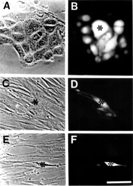
The main cell components of the native immune system are cell barriers (endothelia and epithelia), granulocytes, monocytes/mac-rophages, and natural killer cells. All endo-thelial and epiendo-thelial cells studied express Cxs. Both cell types frequently retain Cx expression and gap junction communication in primary cultures (Figure 1). Exposure to inflammatory mediators reduces gap junc-tion communicajunc-tion between cultured endo-thelial cells. TNF-a and interleukin-1 re-duce dye coupling between human umbilical vein endothelial cells (HUVECs) (42,43). The effect of TNF-a on the expression of Cxs by HUVEC is differential; while Cxs 37 and 40 are reduced, Cx43 remains unchanged (43). Moreover, histamine reduces gap junc-tion communicajunc-tion between high vascular endothelial cells isolated from human ton-sils (Figure 1). In myoendothelial prepara-tions treated with lipopolysaccharides (LPS), TNF-a, or IL-1ß, homocellular coupling re-mains unchanged but the heterocellular cou-pling is drastically reduced (44). Similarly, the heterocellular coupling between rat brain endothelial cells and astrocytes is transiently reduced by TNF-a (45).
trans-fer between lymphocytes and endothelial cells (23) or macrophages P388D1 and IEC-6 epithelial cells has been demonstrated (24-26). In the latter system, gap junction-de-pendent propagation of Ca2+ waves in
re-sponse to mechanical stimulation has also been shown (25), suggesting that these two cell types perform coordinated activities and/ or one regulates the state of the other through
a Ca2+-dependent mechanism mediated by
gap junctions. Polarity of dye movement has been found in studies of gap junction perme-ability between smooth muscle and endothe-lial cells of hamster cheek pouch arterioles (46), suggesting the existence of a direc-tional preference for diffusion of intercellu-lar signals and/or metabolites. It is not known whether gap junctions formed between leu-kocytes and cellular barriers show unidirec-tional permeability preferences.
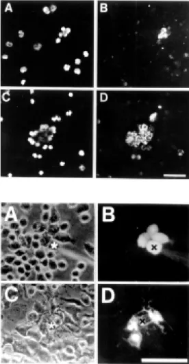
In vertebrates, the main blood cell mem-bers of the native immune response are PMN cells of which the most abundant are neutro-phils. Available information indicates that the expression of Cxs in these cells is induc-ible. Activated human PMN cells form homocellular gap junctions in vitro (15). Moreover, circulating hamster leukocytes do not express Cx43 and after incubation with LPS for 1 h they become reactive to anti-Cx43 antibodies (Figure 2) (14), suggesting that the expression of this protein is induc-ible. In addition, the application of platelet activating factor (PAF) to the hamster cheek pouch induces recruitment and firm adhe-sion of Cx43 positive PMN cells to the endo-thelium of the microcirculation, but fails to induce the expression of Cx43 in isolated leukocytes (47), indicating that PAF-induced Cx43 expression observed in vivo might not result from the direct PAF-hamster leuko-cyte interaction. Similarly, LPS induces for-mation of human PMN aggregates and trans-location of Cx43 towards the plasma mem-brane, but cells remain dye uncoupled. Nev-ertheless, LPS-activated PMN cells in medi-um conditioned by rat brain endothelial cells treated with LPS develop prominent dye cou-pling (15).
Depending on the circulatory region, en-dothelial cells express Cx43 and Cx40 and/ or Cx37 (5). Since these Cxs form gap junc-tions with different permeability and gating properties (5), differences in Cx composi-tion of the homocellular (endothelial cell-endothelial cell) and heterocellular
thelial cell-smooth muscle cell) gap junc-tions formed might explain the dye move-ment polarity found in hamster cheek pouch arterioles (46). During an inflammatory re-sponse, endothelial cells also form gap junc-tions with activated leukocytes (14), sug-gesting that endothelial Cxs are sorted to the apical membrane to form gap junction chan-nels with compatible leukocyte Cxs.
Connective tissues contain a variety of cells with defense and immune functions, such as tissue macrophages and mast cells. The first demonstrations of gap junctional communication between cultured canine and murine macrophage cells were reported two decades ago (10,11). But, it was only during the last decade that Cx43 was detected in several macrophage types, including the murine cell line J774 (48), macrophage foam cells from arteriosclerotic lesions (49), peri-toneal macrophages (14), kidney macrophag-es in inflammatory renal disease (50), Kupffer cells (51), microglia (16) and Langerhans cells (4). Cx43 mRNA has been detected in cultured monocytes/macrophages (52), but not in freshly isolated human monocytes/ macrophages (49). Moreover, it has been recently reported that mast cells express Cxs 32 and 43, but not Cx26 (53).
J774 macrophages (54), human cytes/macrophages or HUVECs and mono-cytes/macrophages (49) do not establish in-tercellular communication in culture. Nev-ertheless, P388D1 or J744 macrophages co-cultured with epithelial cell lines show homocellular dye coupling, as well as het-erocellular dye coupling with epithelial cell lines (25), suggesting that soluble factors present in the co-culture induce macrophag-es to form gap junctions. In support of this possibility, culture medium conditioned with endothelial cells derived from rat brain mi-crocirculation induces dye coupling (Figure 3) and translocation of Cxs from the cyto-plasmic compartment to the plasma mem-brane in J774 cells (Eugenín EA, Garcés G and Sáez JC, unpublished observation).
Mi-croglia, the main immune effector of the central nervous system, also become dye coupled when cultured for a few hours in medium conditioned by rat brain endothelial cells (Eugenín EA, Martínez AD and Sáez JC, unpublished observation). Dye coupling between microglia is also observed after 4-9-h treatment with a calcium ionophore (16) (Figure 3), suggesting that activated macro-phages can establish gap junctional commu-nication.
Structural and functional studies have demonstrated cell junctions equivalent to gap junctions between invertebrate blood cells (hemocytes) (55). These cells establish functional intercellular communication within seconds when they are pushed to-gether (55), suggesting that hemocytes pres-ent a preformed pool of hemichannels for
Figure 2 - Cx43 is not found in circulating PM N cells and its ex-pression is induced by LPS. M ost freshly isolated hamster leukocytes incubated for 3 h at 37oC in culture medium
contain-ing 5% FBS remained as scontain-inglet cells and very few w ere immu-noreactive to Cx43 (B). Nonethe-less, cells treated w ith 1 mg/ml LPS for 3 h formed many aggre-gates and w ere immunoreactive to Cx43 (D). In each situation, the cells show n in (B) and (D) w ere identified by their nuclear staining w ith DAPI in A and C, respectively. Bar: 75 µm.
ready formation of intercellular channels. The structural components of these channels remain unknown, but it is likely that they are proteins homologous to those described to form intercellular channels in Drosophila melanogaster and C. elegans, termed innex-ins (6).
Gap junctions be twe e n ce lls of the spe cific immune syste m
Activation of a specific immune response requires a direct physical interaction between antigen-presenting cells and T-cells, the main cellular effector of the specific immune sys-tem (1). At Langerhans and T-cell inter-phases, gap junction-like structures have been identified both in vitro (19,20) and in vivo
(21). At cell-cell contacts between cultured Langerhans cells and T-cells, at least Cx43 is detected (4). The formation of gap junction channels requires a cell-cell proximity medi-ated by cell adhesion molecules (5). Thus, the anti-vascular cell adhesion molecule-1 (VCAM-1) antibody-induced inhibition of the lymphocyte proliferative response in the allogeneic mixed lymphocyte reaction (56) might be the consequence, at least in part, of the blockade of a gap junction-dependent mechanism.
Lymphocytes (T-cells plus B-cells or just T-cells) treated with either concanavalin A (Con-A) or phytohemagglutinin (PHA) form clusters of variable sizes. Circulating human or bovine lymphocytes treated with PHA express a low resistance pathway that allows the intercellular transfer of electrical stimuli (7,8). Moreover, intercellular transfer of flu-orescein or radiolabeled uridine has been found between mouse spleen lymphocytes, rabbit mesenteric lymphocytes, murine thy-mic lymphocytes and lymph node lympho-cytes (4,27,41,57). Electrical coupling be-tween activated lymphocytes is blocked by an increase in intracellular Ca2+
concentra-tion (9). In addiconcentra-tion, dye coupling is revers-ibly blocked with octanol and prevented with
synthetic peptides homologous to the extra-cellular loop 1 of Cxs (40), supporting the idea that electrical and metabolic coupling between activated lymphocytes occurs through gap junction channels. Consistently, mouse lymphocytes contain Cxs 37 and 43, but not Cxs 32, 33, 40 or 50, and upon treatment with Con-A both Cxs are translo-cated from the plasma membrane to cellular interphases (40). The latter event occurs with-out changes in Cx levels, suggesting that freshly isolated lymph node lymphocytes contain a preformed pool of Cxs. On the other hand, in vivo studies have shown that Cx43 expression by cells of mouse lymph nodes is induced by the administration of antigen (28). Moreover, in situ hybridization studies have shown that follicular dendritic cells and lymphocytes of germinal centers of other secondary lymphoid organs, such as human tonsil and spleen, also express Cx43 (28).
Functional role s of gap junctions in ce lls of the immune syste m
Although in some systems reduced gap junction communication is associated with an increase in tissue function, such as amy-lase secretion by the exocrine pancreas, more frequently it has been demonstrated to cause tissue disfunction (5). Inhibition of gap junc-tional communication of the rat gastric mu-cosa in combination with ischemia-reperfu-sion weakens the barrier function of the gastric mucosa and causes damage to the barrier function (58). Moreover, in long-term cultures of bone marrow the blockade of gap junctions with amphotericin retards stem cell growth (37). In addition, blockade of thymocyte gap junctions with octanol re-duces the secretion of thymulin (13).
com-munication. The latter possibility was re-cently supported by the finding that syn-thetic peptides homologous to the extracel-lular loop 1 of Cxs prevent gap junction formation and drastically reduce the DNA replication of Con-A-treated mouse lympho-cytes (40). Thus, gap junctional communica-tion between proliferating lymphocytes might coordinate their metabolic and cytokine-in-duced responses to allow the appropriate timing of the specific immune response. Simi-larly, the blockade of leukemic cell differen-tiation has been associated with their inter-cellular coupling to stromal cells (34).
The innate and specific immune responses involve homo- and heterocellular contacts essential for their normal functioning. In
many of those events, gap junctional com-munication is established, but their func-tional roles remain speculative except for few cases described above for which direct or indirect evidence has been provided. A putative gap junction role is synchronization of cellular events during the transmigration across cellular barriers. Supporting this view, gap junctions have been observed between metastase-forming leukemia cells and my-eloid sinus endothelium (18), polymorpho-nuclear and endothelial cells (14) and mac-rophages and epithelial cells (24-26).
Clearly, further studies are needed to un-derstand the role of gap junctions in differ-ent physiological and pathophysiological functions of the immune system.
Re fe re nce s
1. Springer T (1990). Adhesion receptors of the immune system. Nature, 346: 425-434.
2. Ben-Baruch A, M ichiel DF & Oppenheim JJ (1995). Signals and receptors involved in recruitment of inflammatory cells. Jour-nal of Biological Chemistry, 270: 11703-11706.
3. Alves LA, Campos de Carvalho AC & Savino W (1998). Gap junctions: a novel route for direct cell-cell communication in the immune system? Immunology Today, 19: 269-275.
4. Sáez JC, Araya R, Brañes M C, Concha M , Contreras JE, Eugenín EA, M artínez AD, Palisson F & Sepúlveda M A (1999). Gap junctions in inflammatory responses: con-nexins, regulation and possible functional roles. In: Peracchia C (Editor), Gap Junc-tions - M olecular Basis of Cell Communi-cation in Health and Diseases. Current Topics in M embranes. Academic Press, San Diego (in press).
5. Bruzzone R, White TH & Paul DL (1996). Connections w ith connexins: the molecu-lar basis of direct intercellumolecu-lar signaling. European Journal of Biochemistry, 238: 1-27.
6. Phelan P, Bacon JP, Davies JA, Stebbings LA, Todman M G, Avery L, Baines RA, Barnes TM , Ford C, Hekimi S, Lee R, Shaw JE, Starich TA, Cutin KD, Sun YA & Wyman RJ (1998). Innexins: a family of invertebrate gap-junction proteins. Trends
in Genetics, 14: 348-349.
7. Hülser DF & Peters JH (1971). Intercellu-lar communication in phytohemagglutinin-induced lymphocyte agglutinates. Euro-pean Journal of Immunology, 1: 494-495. 8. Oliveira-Cast ro GM , Barcinski M A & Cukierman S (1973). Intercellular commu-nication in stimulated human lympho-cytes. Journal of Immunology, 111: 1616-1619.
9. Oliveira-Cast ro GM & Barcinski M A (1974). Calcium-induced uncoupling in communicating human lymphocytes. Bio-chimica et Biophysica Acta, 352: 338-343. 10. Levy JA, Weiss RM , Dirksen EL & Rosen M R (1976). Possible communication be-tw een murine macrophages oriented in linear chains in tissue culture. Experimen-tal Cell Research, 103: 375-385. 11. Porvaznik M & M acVittie TJ (1979).
De-tection of gap junctions betw een the progeny of a canine macrophage colony-forming cell in vitro. Journal of Cell Biol-ogy, 82: 555-564.
12. Krenács T & Rosendaal M (1995). Immu-nohistologic detection of gap junctions in human lymphoid tissue: connexin43 in fol-licular dendritic and lymphoepithelial cells. Journal of Histochemistry and Cytochem-istry, 43: 1125-1137.
13. Alves LA, Campos de Carvalho AC, Lima EOC, Rocha e Souza CM , Dardene M , Spray DC & Savino W (1995). Functional gap junctions in thymic epithelial cells are
formed by connexin 43. European Journal of Immunology, 2: 431-437.
14. Jara PI, Boric M P & Sáez JC (1995). Leu-kocytes express connexin43 after activa-tion w ith lipopolysaccharide and appear to form gap junctions w ith endothelial cells after ischemia-reperfusion. Proceed-ings of the National Academy of Sciences, USA, 92: 7011-7015.
15. Brañes M C, Contreras JE, Bono M R & Sáez JC (1997). Hum an polym orpho-nuclear cells express connexins and form homologous gap junctions. M olecular Bi-ology of the Cell, 8: 417a (Abstract). 16. M artínez AD & Sáez JC (1998). Rat
micro-glia express connexins and upon activa-tion form gap juncactiva-tions. M olecular Biol-ogy of the Cell, 9: 326a (Abstract). 17. Afonso A, Silva J, Lousada S, Ellis AE &
Silva M T (1998). Uptake of neutrophils and neutrophilic components by macro-phages in the inflamed peritoneal cavity of rainbow trout (Oncorhynchus mykiss). Fish and Shellfish Immunology, 8: 319-338.
18. De Bruyn PPH, Cho Y & M ichelson S (1989). Endothelial attachment and plas-malemmal apposition in the transcellular movement of intravascular leukemic cells entering the myeloid parenchyma. Ameri-can Journal of Anatomy, 186: 115-126. 19. Concha M , Figueroa CD & Caorsi I (1988).
lymphocytes. Journal of Pathology, 156: 29-36.
20. Concha M , Vidal A, Garcés G, Figueroa CD & Caorsi I (1993). Physical interaction betw een Langerhans cells and T-lympho-cytes during antigen presentation in vitro. Journal of Investigative Derm atology, 100: 429-434.
21. Brand CU, Hunziker T, Schaffner T, Limat A, Gerber HA & Braathen LR (1995). Acti-vated immunocompetent cells in human skin lymph-derived from irritant contact dermatitis: an immunomorphologic study. British Journal of Dermatology, 132: 39-45.
22. Raine CS, Cannella B, Dujivestijn AM & Cross AH (1990). Homing to central ner-vous system vasculature by antigen-spe-cific lymphocytes. II. Lymphocyte/endo-thelial cell adhesion during the initial stages of autoimmune demyelination. Laboratory Investigation, 63: 476-479. 23. Guinan S, Smith BR, Davies PF & Pober
JS (1988). Cytoplasmic transfer betw een endothelium and lymphocytes: quantita-tion by flow cytometry. American Journal of Pathology, 132: 406-409.
24. El-Sabban M E, M artin CA & Homaidan FR (1998). Signaling betw een immune cells and intestinal epithelial cells in vitro. In: Werner R (Editor), Gap Junctions. IOS Press, The Netherlands, 178-182. 25. M art in CA, Hom aidan FR, Palaia T,
Burakoff R & El-Sabban M E (1998). Gap junctional communication betw een mu-rine macrophages and intestinal epithelial cell lines. Cell Adhesion and Communica-tion,5: 437-449.
26. M art in CA, El-Sabban M E, Zhao L, Burakoff R & Homaidan FR (1998). Adhe-sion and cytosolic dye transfer betw een m acrophages and intestinal epithelial cells. Cell Adhesion and Communication, 5: 83-95.
27. Sellin D, Wallach DFH & Fischer H (1971). Intercellular communication in cell-medi-ated cytotoxicity fluorescein transfer be-tw een H-2d target cells and H-2b
lympho-cytes in vitro. European Journal of Immu-nology, 4: 189-193.
28. Krenács T, Van Dartel M , Lindhout E & Rosendaal M (1997). Direct cell/cell com-munication in the lymphoid germinal cen-ter: connexin43 gap junctions functionally couple follicular dendritic cells to each other and to B lymphocytes. European Journal of Immunology, 27: 1489-1497. 29. Watanabe Y (1985). Fine structure of bone
m arrow st rom a. Act a Haem at ologica Japonica, 48: 1688-1695.
30. Um ezaw a A, Harigaya K, Abe H &
Watanabe Y (1990). Gap-junctional com-munication of bone marrow cells is resist-ant to irradiation in vitro.Experimental He-matology, 8: 1002-1007.
31. Umezaw a A & Hata J (1992). Expression of gap-junctional protein (connexin43 or alpha 1 gap junction) is dow n regulated at the transcriptional level during adipocyte differentiation of H-1/A marrow stromal cells. Cell Structure and Function, 17: 177-184.
32. Ohkaw a H & Harigaya K (1987). Effect of direct cell-cell interaction betw een the KM -102 clonal human marrow stromal cell line and the HL-60 myeloid leukemic cell line on the differentiation and prolifera-tion of the HL-60 line. Cancer Research, 47: 2879-2882.
33. Allen T & Dexter TM (1984). The essential cells of the hematopoietic microenviron-ment. Experimental Hematology, 12: 517-521.
34. Weber M C & Tykocinski M L (1994). Bone marrow stromal cell blockade of human leukemic cell differentiation. Blood, 83: 2221-2229.
35. Rosendaal M , Gregan A & Green CR (1991). Direct cell-cell communication in the blood-forming system. Tissue and Cell, 23: 457-470.
36. Rosendaal M , Green CR, Rahman A & M organ D (1994). Up-regulation of the connexin43+ gap junction netw ork in hae-mopoietic tissue before the grow th of stem cells. Journal of Cell Science, 107: 29-37.
37. Krenács T & Rosendaal M (1998). Con-nexin43 gap junctions in normal, regener-ating, and cultured mouse bone marrow and in human leukemias. Their possible involvement in blood formation. Ameri-can Journal of Pathology, 152: 993-1004. 38. Dorshkind K, Green L, Godw in A & Fletcher WH (1993). Connexin-43-type gap junctions mediate communication be-tw een bone marrow stromal cells. Blood, 82: 38-45.
39. Campbell FR (1982). Intercellular contacts betw een migrating blood cells and cells of the sinusoidal w all of the bone mar-row . An ultrastructural study using tannic acid. Anatomical Record, 20: 365-374. 40. Sáez JC, Sepúlveda M A, Araya R, Sáez
CG & Palisson F (1998). Concanavalin A-activated lymphocytes form gap junctions that increase their rate of DNA replica-tion. In: Werner R (Editor), Gap Junctions. IOS Press, Amsterdam, 372-376. 41. Carolan E & Pitts JD (1986). Some murine
thymic lymphocytes can form gap junc-tions. ImmunologicalLetters, 13: 255-260.
42. Hu VW & Xie HQ (1994). Interleukin-1a suppresses gap junction-mediated inter-cellular communication in human endo-thelial cells. Experimental Cell Research, 213: 218-223.
43. van Rijen HV, van Kempel M J & Jongsma HJ (1998). Tumor necrosis alpha alters the expression of connexin43, connex-in40, and connexin37 in human umbilical vein endothelial cells. Cytokine, 10: 258-264.
44. Hu J & Cotgreave IA (1997). Differential regulation of gap junctions by proinflam-matory mediators in vitro. Journal of Clini-cal Investigation, 99: 2312-2316. 45. Brañes M C, M artínez AD, Recabarren M ,
Couraud PO & Sáez JC (1998). Regulation of gap junctions formed betw een endo-thelial cells (ECs) and ECs and astrocytes. M olecular Biology of the Cell, 9: 325a (Abstract).
46. Little TL, Xia J & Duling BR (1995). Dye tracers define differential endothelial and smooth muscle coupling patterns w ithin the arteriolar w all. Circulation Research, 76: 498-504.
47. Boric M P, Roth A, Jara P & Sáez JC (1997). Gap junction betw een leukocytes and endothelium : expression of con-nexin43 in adherent or activated cells. In: Latorre R & Sáez JC (Editors), From Ion Channels to Cell-to-Cell Conversations. Plenum Press, New York, 249-366. 48. Beyer EC & Steinberg TH (1991).
Evi-dence that gap junction protein connexin-43 is the ATP-induced pore of mouse macrophages. Journal of Biological Chem-istry, 266: 7971-7974.
49. Polacek D, Lal L, Volin M V & Davies PF (1993). Gap junctional communication be-tw een vascular cells. Induction of con-nexin43 messenger RNA in macrophage foam cells of atherosclerotic lesions. American Journal of Pathology, 142: 593-606.
50. Hillis GS, Duthie LA, Brow n PA, Simpson JG, M acLeod AM & Haites NE (1997). Upregulation and co-localization of con-nexin43 and cellular adhesion molecules in inflammatory renal disease. Journal of Pathology, 18: 373-379.
51. Sáez CG, Eugenín E, Hertzberg EL & Sáez JC (1997). Regulation of gap junction in rat liver during acute and chronic CCl4
-induced liver injury. In: Latorre R & Sáez JC (Editors), From Ion Channels to Cell-to-Cell Conversations. Plenum Press, New York, 367-380.
(1991). Interaction of monocytes w ith co-cultures of human aortic w all cells in-volves interleukins 1 and 6 w ith marked increases in connexin43 message. Jour-nal of Clinical Investigation, 8: 1763-1772. 53. Vliagotis H, Hutson AM , M ahmudi-Azer S, Kim H, Rumsaeng V, Oh CK, M oqbel R & M etcalfe DD (1999). M ast cells express connexins on their cytoplasmic mem-brane. Journal of Allergy and Clinical Im-munology, 103: 656-662.
54. Alves LA, Countinho-Silva R, Persechini PM , Spray DC, Savino W & Campos de Carvalho AC (1996). Are there functional gap junctions or junctional hemichannels
in macrophages? Blood, 8: 328-334. 55. Churchill D, Coodin S, Shivers RR &
Caveney S (1993). Rapid de novo forma-tion of gap juncforma-tions betw een insect he-mocytes in vitro: a freeze-fracture, dye transfer and patch clamp study. Journal of Cell Science, 104: 763-772.
56. Lukacs NW, Strieter RM , Evanoff HL, Burdick M D & Kunkel SL (1994). VCAM -1 influences lymphocyte proliferation and cytokine production during mixed lympho-cyte responses. Cellular Immunology, 154: 88-98.
57. Sellin D, Wallach DFH, Weltzein HV, Esch K, Sprenger E & Fisher H (1974).
Intercel-lular communication betw een lympho-cytes in vitro. Fluorescein-permeable junc-tions, their enhancement by lysolecithin and their reduction by synthetic immuno-suppressive lysolecithin analogue. Euro-pean Journal of Immunology, 4: 189-193. 58. Iw ata F, Joh T, Ueda F, Yokoyama Y & Itoh M (1998). Role of gap junctions in inhibiting ischemia-reperfusion injury of rat gastric mucosa. American Journal of Physiology, 275: G883-G888.