Impaired beta-adrenergic response and
decreased L-type calcium current of
hypertrophied left ventricular myocytes
in postinfarction heart failure
1Laboratório de Eletrofisiologia Cardíaca Antonio Paes de Carvalho,
Instituto de Biofísica Carlos Chagas Filho, and 2Departamento de Cardiologia,
Faculdade de Medicina, Universidade Federal do Rio de Janeiro, Rio de Janeiro, RJ, Brasil
R.M. Saraiva2,
N.G.B. Chedid1,
C.C. Quintero H.1,
L.E. Díaz G.1 and
M.O. Masuda1
Abstract
Infarct-induced heart failure is usually associated with cardiac hyper-trophy and decreased ß-adrenergic responsiveness. However, con-flicting results have been reported concerning the density of L-type calcium current (ICa(L)), and the mechanisms underlying the decreased
ß-adrenergic inotropic response. We determined ICa(L) density,
cyto-plasmic calcium ([Ca2+]
i) transients, and the effects of ß-adrenergic
stimulation (isoproterenol) in a model of postinfarction heart failure in rats. Left ventricular myocytes were obtained by enzymatic digestion 8-10 weeks after infarction. Electrophysiological recordings were obtained using the patch-clamp technique. [Ca2+]
i transients were
investigated via fura-2 fluorescence. ß-Adrenergic receptor density was determined by [3H]-dihydroalprenolol binding to left ventricle
homogenates. Postinfarction myocytes showed a significant 25% reduction in mean ICa(L) density (5.7 ± 0.28 vs 7.6 ± 0.32 pA/pF) and a
19% reduction in mean peak [Ca2+]
i transients (0.13 ± 0.007 vs 0.16 ±
0.009) compared to sham myocytes. The isoproterenol-stimulated increase in ICa(L) was significantly smaller in postinfarction myocytes
(Emax: 63.6 ± 4.3 vs 123.3 ± 0.9% in sham myocytes), but EC50 was not
altered. The isoproterenol-stimulated peak amplitude of [Ca2+] i
tran-sients was also blunted in postinfarction myocytes. Adenylate cyclase activation through forskolin produced similar ICa(L) increases in both
groups. ß-Adrenergic receptor density was significantly reduced in homogenates from infarcted hearts (Bmax: 93.89 ± 20.22 vs 271.5 ±
31.43 fmol/mg protein in sham myocytes), while Kd values were similar. We conclude that postinfarction myocytes from large infarcts display reduced ICa(L) density and peak [Ca2+]i transients. The response
to ß-adrenergic stimulation was also reduced and was probably related to ß-adrenergic receptor down-regulation and not to changes in aden-ylate cyclase activity.
Correspondence R.M. Saraiva Rua Abélia, 225/101 Ilha do Governador 21940-010 Rio de Janeiro, RJ Brasil
Fax: +55-21-3393-7683 E-mail: rsaraiva.ntg@terra.com.br
Research supported by CNPq, CAPES-MEC, FAPERJ, CEPG-UFRJ and MCT-PRONEX.
Received June 5, 2002 Accepted January 6, 2003
Key words
·Ca2+ channel ·Heart failure ·Hypertrophy ·Infarction
Introduction
After myocardial infarction, the acute loss of myocytes leads to an increased load to the heart and to the onset of a cascade of biochemical signaling processes that induce the remodeling of the infarcted zone and of the remote noninfarcted myocardium. Ven-tricular remodeling is complex and includes hypertrophy, which counterbalances the in-creased wall stress and attenuates progres-sive dilation. Nevertheless, heart failure is often the final result of this process after large infarcts (1,2).
The model of chronic infarct in rats is characterized by the presence of hypertro-phy and, in the case of large infarcts, of heart failure (3-8), and represents a clinically rel-evant model of left ventricle dysfunction (9). The decrease in the mechanical performance in this model includes not only the loss of myocardium caused by the infarct, but also decreased contractility and response to ß-adrenergic stimulation of the remaining hy-pertrophied myocardium (3,5,8,10). How-ever, the mechanisms underlying the reduc-tions of inotropism and the response to ß-adrenergic stimulation are not completely understood.
The changes in cardiomyocyte function can be traced to several steps in excitation-contraction coupling and calcium handling. Among them, alterations in L-type calcium current (ICa(L)), cytoplasmic calcium ([Ca2+]i)
transients during contraction, and ß-adrener-gic modulation have been studied, but the data in the literature are quite controversial. L-type calcium currents have been mostly described as unaltered (3,11-14), but also as reduced (15,16) in postinfarction hypertro-phied myocytes isolated from the left ven-tricle (postinfarction myocytes). On the other hand, dihydropyridine (DHP)-binding sites have always been shown to be reduced (14,17,18), even in a study in which ICa(L)
density was found to be maintained (14). The peak amplitude of [Ca2+]
i transients has
been shown to be reduced (3,11,19) or main-tained (5,20).
The effect of ß-adrenergic stimulation on the isometric contraction of isolated papil-lary muscles from infarcted rats has been shown to be reduced (5,8,21). However, the effect of ß-adrenergic stimulation on ICa(L) in
postinfarction myocytes has been described either as reduced (14) or maintained (15). Also, the increase in the peak amplitude of [Ca2+]
i transients induced by ß-adrenergic
stimulation has been shown to be attenuated (5) or augmented (22) in postinfarction myo-cytes. Binding experiments showed even more diverse results, i.e., maintained (22-25), reduced (6,8,18) or increased (26) ß-adrenergic receptor density.
These conflicting results could reflect the wide variability among the models used by the different investigators. There are im-portant differences in the size of the in-farcted area and in the times after infarction that could lead to differences in the severity of hypertrophy and heart failure. In addition, in the case of ICa(L) density, the use of a small
number of cells as frequently occurs in elec-trophysiological experiments may also limit adequate statistical analysis (27) and thus contribute to these controversial results.
Thus, we propose to reevaluate the follow-ing parameters in a well-defined model of large chronic infarction in rats, with cellular hypertrophy and clear signals of heart failure, 8 to 10 weeks after surgery: 1) ICa(L) density in
a large number of cells to permit adequate statistical analysis, 2) [Ca2+]
i transients, 3) the
effect of ß-adrenergic stimulation on these two parameters, 4) the effect of forskolin (an aden-ylate cyclase activator) on ICa(L), and 5) the
density of ß-adrenergic receptors.
Material and Methods
Reagents
Darmstadt, Germany); l-isoprenaline hydro-chloride, tetraethylammonium chloride (TEA), 4-aminopyridine (4-AP), HEPES, EGTA, Tris, phenylmethylsulfonyl fluoride (PMSF), polyethylenimine, cesium chloride, alprenolol and forskolin (Sigma, St. Louis, MO, USA); collagenase type 2 (Worthing-ton, Lakewood, NJ, USA); EDTA (Reagen, Rio de Janeiro, RJ, Brazil); fura 2-AM (Mo-lecular Probes, Eugene, OR, USA), and [3
H]-dihydroalprenolol ([3H]-DHA, 120 Ci/mmol;
New England Nuclear Life Science Prod-ucts, Boston, MA, USA).
Experimental infarction
Myocardial infarction was produced in Wistar rats of both sexes weighing 200-250 g by ligature of the left coronary artery ac-cording to protocols that established the effi-cacy of the method to produce large infarcts (16,28). Sham-operated rats were subjected to the same surgical procedure, with the exception of left coronary artery ligature. Eight to ten weeks after surgery, age- and sex-matched rats were sacrificed under ether anesthesia and hearts rapidly removed and treated according to the specific experiments to be performed. This investigation conforms to the Guide for the Care and Use of Labora-tory Animalspublished by the US National Institutes of Health (NIH Publication No. 85-23, revised 1996) as attested by the com-petent institutional board.
Criteria for inclusion in the myocardial infarction group
Only hearts from infarcted rats that at visual inspection had large infarctions were assigned to the myocardial infarction group. No precise quantification of the infarction was performed since all experiments required a rapid manipulation of the heart soon after animal sacrifice. The efficacy of this visual inspection to include only hypertrophied, failing hearts in this study was confirmed in
a group of 13 infarcted and 13 sham-oper-ated animals in which several parameters associated with cardiac hypertrophy and heart failure were evaluated (Table 1).
Cell isolation
Ventricular myocyte isolation was per-formed as reported (16). Briefly, after rats were sacrificed, the hearts were rapidly re-moved and attached to a modified Langen-dorff apparatus for coronary perfusion with Tyrode solution (132 mM NaCl, 1.25 mM
CaCl2, 1.2 mM MgCl2, 4 mM KCl, 10 mM
HEPES, and 5 mM glucose, pH 7.35) satu-rated with oxygen. When blood was com-pletely washed out the perfusion solution was changed to a calcium-free Tyrode solu-tion until the heart stopped beating. Colla-genase (0.5 mg/ml) was then added and per-fusion was maintained until the heart ac-quired a soft consistency (usually within 6 to 10 min). Then, washout of the enzyme was performed by perfusing the heart with the same calcium-free Tyrode solution. Next, fragments of the remaining intact left ven-tricular free wall and intervenven-tricular sep-tum were cut and kept in the calcium-free external solution (142 mM NaCl, 1.0 mM
MgCl2, 4 mM KCl, 10 mM HEPES, and 10
mM glucose, pH 7.4), to which CaCl2 was
progressively added until the normal con-centration of 1.25 mM to prevent cell death by calcium overload. Isolated myocytes were obtained by gently shaking the myocardium fragments in the external solution. Only qui-escent rod-shaped cells with smooth bor-ders, sharp striations and no vacuolization were selected for study.
Voltage-clamp studies
an inverted microscope (Nikon Diaphot-TMD, Tokyo, Japan) and continuously superfused with the external solution (see composition above) to which 2 mM 4-AP had been added.
Cell currents were measured using a patch-clamp amplifier (Axopatch 1D; Axon Instruments, Foster City, CA, USA) and mi-croelectrodes of 4-5 MW tip resistance (when filled with internal solution) made from 1.2-mm outer diameter glass capillaries with a three-stage pipette puller (Sutter Instruments Co., Novato, CA, USA). For data acquisition and analysis, a PC computer connected through a Digidata 1200 interface (Interface Axon Instruments) to the patch-clamp am-plifier was used in conjunction with the pCLAMP software version 6.0.3 (Axon In-struments).
The pipette solution contained 100 mM
CsCl, 20 mM NaCl, 0.5 mM CaCl2, 2.0 mM
MgCl2, 11 mM EGTA-Cs, 10 mM HEPES,
and 20 mM TEA-Cl, pH 7.2. Cesium, 4-AP and TEA were used in order to minimize potassium currents. ICa(L) was elicited by 12
depolarizing clamp steps from a holding po-tential of -90 mV, with a pre-pulse to -40 mV for 200 ms to inactivate Na+ and T-type
calcium currents (30), and with test poten-tials ranging from -60 to +50 mV for 500 ms. The interpulse interval was 4 s to assure complete recovery from ICa(L) inactivation.
ICa(L) was measured as the peak inward
cur-rent with reference to the curcur-rent at the end of the test pulse and normalized by dividing the current amplitude by the cell capaci-tance, measured as previously described (16). Activation curves were plotted as Gtest/Gmax
ratio against voltage. Conductance for each test potential (Gtest) was obtained as
previ-ously described (16). The steady-state acti-vation curves were obtained by mathemati-cal adjustment of the data according to Boltzman equation (y = A/{1b + exp[(x -V0.5)/s]}), where V0.5 is the half-activation
potential and s is the slope factor. Current decay was biphasic and time constants were
measured using the following equation: f(t) = a + b exp-t/t
fast + c exp-t/tslow.
Fluorescence measurements of [Ca2+] i
Isolated myocytes placed in a 1.0-ml su-perfusion chamber mounted on the stage of a Nikon inverted microscope (Diaphot 300) equipped for fluorescence and photometry were loaded with fura 2-AM, the membrane permeant form of an UV-excitable, ratio-metric calcium indicator. Fura 2-AM was added to the external solution to a final concentration of 4 µM at room temperature and kept for 20 min. The cells were then superfused with the external solution for 5 min at 0.5 ml/min to allow complete dye washout. Field stimulation (SEN 3201 stimu-lator, Nihon Kohden, Tokyo, Japan) was applied at 0.5 Hz through platinum wire electrodes attached to the bottom and the sides of the chamber. The area for fluores-cence measurement was restricted to the minimum necessary to completely include the selected cell in order to minimize back-ground fluorescence. For dye excitation, a high-speed dual-wavelength scanning illu-minator (xenon arc lamp, 75 W) capable of switching between 340 and 380 nm at a speed of 650 ratios/s (Delta Scan, Photon Technology International, South Brunswick, NJ, USA) was used. Excitation light was directed at myocytes only during data acqui-sition to minimize photobleaching. Emitted light was detected by a photomultiplier tube after a 520-nm filter and recorded at 100 points/ s using a computer-based data acquisition sys-tem (Photon Technology International). The ratio of the background-subtracted fluores-cence signals (340/380) expressed in arbitrary units was used to estimate [Ca2+]
i.
Peak amplitude, mean rate of increase to the peak (RtPCa) and mean decay rate to 50% amplitude (RD50) of the [Ca2+]i
amplitude of [Ca2+]
i transients represents the
mean of 10 successive cycles and RtPCa and RD50 were measured in one representative
transient for a given experimental condition.
Membrane preparation
For this purpose, after sacrifice the hearts were rapidly removed and transferred to ice-cold Tyrode solution. Atria and connective tissue were discarded and the right ventricle, the scar tissue and the remaining left ven-tricle including the interventricular septum were weighed separately. The scar tissue was discarded and left and right ventricles were processed separately. The following steps were all performed at 4ºC. Ventricular tissue was minced and suspended in ice-cold buffer A (10 mM Tris-HCl, 1 mM EDTA,
0.1 mM PMSF, and 5 mM MgCl2, pH 7.5)
and then homogenized with three 15-s strokes at maximal velocity with a Tissumizer (SDT Tissumizer, Tekmar Company, Cincinnati, OH, USA). Each stroke was separated by a 60-s resting interval. The suspension was filtered through two layers of cheese-cloth and centrifuged at 4ºC for 15 min at 48,000 g. The resulting pellet was resuspended in buffer B (similar to buffer A, except for a higher Tris-HCl concentration of 50 mM) using a Teflon-glass homogenizer. Next, the suspension was again centrifuged for 30 min at 48,000 g. This procedure was repeated once again and the final pellet was resus-pended and stored at -70ºC. Protein concen-tration was determined by the method of Lowry et al. (31) using bovine serum albu-min as standard.
Radioligand binding studies
Left ventricular homogenates of three groups of infarcted and sham-operated ani-mals (total of 20 infarcted and 20 sham animals) were used in the binding experi-ments. ß-Adrenergic receptor density was determined by [3H]-DHA
saturation-bind-ing assays. Fixed quantities of protein (100 µg) were incubated in buffer B with increas-ing concentrations of [3H]-DHA (5 to 40
nM) at 25ºC for 1 h. The reaction was stopped by rapid vacuum filtration using a Brandel cell harvester (Semat, Herts, UK) through Whatman GF/B glass fiber filters (Whatman, Madstone, UK) pre-soaked in 0.3% aqueous polyethylenimine followed by three fast washes with 5 ml ice-cold 10 mM phosphate buffer, pH 7.4. The filters were dried and the radioactivity trapped in each filter disk was determined by liquid scintillation spectrom-etry (Packard Instruments Co., Meriden, CT, USA) with a counting efficiency of 45%. Nonspecific binding was defined as the bound radioactivity in the presence of nonlabeled alprenolol (50 µM). Specific [3H]-DHA
bind-ing activity was estimated by subtractbind-ing the nonspecific binding activity from the total radioligand bound. Estimates of maximal bound (Bmax) and dissociation constant (Kd)
were obtained from least square curve fitting according to the rectangular hyperbolic mo-del (32) using GraphPad Prism 3.02 soft-ware (San Diego, CA, USA). All experi-ments were performed in duplicate.
Statistical analysis
Data are reported as means ± SEM. Sta-tistical significance was determined using the unpaired Student t-test for the compari-son of variables between sham and postin-farction groups. Significance between re-gression lines was tested by analysis of vari-ance, with P<0.05 being considered statisti-cally significant.
Results
Experimental model
large infarct in rats, 8-10 weeks after sur-gery. Hypertrophy was quantified in 13 in-farcted rats in comparison to 13 sham-oper-ated animals. The presence of hypertrophy in infarcted rats was demonstrated by a sig-nificant increase in heart weight (106%) and heart to body weight ratio (107%) in postin-farction compared to sham animals (Table 1). Wet and dry lung/body weight ratios were also significantly higher in postinfarc-tion animals (81 and 134% increases, re-spectively, Table 1). Right ventricular hy-pertrophy was also detected: the right ven-tricle/body weight ratio in postinfarction ani-mals was increased by 123% compared to sham animals (Table 1). The extent of in-crease in both wet and dry lung and right ventricle weights observed here corresponds to that reported for large infarcts in rats with reduced ventricular function and increased left ventricular diastolic pressure, which are clear signs of heart failure (3,4,6,7,10,17,26). As expected, we also detected ascites, a typical sign of heart failure in most of the postinfarction animals. The postinfarction myocytes were indeed hypertrophied, with a 38.5% higher mean capacitance than ob-served in sham myocytes: 216.4 ± 7.8 pF, N = 60, in postinfarction myocytes vs 156.2 ± 5.5, N = 69, in sham myocytes (P<0.001).
L-type calcium current
Whole-cell experiments were performed to characterize the ICa(L) density and kinetics.
Table 1. Characteristics of cardiac hypertrophy and heart failure in rats submitted to induction of a large infarct, compared to sham-operated animals.
Sham Postinfarction
Body weight (g) 252.50 ± 11.90 252.70 ± 11.30 Heart weight (g) 0.97 ± 0.05 2.0 ± 0.08*
Scar weight (g) - 0.23 ± 0.03
Organ/body weight ratio (mg/g)
Heart 3.89 ± 0.12 8.05 ± 0.35*
Wet lung 7.35 ± 0.26 13.34 ± 0.85*
Dry lung 2.50 ± 0.12 5.84 ± 0.40*
Right ventricle 0.66 ± 0.04 1.47 ± 0.09*
*P<0.001 compared to sham-operated animals, N = 13 in each group (unpaired Student t-test).
ICa(L)
(pA)
ICa(L)
(pA/pF)
0
1
2
3
4
5
6
7
8
9
-60 -40 -20 0 20 40 60
2500
2000
1500
1000
500
50 100 150 200 250 300 350
Cell capacitance (pF) A
B
Voltage (mV)
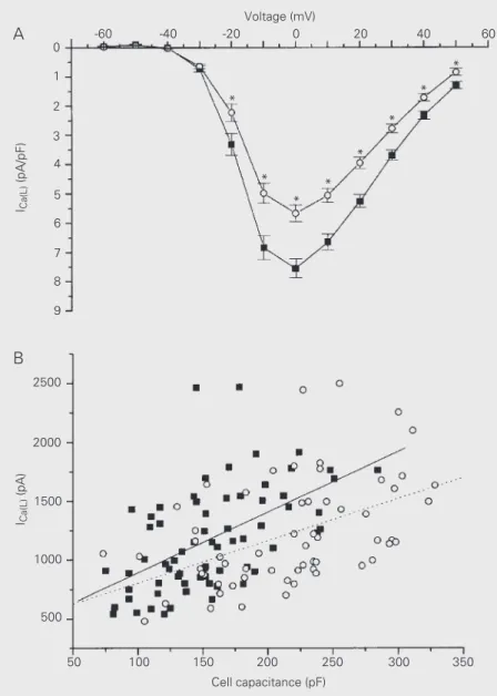
Figure 1. A, L-type calcium current (ICa(L)) versus voltage
plots of sham (squares; N = 69) and postinfarction myo-cytes (circles; N = 60). Mean peak ICa(L) density is
signifi-cantly reduced in postinfarction myocytes. *P<0.05 com-pared to sham (unpaired Student t-test). B, Peak ICa(L)
versus cell capacitance plots of sham (squares) and postinfarction myocytes (circles). There is a linear rela-tionship between calcium current and cell capacitance in both groups (r = -0.48, P<0.001, linear regression, for postinfarction myocytes, N = 60; r = -0.54, P<0.001, linear regression, for sham myocytes, N = 69). Slope values obtained for regression lines did not differ (-3.59 ± 0.87 for postinfarction myocytes and -5.15 ± 0.97 for sham myocytes, P = 0.24).
* *
* *
* *
Mean peak ICa(L) at 0 mV was similar in
myocytes from both groups: 1,182 ± 67 pA (N = 60) for postinfarction myocytes vs 1,138 ± 51 pA (N = 69) for sham myocytes (P = 0.59). However, after normalization for cell capacitance, mean peak ICa(L) density was
25% lower in postinfarction myocytes: 5.7 ± 0.28 vs 7.6 ± 0.32 pA/pF for sham myocytes (P<0.001) (Figure 1A).
Comparison of the calcium current den-sities between the two experimental groups assumes that the relationship between the peak calcium current magnitude and cell capacitance is linear and that this relation-ship does not change in hypertrophy. To check this point, we plotted the peak calcium current (at 0 mV) against cell capacitance for each experimental group. As shown in Fig-ure 1B, we found linear relationships in both groups, and the slope values obtained for the regression lines did not differ significantly. The regression line for postinfarction myo-cytes was shifted towards lower calcium currents. This point was further corrobo-rated by additional statistical analysis using the extracted residuals from the regression of peak calcium currents. The extracted re-siduals were used as a new variable in ANOVA and represented variation in peak calcium currents independent of myocyte capacitance. We found that postinfarction myocytes had lower ICa(L) values than those
from sham rats when cell size influence was removed from the analysis (ANOVA, P<0.05).
In contrast, the calcium current steady-state activation curves were similar for both groups: values for V0.5 were -15.8 ± 0.3 and
-15.2 ± 0.5 mV for sham (N = 69) and postinfarction (N = 60) myocytes, respec-tively (P = 0.27). Slope factors were 5.3 ± 0.3 and 5.5 ± 0.4 mV for sham and postinfarc-tion myocytes, respectively (P = 0.69). The time course of ICa(L) inactivation was biphasic.
Fast (tfast) and slow (tslow) time constants were determined at maximal current density, measured at 0 mV, and also did not differ
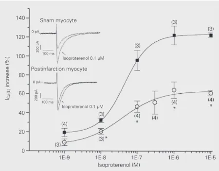
Figure 2. Dose-response curves for isoproterenol on L-type calcium current (ICa(L)). Note the
smaller increase of ICa(L) density in postinfarction (circles) than in sham myocytes (squares):
Emax was 63.6 ± 4.3% in postinfarction and 123.3 ± 0.9% in sham myocytes, P<0.001. The
EC50 was similar for both groups: 38 ± 20 nM in postinfarction and 44 ± 2 nM in sham
myocytes (P>0.5, sigmoidal curve fit using logistical equation). (N), tested cell number for each point; *P<0.05 (unpaired Student t-test). Inset shows ICa(L) records in sham and
postinfarction myocytes. ICa(L) was recorded in whole-cell voltage clamp mode before and
during exposure to 0.1 µM isoproterenol in the same myocyte. Test voltage was 0 mV for all data.
ICa(L)
increase (%)
140
120
100
80
60
40
20
0
Sham myocyte
Isoproterenol 0.1 µM
Isoproterenol 0.1 µM
Postinfarction myocyte
(4)
(3)
(3)
(3)
(4)
*
(4)
*
(4) (4)
*
(3)*
(3)
0 pA
200 pA
100 ms
200 pA
100 ms 0 pA
1E-9 1E-8 1E-7 1E-6 1E-5
Isoproterenol (M)
significantly. tfast was 24.0 ± 1.2 and 26.1 ± 1.4 ms, for sham (N = 57) and postinfarction (N = 49) myocytes, respectively (P = 0.25), and tslow was 134.8 ± 11.7 and 134.8 ± 12.4 ms, for sham and postinfarction myocytes, respectively (P = 0.99).
ICa(L) ß-adrenergic sensitivity
In order to evaluate the ICa(L) response to
ß-adrenergic stimulation we exposed the cells to different concentrations of isoproterenol. A given cell was exposed to only one isopro-terenol concentration. Therefore, each con-centration value was tested in a different group of cells. The ICa(L) increase induced by
isoproterenol was less prominent in postin-farction myocytes, as shown in Figure 2 (inset). Dose-response curves for isoprote-renol were constructed with the average
maximal ICa(L) density increases obtained for
each concentration. After sigmoidal fit, a clear reduction in the effect of ß-adrenergic stimulation on ICa(L) could be seen in
postin-farction myocytes (Figure 2). The maximum effect (Emax) was reduced by 48% in
postin-farction versus sham myocytes. Isoprote-renol potency was similar for both groups of
cells, as demonstrated by similar dose for 50% of maximum effect (EC50) found in
both groups of cells (Figure 2).
Effect of forskolin on ICa(L) density
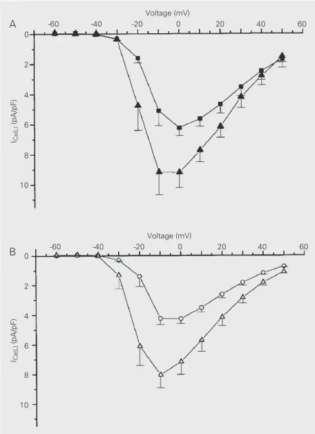
To investigate the effect of hypertrophy on adenylate cyclase activity, forskolin, a direct activator of adenylate cyclase, was applied to the external solution. Forskolin induced simi-lar increases in ICa(L) in both groups (Figure 3),
suggesting that the ß-adrenergic pathway down-stream of the adenylate cyclase step is pre-served in postinfarction myocytes.
[Ca2+]
i transients and the effect of
isoproterenol
The mean resting [Ca2+]
i was 0.40 ± 0.01
(arbitrary units) for both postinfarction (N = 34) and sham (N = 33) myocytes. The mean peak amplitude of [Ca2+]
i transients (Figure
4A) and RtPCa (Figure 4B) upon stimulation at 0.5 Hz were significantly lower in postin-farction myocytes. There was no difference in RD50 between the two groups (Figure 4B).
Figure 4F, left (control), shows records of fluorescence ratios (340/380) versus time ob-tained from representative postinfarction and sham myocytes. Thus, upon stimulation at 0.5 Hz, the [Ca2+]
i transient increased more slowly
and to lower values in postinfarction myocytes compared to sham myocytes.
Figure 4C, D and E show that 1 µM isoproterenol significantly increased all three parameters, mean peak amplitude of [Ca2+]
i
transients, RtPCa, and RD50, both in
postin-farction and sham myocytes.
Figure 4C also shows that the mean peak amplitude of the [Ca2+]
i transient in the
pres-ence of isoproterenol (1 µM) was lower in postinfarction than in sham myocytes. How-ever, the relative increases of the mean peak amplitudes of [Ca2+]
i transients induced by
isoproterenol were not significantly differ-ent between the two groups: 76.5 ± 11.0% (N = 8) in postinfarction and 86.8 ± 10.5% ICa(L)
(pA/pF)
0
2
4
6
8
10
ICa(L)
(pA/pF)
0
2
4
6
8
10
-60 -40 -20 0 20 40 60
Voltage (mV)
-60 -40 -20 0 20 40 60
Voltage (mV) A
B
Figure 3. Effect of forskolin on L-type calcium current (ICa(L)). Forskolin (10 µM) added to the
extracellular saline provoked similar increases in ICa(L) density in both sham (A, filled squares in the absence and filled triangles in the presence of forskolin) and postinfarction myocytes (B, open circles in the absence and open triangles in the presence of forskolin). There was
no significant difference in the mean increase of ICa(L) induced by forskolin at any voltage in
Figure 4. Cytoplasmic calcium ([Ca2+]i) transient parameters obtained from isolated ventricular myocytes loaded
with fura 2-AM. A, Peak amplitudes of [Ca2+]i transients measured from the resting level: 0.13 ± 0.007, N = 34, in
postinfarction (MI) and 0.16 ± 0.009, N = 33, in sham myocytes (P = 0.01, unpaired Student t-test). B, Rates of increase to peak (RtPCa), and of decay to 50% amplitude (RD50) of the [Ca2+]i transients: RtPCa: 0.87 ± 0.0067,
N = 34, in MI and 1.15 ± 0.075 units/s, N = 33, in sham (P<0.001) and RD50: 0.34 ± 0.028, N = 34, in MI and 0.38
± 0.022 units/s, N = 33, in sham myocytes (P = 0.27, unpaired Student t-test). C, Peak transient amplitudes,
D, RtPCa, and E, RD50 of the [Ca2+]i transients under control condition and in the presence of 1 µM isoproterenol in
MI and sham myocytes. Isoproterenol induced an increase in peak amplitudes of the [Ca2+]i transients from 0.12 ±
0.02 to 0.23 ± 0.01, N = 8, in MI and from 0.16 ± 0.01 to 0.32 ± 0.04, N = 7, in sham. RtPCa was increased in MI from 0.97 ± 0.13 to 1.94 ± 0.18 units/s, N = 8, and in sham from 1.36 ± 0.40 to 3.43 ± 0.63 units/s, N = 7; isoproterenol-induced increase in RD50 was from 0.38 ± 0.07 to 0.75 ± 0.09 units/s, N = 8, in MI and from 0.45 ±
0.07 to 0.89 ± 0.13 units/s, N = 7, in sham. Measurements of [Ca2+]i transients were always performed first under
control condition and then in the presence of the drug in the same cell. Each cell was exposed only once to isoproterenol to avoid desensitization. F, Sample records from one MI and one sham cell, bathed in control saline (control) and at 3rd minute of exposure to 1 µM isoproterenol. *P<0.05 for comparisons between MI and sham, and +P<0.05 for comparisons between isoproterenol and control (unpaired Student t-test). Iso, isoproterenol.
Peak transient amplitude
(arbitrary units)
0.16
0.12
0.08
0.04
0.00
Peak transient amplitude
(arbitrary units)
0.30
0.24
0.18
0.12
0.06
0.00 RtPCa (arbitrary units/s)
4
3
2
1
0 RD
50
(arbitrary units/s)
1.0
0.8
0.6
0.4
0.0 0.2
Arbitrary units/s
1.2
1.0
0.8
0.6
0.0 0.4
0.2
MI Sham MI Sham
RtPCa RD50
* *
A B
Control Iso
Control
Iso ControlIso
MI Sham MI Sham MI Sham
C D E
MI
340/380
0.8 0.7 0.6 0.5
0.3 0.4
0.8 0.7 0.6
0.5
0.3 0.4
Sham 340/380
0.8 0.7 0.6 0.5
0.4
0.8 0.7 0.6 0.5
0.4
1 s 1 s
1 s 1 s
F
Control Iso
340/380
340/380
* *+
+
* *+
+
+
(N = 7) in sham myocytes (P = 0.52). The relative increases in RtPCa and RD50
in-duced by isoproterenol were also similar in both groups: 109.0 ± 24.4% in postinfarc-tion and 137.8 ± 26.5% in sham myocytes (RtPCa), P = 0.48, and 89.0 ± 30.4% in postinfarction and 91.7 ± 16.0% in sham myocytes (RD50), P = 0.94. Figure 4F shows
the effect of 1 µM isoproterenol on represen-tative postinfarction and sham myocytes.
ß-Adrenergic receptor density
The [3H]-DHA binding was significantly
lower in homogenates of left ventricles of myocardial infarction hearts as shown by the saturation curves in Figure 5. The calculated Bmax of [3H]-DHA for myocardial infarction
corresponded to 35% of the value found for sham myocytes, indicating that the maximal number of radioligand binding sites was de-creased in myocardial infarction hearts. Scatchard analysis of the data (Figure 5, inset)
indicated that the equilibrium dissociation con-stants were the same for both membrane prepa-rations, suggesting a noninteracting single population of DHA binding sites in both post-infarction and sham myocytes.
Discussion
This is the first study in which calcium current densities, intracellular calcium tran-sients and binding to ß-adrenergic receptors were evaluated simultaneously in a well-defined model of cardiac hypertrophy. We were able to show decreased cardiac ß-adre-nergic response, impairment of ICa(L) under
basal or stimulated conditions, and decreased [Ca2+]
i transients upon stimulation in the
model of hypertrophied failing heart, 8-10 weeks after extensive myocardial infarction.
L-type calcium channel
The linear correlation between peak ICa(L)
and cell capacitance for both postinfarction and sham myocytes with almost parallel re-gression lines validates the procedure of com-paring current densities normalized by cell capacitance in this model. The analysis of the extracted residuals from the regression of peak calcium currents showed that the decrease in peak calcium current in myocar-dial infarction is independent of cell size.
In contrast to most studies, in which ICa(L)
density is evaluated in a small number of cells, in the present investigation, by analyzing a larger number of cells, we confirmed that ICa(L)
density is indeed reduced (Figure 1A) in post-infarction myocytes, in agreement with the reduction in DHP binding sites described in homogenates from infarcted rats (14,17,18).
In contrast, several investigators reported preservation of ICa(L) density in
postinfarc-tion myocytes (3,11-14). What could underly this discrepancy? In some cases, the degree of hypertrophy is clearly different: the in-crease in mean capacitance of myocardial infarction cells in the present study (38.5%)
Specific binding (fmol/mg)
300
200
100
0
0 10 20
Free lingand (nM)
0.02
0.01
0.00
B/F
0 100 200 300
B (fmol/mg)
Figure 5. Saturation binding isotherm of [3H]-DHA binding to left ventricle homogenates in
sham (squares) and myocardial infarction (MI) rat heart (circles). Inset shows Scatchard analysis of the data. Bmax was 93.89 ± 20.22 in MI and 271.5 ± 31.43 fmol/mg protein in
was larger than the 11% (3) and 12% (14) values reported by others. The smaller hy-pertrophy inferred from the capacitance measurements in the study by Holt et al. (3) was confirmed by the smaller increase in the heart weight/body weight ratio in myocar-dial infarction, which was 67% in compari-son to the 107% increase found in our mo-del. Thus, the degree of cardiac hypertrophy possibly related to the intensity of left ven-tricular overload could be one variable asso-ciated with the decreased ICa(L) density in
postinfarction myocytes. Additionally, Qin et al. (12), Holt et al. (3) and Wasserstrom et al. (13), who did not find alterations in peak ICa(L) density, were all working with
myocar-dial infarction rats earlier after experimental infarction, i.e., 3-4 weeks (12) and 6 weeks (3,13), when compared to the present model. Thus, time elapsed after myocardial infarc-tion, which determines the degree of pro-gression of heart failure, could be another important parameter. Accordingly, Aimond et al. (15) reported decreased ICa(L) density
(by 45%) in postinfarction myocytes 4-6 months after infarction in a model with clear signs of left ventricular dysfunction evalu-ated by echocardiography and hemodynam-ic parameters.
Calcium transients
In the present study, mean peak amplitudes of [Ca2+]
i transients and RtPCa (a parameter
that estimates the velocity of calcium release) were reduced in postinfarction myocytes, in agreement with a report by Holt et al. (3), who also reported decreased fractional shortening and increased times to peak tension and recov-ery. Anand et al. (33), on the other hand, reported unchanged [Ca2+]
i transients in a
model with signs of heart failure. However, in this case the degree of left ventricular impair-ment was clearly less severe, a fact that may explain the different results.
Our observations of decreased ICa(L) and
peak calcium transient conform with
previ-ous reports of a linear relation between the magnitudes of ICa(L) and of Ca2+ release, and
with the proposals that reductions of at least 20% in ICa(L) density can disturb intracellular
calcium handling (27).
Thus, in our model the decreased con-tractility reported for the whole heart and papillary muscle (6,8) may be associated with the reduced trigger represented by the reduced ICa(L) density (Figure 1A), leading to
decreased calcium mobilization during sys-tole. However, we cannot rule out a contri-bution of other mechanisms to the reduction of the peak amplitude of [Ca2+]
i transients,
such as a smaller calcium store in sarcoplas-mic reticulum (34) and a decrease in the ability of ICa(L) to activate sarcoplasmic
reticu-lum Ca2+ release (11).
On the other hand, RD50, which estimates
the rate of decay of the [Ca2+]
i transient
fol-lowing a contraction, was found to be unal-tered in postinfarction myocytes, as also shown by Prahash et al. (22), possibly indicating that the mechanisms of calcium clearance are preserved or that these cells develop some kind of compensatory mechanism.
Depressed ß-adrenergic response in postinfarction myocytes
We found a significant decrease in the Emax of isoproterenol on ICa(L) in
postinfarc-tion myocytes compared to sham myocytes, while the EC50 was unchanged. The Emax of
isoproterenol obtained for sham myocytes was similar to values previously reported for adult rat hearts (36), while the present study is the first report of Emax and EC50 in
postin-farction rat myocytes. The maintenance of EC50 and the decrease in Emax in
postinfarc-tion myocytes suggest that the reduced effect of isoproterenol on these cells is more likely to be related to a decreased ß-adrenergic receptor density rather than to a change in affinity of the ß-adrenergic receptor. Fur-thermore, the isoproterenol-stimulated mean peak amplitude of the [Ca2+]
i transients was
lower in postinfarction myocytes (Figure 4) although the relative increases induced by 1 µM isoproterenol were the same for sham and myocardial infarction rats.
The functional evidence cited above for decreased ß-adrenergic receptor density with maintained affinity of the receptor for the agonist is in good agreement with the signifi-cant reduction in the calculated Bmax of [3
H]-DHA binding to the postinfarction left ventric-ular tissue preparation (Figure 5), without changes in Kd. Taking into account the in-crease in right ventricle/body weight (123%) and in wet lung/body weight (81%) ratios in the present study, the magnitudes of hypertro-phy and heart failure in our model were higher than in studies that reported preservation of ß-adrenergic receptor density (4,23), and similar to studies that also reported a decrease in ß-adrenergic receptor density (6,8). Thus, whether or not ß-adrenergic receptor density is decreased in a given model of myocardial infarction seems to depend also on the severity of heart failure.
This decreased ß-adrenergic receptor den-sity could be an additional cause for the decreased basal ICa(L) density in this model
since it is proposed that G protein-coupled receptors, including ß-adrenergic receptor, exert a basal amount of intrinsic activity
leading to basal steady-state adenylate cy-clase activity (37).
The possibility that the reduction shown in the present study could be related to de-creased ß-adrenergic receptor density lim-ited to the scar and neighboring tissue (4,24) is not likely since we removed the scars before homogenization of the left ventricles. Forskolin stimulation induced similar ef-fects on ICa(L) density in sham and
postinfarc-tion myocytes, in agreement with previous studies that evaluated chronotropic and ino-tropic responses to forskolin (4,22). This find-ing, taken together with the decreased ß-adre-nergic receptor density, limits the possible altered steps in the ß-adrenergic pathway in postinfarction myocytes to the receptor-G pro-tein complex and suggests the interpretation that the decrease in ß-adrenergic receptor den-sity is a possible mechanism underlying the reduced ß-adrenergic stimulation response in postinfarction myocardium.
We propose that in the cardiac hypertro-phy model induced by large infarcts in rats with clear signs of heart failure, evaluated 8-10 weeks after infarction, ICa(L) density and
mean peak amplitude of [Ca2+]
i transients
are both reduced, events that may contribute to the decreased contractility of postinfarc-tion myocardium. The decrease of the re-sponse of ICa(L) to ß-adrenergic stimulation in
postinfarction myocardium is not related to changes in adenylate cyclase function or sensitivity of ß-adrenergic receptors to the agonist, with down-regulation of ß-adrener-gic receptors being an important mechan-ism. The controversial reports concerning ICa(L) density and ß-adrenergic effects on
hy-pertrophied myocytes in healed infarction are probably related to differences in the severity of heart failure in different studies.
Acknowledgments
References
1. Sutton MGJ & Sharpe N (2000). Left ventricular remodeling after myocardial infarction. Pathophysiology and therapy. Circulation, 101: 2981-2988.
2. Swynghedauw B (1999). Molecular mechanisms of myocardial re-modelling. Physiological Reviews, 79: 215-262.
3. Holt E, Tonnessen T, Lunde PK, Semb SO, Wasserstrom JÁ, Sejersted OM & Christensen G (1998). Mechanism of cardiomyo-cyte dysfunction in heart failure following myocardial infarction in rats. Journal of Molecular and Cellular Cardiology, 30: 1581-1593. 4. Kompa AR, Gu X-H, Evans BA & Summers RJ (1999).
Desensitiza-tion of cardiac ß-adrenoceptor signaling with heart failure produced by myocardial infarction in the rat. Evidence for the role of Gi but not Gs or phosphorylating proteins. Journal of Molecular and Cellular Cardiology, 31: 1185-1201.
5. Litwin SE & Morgan JP (1992). Captopril enhances intracellular calcium handling and ß-adrenergic responsiveness of myocardium from rats with postinfarction failure. Circulation Research, 78: 797-807.
6. Sanbe A & Takeo S (1995). Diminished responsiveness to cardiac ß1-adrenoceptor agonists in rats with chronic heart failure following
myocardial infarction. Biological and Pharmaceutical Bulletin, 18: 1362-1366.
7. Sethi R, Dhalla KS, Beasmish RE & Dhalla NS (1997). Differential changes in left and right ventricular adenyl cyclase activities in congestive heart failure. American Journal of Physiology, 272: H884-H893.
8. Warner AL, Bellah KL, Raya TE, Roeske WR & Goldman S (1992). Effects of ß-adrenergic blockade on papillary muscle function and the ß-adrenergic receptor system in noninfarcted myocardium in compensated ischemic left ventricular dysfunction. Circulation, 86: 1584-1595.
9. Hasenfuss G (1998). Animal models of human cardiovascular dis-ease, heart failure and hypertrophy. Cardiovascular Research, 39: 60-76.
10. Mill JG, Novaes MAS, Galon M, Nogueira JB & Vassallo DV (1998). Comparison of the contractile performance of the hypertrophied myocardium from spontaneous hypertensive rats and normotensive infarcted rats. Canadian Journal of Physiology and Pharmacology, 76: 387-394.
11. Gómez AM, Guatimosim S, Dilly KW, Vassort G & Lederer WJ (2001). Heart failure after myocardial infarction. Altered excitation-contraction coupling. Circulation, 104: 688-693.
12. Qin D, Zhang Z, Caref EB, Boutjdir M, Jain P & El-Sherif N (1996). Cellular and ionic basis of arrhythmias in postinfarction remodeled ventricular myocardium. Circulation Research, 79: 461-473. 13. Wasserstrom JÁ, Holt E, Sjaastad I, Lunde PK, Ødegaard A &
Sejersted OM (2000). Altered E-C coupling in rat ventricular myo-cytes from failing hearts 6 wk after MI. American Journal of Physiol-ogy, 279: H798-H807.
14. Zhang XQ, Moore RL, Tillotson DL & Cheung JY (1995). Calcium currents in postinfarction rat cardiac myocytes. American Journal of Physiology, 269: C1464-C1473.
15. Aimond F, Alvarez JL, Rauzier J-M, Loronte P & Vassort G (1999). Ionic basis of ventricular arrhythmias in remodeled rat heart during long-term myocardial infarction. Cardiovascular Research, 42: 402-415.
16. Santos PEB, Barcellos LC, Mill JG & Masuda MO (1995). Ventricular action potential and L-type calcium channel in infarct-induced hyper-trophy in rats. Journal of Cardiovascular Electrophysiology, 6:
1004-1014.
17. Dixon IMC, Lee SL & Dhalla NS (1990). Nitrendipine binding in congestive heart failure due to myocardial infarction. Circulation Research, 66: 782-788.
18. Gopalakrishnan M, Triggle DJ, Rutledge A, Kwon YW, Bauer JÁ & Fung H-L (1991). Regulation of K+ and Ca2+ channels in
experimen-tal cardiac failure. American Journal of Physiology, 261: H1979-H1987.
19. Zhang XQ, Moore RL, Tenhave T & Cheung JY (1995). [Ca2+]i
tran-sients in hypertensive and postinfarction myocytes. American Jour-nal of Physiology, 269: C632-C640.
20. Tajima M, Weinberg EO, Bartunek J, Jin H, Yang R, Paoni NF & Lorell BH (1999). Treatment with growth hormone enhances con-tractile reserve and intracellular calcium transients in myocytes from rats with postinfarction heart failure. Circulation, 99: 127-134.
21. Qi X & Rouleau JL (1996). Beta-adrenergic responsiveness of papil-lary muscles in the rat postinfarction model. Canadian Journal of Physiology and Pharmacology, 74: 1166-1170.
22. Prahash AJ, Gupta S & Anand IS (2000). Myocyte response to beta-adrenergic stimulation is preserved in the noninfarcted myocardium of globally dysfunctional rat hearts after myocardial infarction. Circu-lation, 102: 1840-1846.
23. Chasteney EA, Liang C-S & Hood WB (1992). Beta-adrenoceptor and adenylate cyclase function in the infarct model of rat heart failure.
Proceedings of the Society for Experimental Biology and Medicine, 200: 90-94.
24. van Veldhuisen DJ, Brode OE, van Gilst WH, Schulze C, Hegeman H, Anthonio RL, Scholtens E, Graeff PA, Wesseling H & Lie KI (1995). Relation between myocardial ß-adrenoceptor density and hemody-namic and neurohumoral changes in a rat model of chronic myocar-dial infarction: effects of ibopamine and captopril. Cardiovascular Research, 30: 386-393.
25. Yamamoto J, Ohyanagi M, Morita M & Iwasaki T (1994). ß-Adreno-ceptor-G protein-adenylate cyclase complex in rat hearts with is-chemic heart failure produced by coronary artery ligation. Journal of Molecular and Cellular Cardiology, 26: 617-626.
26. Clozel JP, Holck M, Osterrieder W, Burkard W & Prada M (1987). Effects of chronic myocardial infarction on responsiveness to iso-prenaline and the state of myocardial beta adrenoceptors in rats.
Cardiovascular Research, 21: 688-695.
27. Hart G (1994). Cellular electrophysiology in cardiac hypertrophy and failure. Cardiovascular Research, 28: 933-946.
28. Selye H, Bajusz E, Grasso S & Mendell P (1960). Simple techniques for the surgical occlusion of coronary vessels in the rat. Angiology, 1: 398-407.
29. Hamill OP, Marty A, Neher E, Sakmann B & Sigworth FJ (1981). Improved patch-clamp techniques for high resolution current record-ing from cells and cell-free membrane patches. Pflügers Archiv, 391: 85-100.
30. Huang B, Qin D, Deng L, Boutjdir M & El-Sherif N (2000). Reexpression of T-type Ca2+ channel gene and current in
postinfarc-tion remodeled rat left ventricle. Cardiovascular Research, 46: 442-449.
31. Lowry CM, Rosebrough NJ, Farr AL & Randall RJ (1951). Protein measurement with the Folin phenol reagent. Journal of Biological Chemistry, 193: 265-272.
33. Anand IS, Liu D, Chugh SS, Prahash AJC, Gupta S, John R, Popescu F & Chandrashekhar Y (1997). Isolated myocyte contractile function is normal in postinfarct remodeled rat heart with systolic dysfunc-tion. Circulation, 96: 3974-3984.
34. Zarain-Herzberg A, Afzal N, Elimban V & Dhalla NS (1996). De-creased expression of cardiac sarcoplasmic reticulum Ca(2+)-pump
ATPase in congestive heart failure due to myocardial infarction.
Molecular and Cellular Biochemistry, 163-164: 285-290.
35. Mukherjee R & Spinale FG (1998). L-type calcium channel
abun-dance and function with cardiac hypertrophy and failure: a review.
Journal of Molecular and Cellular Cardiology, 30: 1899-1916. 36. Scamps F, Mayoux E, Charlemagne D & Vassort G (1990). Calcium
current in single cells isolated from normal and hypertrophied rat heart. Effects of ß-adrenergic stimulation. Circulation Research, 67: 199-208.
![Figure 4. Cytoplasmic calcium ([Ca 2+ ] i ) transient parameters obtained from isolated ventricular myocytes loaded with fura 2-AM](https://thumb-eu.123doks.com/thumbv2/123dok_br/15808315.650535/9.918.86.671.144.819/figure-cytoplasmic-transient-parameters-obtained-isolated-ventricular-myocytes.webp)
![Figure 5. Saturation binding isotherm of [ 3 H]-DHA binding to left ventricle homogenates in sham (squares) and myocardial infarction (MI) rat heart (circles)](https://thumb-eu.123doks.com/thumbv2/123dok_br/15808315.650535/10.918.85.538.637.959/figure-saturation-binding-isotherm-ventricle-homogenates-myocardial-infarction.webp)
