Effe ct o f actino m ycin D o n sim ian
ro tavirus (SA1 1 ) re plicatio n in ce ll
culture
Laboratório de Virologia, Departamento de Microbiologia, Centro de Ciências Biológicas, Universidade Estadual de Londrina, Londrina, PR, Brasil
C.C. Stefanelli, J.G. Castilho, M.V.J. Botelho, R.E.C. Linhares and C.M. Nozawa
Abstract
Rotaviruses are the major cause of viral diarrhea in humans and animals. Actinomycin D (Act D) is an antibiotic that intercalates DNA and therefore inhibits DNA-dependent transcription. The current study was carried out to assess the influence of Act D on the replication of simian rotavirus (SA11) in cell culture. Virus-infected MA-104 cell cultures were studied in the presence of Act D at concentrations of 1.25 and 2.5 µg/ml. Treatment of rotavirus-infected cells with 2.5 µg/ml Act D 48 h post-infection reduced the cytoplasmic metachromasia after staining with acridine orange by 25%. Viral RNA labeled with 3H-uridine in the presence of the drug was separated by
polyacryl-amide gel electrophoresis. Viral RNA replication was not affected by Act D, but increased 3H-uridine uptake was demonstrable by infected cells in the presence of the drug. This possibly was due to the inhibition of cellular RNA synthesis by Act D, which thus enhances incorporation of the radionuclide into the viral RNA. Act D reduced the number of infected cells presenting virus-specific fluorescence 48 h post-infection by more than 50%. These data suggest that Act D may have complexed with viral RNA and prevented newly syn-thesized mRNA from being translated, but may not have prevented early replication.
Co rre spo nde nce
R.E.C. Linhares
Departamento de Microbiologia CCB, UEL
86051-990 Londrina, PR Brasil
Fax: + 55-43-371-4207 E-mail: relin@ uel.br Research supported by CAPES, CNPq and CPG/UEL. Part of a Master’s thesis presented by C.C. Stefanelli to the
Departamento de Microbiologia, UEL, Londrina, PR, Brasil. C.C. Stefanelli was the recipient of a CAPES fellowship.
Received June 5, 2001 Accepted February 14, 2002
Ke y wo rds
·Rotavirus ·Actinomycin D ·Replication ·Cell culture
Rotaviruses are classified as a genus within the family Reoviridae. These viruses are the major causal agents of acute diarrhea in mammals and fowl. The virions are nonenveloped 75 nm in diameter, and the genome consists of eleven segments of double-stranded RNA that encode six struc-tural (VP1-VP4, VP6 and VP7) and five nonstructural (NSP1-NSP5) proteins (1). They have a triple-layered double capsid and replicate in the cytoplasm. Virion uncoating is followed by penetration into susceptible host cells and productive infection depends
on the function of viral transcriptase in the synthesis of mRNA and double-stranded RNA segments of the progeny. The minus RNA strands act as templates for the synthe-sis of full-length plus strands (2,3). Both newly synthesized and pre-existing plus strand RNA act as templates for minus strand syn-thesis.
antimicrobial agent, but is used as an antitu-mor agent (6). The structure of Act D consists of a phenoxazone ring linked to two identical cyclic pentapeptides. It binds tightly to double-stranded DNA but not to single-stranded DNA or RNA, double-single-stranded RNA, or RNA-DNA hybrids (7). Act D in-hibits the replication of DNA- and RNA-containing viruses.
Vaccinia virus double-stranded DNA rep-lication was 99% inhibited by Act D at 0.1 µg/ml; however, the replication of mengo-virus single-stranded RNA was not affected by the drug at 1.0 µg/ml (8). At 1-10 µg/ml the drug inhibited by 99% the replication of fowl plague virus segmented single-stranded RNA. In contrast, Newcastle disease virus single-stranded RNA synthesis was not af-fected (8,9). Reovirus segmented double-stranded RNA replication was inhibited by up to 98% in the presence of 0.1-5.0 µg/ml of the drug in L cells (10). Scheiber et al. (11) demonstrated that human coronavirus, strain 229 E, single-stranded RNA was not af-fected by Act D. However, experiments with two feline strains of coronavirus with differ-ent tropism and virulence revealed that only the virulent strain was inhibited by the drug (12). Fish pancreatic necrosis virus seg-mented double-stranded RNA replication was inhibited by the drug at very low concentra-tions (13). Poliovirus, as well as several retrovirus single-stranded RNA were also shown to be inhibited by the drug (14-16). Therefore, Act D has been used as a tool for studying the in vitro replicative process of several viruses and its interaction with func-tions encoded by the host cell (11,17).
The present study assessed simian rota-virus (strain SA11) replication in MA-104 cells maintained with Act D at concentra-tions that inhibit cellular transcription.
MA-104 cell cultures (Rhesus monkey kidney fetal cells) were used at 70% conflu-ence throughout the experiments. Simian rotavirus (SA11) pretreated and maintained in cell culture with crystalline trypsin (Gibco,
Gaithersburg, MD, USA) at 30 and 10 µg/ ml, respectively, was used at multiplicity of infection of approximately 1. A stock solu-tion of Act D (Sigma, St. Louis, MO, USA) was prepared at 2.5 mg/ml in sodium phos-phate-buffered saline (PBS), pH 7.3, and maintained at 4ºC.
The Act D cytotoxicity test was carried out by submitting cell cultures to drug-con-taining medium at the concentrations of 0.1, 0.2, 0.3, 0.4, 0.5, 1.25, 2.5 and 5.0 µg/ml at 37ºC, for 7 days. Cells were observed daily for morphological changes.
The effect of Act D on viral RNA synthe-sis was monitored by acridine orange stain-ing and 3H-uridine incorporation. Cell cul-tures grown in 10 x 100 mm glass tubes were inoculated in triplicate and, after adsorption for 1 h at 37ºC, inocula were removed, and the cultures were refed with fresh medium containing Act D at final concentrations of 1.25 and 2.5 µg/ml, and 5 µCi/ml of 3 H-uridine. Infected untreated and mock-infected cell cultures with and without the drug were used in parallel as controls. At intervals of 24, 36, 48, 60 and 72 h post-infection cul-tures were harvested and submitted to three cycles of freezing and thawing. The cell homogenates were concentrated by dialysis to one third of the original volume and sub-mitted to polyacrylamide gel electrophoresis (17). A human strain was used for control. The strips of the gel between the limits of the largest and the smallest segments of viral RNA (3.30-0.66 kb) were cut out and dis-solved in 2.0 ml of hydrogen peroxide/am-monium hydroxide solution (19:1) (18). One milliliter of aqueous scintillation solution (Sigma) was added and counting was per-formed with a beta counter (Beckman LS 6000 SC).
ac-cording to Rovozzo and Burke (19). Cultures were observed under ultraviolet light and results were expressed as the mean counts of cells with cytoplasmic metachromasia in ten fields.
Viral protein synthesis was monitored by immunofluorescence staining. Briefly, cell cultures grown on coverslips in Leighton tubes were inoculated as above. After ad-sorption, excess inoculum was removed and the monolayers were washed with PBS and refed with fresh maintenance medium con-taining 1.25 or 2.5 µg/ml of the drug. In-fected monolayers in the absence of drug and noninfected monolayers with and with-out the drug were maintained as controls. At intervals of 48, 52, 56, and 72 h post-infec-tion, coverslips were removed and washed with PBS and monolayers were fixed with cold acetone for 20 min. Coverslips were overlaid with mouse anti-porcine rotavirus monoclonal antibody directed at VP6, di-luted at 1:40. A FITC conjugate goat anti-mouse immunoglobulin (Sigma) diluted at 1:40 was used. Results are reported as the mean counts of fluorescent cells showing specific fluorescence in ten fields. MA-104 cell cultures treated with 0.1 µg/ml Act D did not show any detectable morphological al-terations, whereas at higher concentrations morphological changes were observed 72 h post-treatment.
The treatment of virus-infected cultures with 2.5 µg/ml Act D reduced by 25% the number of cells presenting cytoplasmic metachromasia at 48 h post-infection,
whereas at 1.25 µg/ml the reduction was under 5%. The kinetics of 3H-uridine uptake (Figure 1) demonstrated that viral RNA syn-thesis was not affected; however, increased counts were observed in infected cells in the presence of the drug.
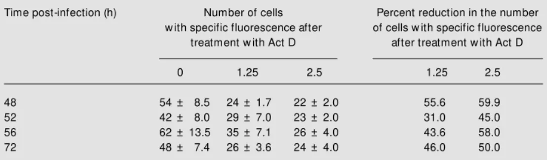
The immunofluorescence assay demon-strated a reduction of the number of fluores-cent cells when the drug was used at the concentrations of 1.25 or 2.5 µg/ml (Table 1). At 48 h post-infection, 54 fluorescent cells were detected in infected cultures in the absence of drug as compared to 24 and 22 cells in infected cultures treated with the
3H
-u
ri
d
in
e
u
p
ta
k
e
3000
2500
2000
1500
1000
500
0
24 36 48 60 72
Time post-infection (h)
Figure 1. Effect of actinomycin D on rotavirus RNA synthesis. Kinetics of 5 µCi/ml 3H-uridine uptake by SA11-infected M A-104 cells in the presence of acti-nomycin D at the concentrations of 1.25 (circles) and 2.5 µg/ml (triangles) and by infected un-treated cells (lozenges). At the times post-infection indicated cell homogenates w ere submit-ted to PAGE, and the labeled viral RNA w as eluted and radio-activity counted. Data are re-ported as means ± SEM for 3 experiments.
Table 1. Reduction of fluorescent cells upon actinomycin D (Act D) treatment.
Time post-infection (h) Number of cells Percent reduction in the number
w ith specific fluorescence after of cells w ith specific fluorescence
treatment w ith Act D after treatment w ith Act D
0 1.25 2.5 1.25 2.5
48 54 ± 8.5 24 ± 1.7 22 ± 2.0 55.6 59.9
52 42 ± 8.0 29 ± 7.0 23 ± 2.0 31.0 45.0
56 62 ± 13.5 35 ± 7.1 26 ± 4.0 43.6 58.0
72 48 ± 7.4 26 ± 3.6 24 ± 4.0 46.0 50.0
drug at concentrations of 1.25 and 2.5 µg/ ml, respectively, corresponding to reduc-tions of 55.6 and 59.9%. At 52 h post-infection, infected cultures in the absence of drug showed 42 fluorescent cells in compar-ison to 29 and 23 in cultures treated at concentrations of 1.25 and 2.5 µg/ml, re-spectively, representing reductions of 31 and 45%. Similar results were obtained at 56 and 72 h post-infection with reduction of fluo-rescent cells of 43.6 and 58%, and 46 and 50%, for 1.25 and 2.5 µg/ml Act D, respec-tively.
Act D has been used to study the interac-tion of funcinterac-tions encoded by the host cell in the replication process of several viruses (10,15,16). In the current study we demon-strated an increase of 3H-uridine uptake by cells infected with simian rotavirus in the presence of Act D in comparison to un-treated cells. It is reasonable to expect that at the drug concentrations used, inhibition of most of the synthesis of cell RNA was achieved, favoring the uptake of radioactiv-ity into viral RNA. Shatkin (10) showed an increase in 14C-uridine uptake in viral RNA in L929 cells (mouse fibroblast cells) in-fected with reovirus and treated with Act D, with 92% inhibition of the synthesis of L929 cell RNA. Barry et al. (9) demonstrated that Act D at concentrations of 1.0 to 10.0 µg/ml inhibited the replication of influenza virus (segmented single-stranded RNA). Similar results were obtained for fowl plague virus. However, Act D did not interfere with the replication of Newcastle disease virus non-segmented single-stranded RNA (8). The sites of replication of these viruses in the host cells are different. Fowl plague virus repli-cates in the nucleus and Newcastle disease virus in the cytoplasm, and, in addition, fowl plague virus uses cellular RNA as primer for the initiation of transcription.
The reduction in the number of cells presenting cytoplasmic metachromasia in the present study could represent an inhibition of viral RNA synthesis, or more likely, the
Re fe re n c e s
1. Estes M K (1996). Rotaviruses and their replication. In: Fields BN, Knipe DM & How ley PM (Editors), Virology. Vol. 2. 3rd edn. Lippincott-Raven Publishers, Phila-delphia, PA, USA, 1625-1655.
2. Patton JT (1994). Rotavirus replication. Current Topics in M icrobiology and
Im-munology, 185: 107-124.
3. Chen D, Zeng CY, Wentz M J, Gorziglia M , Estes M K & Ramig RF (1994). Template-dependent, in vitro replication of rota-virus RNA. Journal of Virology, 68: 7030-7039.
4. Betzel C, Rachev R, Dolashka P & Genov N (1993). Actinomycins as proteinase in-hibitors. Biochimica et Biophysica Acta, 1161: 47-51.
5. Jeeninga RE, Huthoff HT, Gultyaev AP & Berkhout B (1998). The mechanism of actinomycin D-mediated inhibition of HIV-1 reverse transcription. Nucleic Acids
Re-search, 26: 5472-5479.
6. Yang X-L & Wang AH-J (1999). Structural studies of atom-specific anticancer drugs acting on DNA. Pharmacology and
Thera-peutics, 83: 181-215.
7. Reich E, Franklin RM , Shatkin AJ & Tatum EL (1961). Effect of actinomycin-D on cel-lular nucleic acid synthesis and virus pro-duction. Science, 134: 556.
8. Barry RD (1964). The effect of
actinomy-cin D and ultraviolet irradiation on the production of fow l plague virus. Virology, 24: 563-569.
9. Barry RD, Ives DR & Cruickshank JG (1962). Participation of deoxyribonucleic acid in the multiplication of influenza vi-rus. Nature, 194: 1139-1140.
10. Shatkin AJ (1965). Actinomycin and the differential synthesis of reovirus and L cell RNA. Biochemical and Biophysical
Re-search Communications, 19: 506-510.
11. Scheiber SS, Kamahora T & Lai M M C (1989). Sequence analysis of the nucleo-capsid protein gene of human coronavi-rus 229E. Virology, 169: 142-151. 12. Lew is EL, Harbour DA, Beringer JE &
Grinsted J (1992). Differential in vitro inhi-bition of feline enteric coronavirus and feline infectious peritonites virus by acti-nomycin D. Journal of General Virology, 73: 3285-3288.
13. Becht H (1994). Birnaviruses-animal. In: Webster RG & Granoff A (Editors),
Ency-clopedia of Virology. Academic Press,
London, UK, 143-149.
14. Kean KM , Agut H, Fichot O & Girard M (1989). Substitution in the poliovirus repli-case gene determines actinomycin D sen-sitivity of viral replication at elevated tem-perature. Virus Research, 12: 19-32. 15. Guo J, Wu T, Bess J, Henderson LE &
Levin JG (1998). Actinomycin D inhibits human immunodeficiency virus type 1 minus-strand transfer in in vitro and en-dogenous reverse transcriptase assays.
Journal of Virology, 72: 6716-6724.
16. Vogel U & Scholtissek C (1995). Inhibi-tion of the intracellular transport of influ-enza viral RNA by actinomycin D.
Ar-chives of Virology, 140: 1715-1723.
17. Herring AJ, Inglis NF, Ojeh CK, Snodgrass DR & M enzies J (1982). Rapid diagnosis of rotavirus infection by direct detection of viral nucleic acid in silver stained polyacrylamide gels. Journal of
Clinical M icrobiology, 16: 473-477.
18. Bonner M & Laskey RA (1974). A film detection method for tritium-labelled pro-teins and nucleic acids in polyacrylamide gels. European Journal of Biochemistry, 46: 83-88.
19. Rovozzo GC & Burke CN (1973). A M anual
of Basic Virological Techniques. 1stedn.
Prentice Hall Inc., Englew ood Cliffts, NJ, USA, 150-163.
20. M izutani T, Inagaki H, Tada M , Hayasaka D, M urphy M , Fujiw ara T, Hamada J, Kariw a H & Takashima I (2000). The mech-anism of actinom ycin D-m ediated in-crease of Borna disease virus (BDV) RNA in cells persistently infected by BVD. M