A morphometric study of the lumbar
spinous process in the Chinese population
B. Cai
1*, B. Ran
2*, Q. Li
1*, Z.H. Li
3, F.N. Li
1,4, M. Li
1and W.J. Yan
5 1Department of Orthopedics, Changhai Hospital Affiliated to the Second Military Medical University, Shanghai, China 2
Department of Orthopedics, First Affiliated Hospital of Xuzhou Medical University, Xuzhou, China 3
Department of Orthopedics, First Affiliated Hospital of PLA General Hospital, Beijing, China 4Third Affiliated Hospital of Second Military Medical University, Shanghai, China 5Department of Orthopedic Oncology, Changzheng Hospital, The Second Military Medical University, Shanghai, China
Abstract
Our goal was to analyze the anatomical parameters of the lumbar spine spinous process for an interspinous stabilization device designed for the Chinese population and to offer an anatomical basis for its clinical application. The posterior lumbar spines (T12-S1) of 52 adult cadavers were used for measuring the following: distance between two adjacent spinous processes (DB),
distance across two adjacent spinous processes (DA), thickness of the central spinous processes (TC), thickness of the superior margin of the spinous processes (TS), thickness of the inferior margin of the spinous processes (TI), and height of the spinous processes (H). Variance and correlation analyses were conducted for these data, and the data met the normal distribution and homogeneity of variance. DB decreased gradually from L1-2to L5-S1. DA increased from T12-L1to L2-3and then
decreased from L2-3to L4-5. The largest H in males was noted at L3(25.45±5.96 mm), whereas for females the largest H was
noted at L4 (18.71±4.50 mm). Usually, TS of the adjacent spinous process was lower than TI. Based on the anatomical
parameters of the lumbar spinous processes obtained in this study, an ‘‘H’’-shaped coronal plane (posterior view) was proposed as an interspinous stabilization device for the Chinese population. This study reports morphometric data of the lumbar spinous processes in the Chinese population, which provides an anatomical basis for future clinical applications.
Key words: Lumbar spine; Morphometry; Spinal stenosis; Spinous process
Introduction
Lumbar spinal stenosis comprises a narrowing of the spinal canal, with subsequent neural compression, and is frequently associated with symptoms of neurogenic claudi-cation. This condition occurs as a result of age-related spinal degeneration, particularly in the intervertebral disc and ligamentum flavum. Patients who exhibit mild to moderate symptoms of lumbar spinal stenosis should undergo multi-modal conservative treatment. In patients with severe symptoms, decompression surgery is indicated if conserva-tive treatment proves ineffecconserva-tive after 3-6 months (1-3). However, there are some drawbacks with surgical treat-ments, such as secondary instability and back pain in laminectomy without fusion, and a considerable amount of morbidity and complication in rigid arthrodesis (3-5).
Recently, there has been an increased popularity of the procedure of nonfusion stabilization of the lumbar spine, which maintains or restores intersegmental motion to the
magnitude of the intact spine and has no negative effects on the segments adjacent to the stabilized one (6). A biomechanical study of an interspinous stabilization spinal implant indicates that it offers nonrigid fixation and can return a partially destabilized specimen back to the intact condition in terms of motion in flexion/extension and axial rotation (7). Another biomechanical evaluation of an interspinous stabi-lizing device called a ‘‘locker’’ indicates that such a locker shows a significant stabilizing effect on the spinal motion segment both in the intact and destabilized spine, without any significant effect on adjacent segments (8). Dynamic stabilization using interspinous implants is less invasive in terms of its simple surgical procedure and the shorter operation time, and it can modify surgical procedures (2). Nevertheless, complications may also occur in interspinous implants such as implant migration and spinous fracture (9-11). Establishing clear indications and developing
Correspondence: Ming Li,[email protected].; Wangjun Yan,[email protected]..
*These authors contributed equally to this study.
sophisticated implants may prevent these complications (2,11). Therefore, it is important to clarify the exact anatomy of the spinous process and provide a snugly fitting device.
To our knowledge, there have been few reports of spinous process morphometry in the Chinese population. In the present work, the anatomical parameters of the lumbar spine spinous process were measured, for the purpose of designing interspinous stabilization devices for the Chinese population. The results of this study will be helpful by offering an anatomical basis for clinical applications.
Material and Methods
Material
A total of 52 adult cadavers were used. The study group comprised 30 men and 22 women. Exclusion criteria included pathological changes such as congenital vertebral anomalies, trauma, tumors, and sacralization. For inclusion of cadavers in the study, written informed consent was obtained from family members or legal guardians. In addition, all human studies were approved by the China Ethics Committee and performed in accordance with its ethical standards.
Measuring parameters
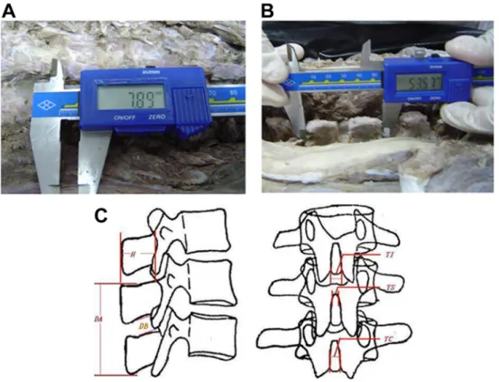
The cadavers were placed in a prone position for numbering the vertebra. The posterior lumbar spine (T12
-S1) was exposed, and the spinous process, vertebral plate,
and articular process were revealed (Figure 1A and B). A digital caliper was used for measurement. Three measure-ments were made for each distance. The main measuring parameters were as follows (Figure 1C): 1) distance between the two adjacent spinous processes (DB), L1-2,
L2-3, L3-4, L4-5, and L5-S1 were determined; 2) distance
across the two adjacent spinous processes (DA), T12-L1,
L1-2, L2-3, L3-4, and L4-5were measured;3) thickness of the
central spinous processes (TC);4) thickness of the superior margin of the spinous processes (TS), L2, L3, L4, L5, and S1
were measured;5) thickness of the inferior margin of the spinous processes (TI), L1-L5were measured; and6) height
of spinous processes (H), L1-L5were measured.
Statistical analysis
The results are reported as means±SD. P,0.05 was considered to be statistically significant. Statistical evalua-tion was performed using the SPSS version 12.0 software (SPSS Inc., USA). One-sample Kolmogorov-Smirnov test and one-way analysis of variance were used to evaluate the
normal distribution and the homogeneity of variance among data. The influence of gender on the interspinous distance was analyzed using Pearson’s correlation analysis.
Results
Statistics
A one-sample Kolmogorov-Smirnov test confirmed the approximately normal distribution among data in each group, and one-way analysis of variance indicated the homogeneity of variance among data in each group. There were no statistically significant differences among TC and TI in males and TC, TI, and H in females by variance analysis, respectively (Table 1).
Anatomical parameters of the lumbar spine spinous process
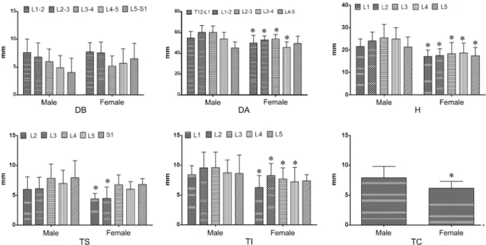
The anatomical parameters of the lumbar spine spinous process in males and females indicated a similar variation trend (Figure 2). The DB decreased gradually from L1-2(7.61±2.44 mm) to L5-S1(4.03±2.57 mm). The
DA increased from T12-L1 (54.63±6.50 mm) to L2-3
(60.18±6.11 mm), then decreased from L2-3 to L4-5
(45.07±5.89 mm). The largest H in males was noted at L3 (25.45±5.96 mm), while it was noted at L4
(18.71±4.50 mm) in females. Usually, the TS values for the adjacent spinous processes were lower than the TI values. For example, the TS of L2(5.97±2.11 mm) was
lower than the TI of L1(8.42±1.52 mm), and the TS of
L3 (6.12±1.89 mm) was lower than the TI of L2
(9.57±2.63 mm).
Comparisons of each parameter between males and females are shown in Figure 2. The DB values showed no statistically significant difference between males and females. There was, however, a statistically significant difference between the DA of males and females, except
for L4-5. The TS of L2 and L3 showed a statistically
significant difference between males and females, and the other segments did not. Except for L5, there was a
statistically significant difference in the TI of L1-L4between
males and females. In addition, a statistically significant difference was found in the TC and H values between males and females. Compared to males, the female spinous process was shorter, thinner, and lower.
Relevance of gender for each parameter
According to Hinkle et al. (12), a Pearson coefficient of 1.0-0.9 means very high relevance, 0.9-0.7 means high relevance, 0.7-0.5 means moderate relevance, 0.5-0.3 means low relevance, and less than 0.3 means no relevance. The relevance of each parameter to gender is shown in Table 1. The L1-2, L2-3, and L3-4of DA were of
moderate relevance in males, while L4-5 and L5-S1of DA
were of low relevance in males (P,0.05). The L1-2, L2-3, L3-4,
L4-5, and L5-S1 of DA were of low relevance in females
(P,0.05). Moreover, L1-2of TI was of low relevance in males
(P,0.05). For other parameters, there was no relevance found to either males or females.
Design of the interspinous stabilization device
Based on the anatomical parameters of the lumbar spine spinous processes obtained in this study, we proposed an H-shaped coronal plane (posterior view, Figure 3) for an interspinous stabilization device for Chinese patients. Two grooves were located at the upper and lower ends of the spinous process, the width of the inferior groove (WIG) and the superior groove (WSG) matched with two adjacent spinous processes. This device has a kidney shape when viewed laterally (Figure 3). The sagittal diameter (SD) is a little smaller than the length of the spinous process, around 25 mm, the central height (CH) ranges from 3 to 13 mm, the wing height is about 30-40 mm, and the WSG is about
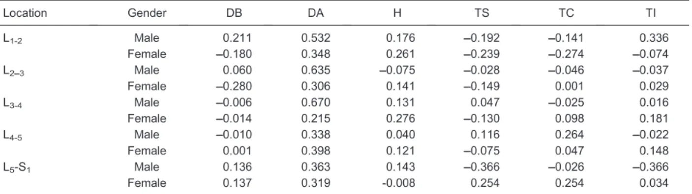
Table 1. Relevance of each parameter with gender.
Location Gender DB DA H TS TC TI
L1-2 Male 0.211 0.532 0.176 ––0.192 ––0.141 0.336
Female ––0.180 0.348 0.261 ––0.239 ––0.274 ––0.074
L2––3 Male 0.060 0.635 ––0.075 ––0.028 ––0.046 ––0.037
Female ––0.280 0.306 0.141 ––0.149 0.001 0.029
L3-4 Male ––0.006 0.670 0.131 0.047 ––0.025 0.016
Female ––0.014 0.215 0.276 ––0.130 0.098 0.181
L4-5 Male ––0.010 0.338 0.040 0.116 0.264 ––0.022
Female 0.001 0.398 0.121 ––0.075 0.047 0.148
L5-S1 Male 0.136 0.363 0.143 ––0.366 ––0.026 ––0.366
Female 0.137 0.319 -0.008 0.254 0.254 0.034
5-10 mm, whereas the WIG is 3-6 mm less than that of the WSG.
Discussion
Until recently, numerous interspinous implants have been introduced and have shown favorable outcomes in the treatment of degenerative disc disease, herniated nucleus pulposus, lumbar spinal stenosis, lumbar instabil-ity, and degenerative spondylolisthesis (8,9,13-15). Nevertheless, complications still occur in interspinous
implants. Thus, selection of an optimal size is mandatory to avoid unwanted complications, and the size of the device should be carefully evaluated (2,11). In the present work, we measured the anatomical parameters of the lumbar spinous processes in the Chinese population using 52 adult cadavers. Based on the anatomical parameters of the lumbar spinous processes obtained in this study, we proposed a design for an interspinous stabilization device for the Chinese population.
Data obtained in each group were of normal distribu-tion and homogeneity of variance, which could reflect a common Chinese population. According to the measure-ment data, several characteristics were found:1) the DB decreased gradually from L1-2 to L5-S1; 2) the DA
increased from T12-L1to L2-3, and then decreased from
L2-3 to L4-5; 3) the largest H in males was noted at L3,
whereas it was noted at L4in females; and4) the TS of the
adjacent spinous process was lower than that of the TI. The middle sections of the DA, H, and TI were found to be larger than those of the upper and lower ends. Compared to males, the female spinous processes were shorter, thinner, and lower. The difference in size of the lumbar spinous processes between males and females probably reflects the difference in average physical size between the genders (16). Ihm et al. (2) investigated the morphometry of the spinous process for interspinous device implantation in Korean patients. They found that the interspinous distance decreased from L1-2 to L5-S1, and the height
increased from L1to L2and gradually decreased below L3.
Figure 2.Comparison of each parameter (DB, DA, H, TS, TI, and TC) between males and females. DB: distance between two adjacent spinous processes; DA: distance across two adjacent spinous processes; H: height of the spinous processes; TS: thickness of the superior margin of the spinous processes; TI: thickness of the inferior margin of the spinous processes; TC: thickness of the central spinous processes. *P,0.05 femalevsmale (one-way ANOVA).
The tendency for variation of the interspinous distance was similar to the results obtained in our research, but the results for height were not similar. The largest value for height obtained in our study was at L3 and gradually
decreased below L3.
Based on the results of this study for the anatomical parameters of the lumbar spinous processes, an H-shaped coronal plane (posterior view) was proposed for the design of an interspinous stabilization device for the Chinese population. As described previously, implant subsidence is a naturally occurring process that is observed during aging and after spine surgery (16,17). A device can migrate if a loosely fitted implant is used when considering future subsidence (11). Dynamic implants allow normal (natural) subsidence to occur, which can stabilize the spine effectively by preventing translation, rotation, and angular deformation (17). The effect of aging and subsidence should be considered carefully when using interspinous implants.
We acknowledge that there are several limitations to this study. The study may not capture all the anatomical variations because of the small sample size. A larger sample size may result in narrower standard deviations. This study was performed on cadaver specimens. A larger sample from live patients would be better. Future work is needed for morphometric studies of the human lumbar spine by computed tomography for Chinese populations. In conclusion, despite these limitations, our study still has value in terms of reporting on the anatomical parameters of the lumbar spinous process, for the design of an interspinous stabilization device for the Chinese population and by offering an anatomical basis for clinical applications.
Acknowledgments
We wish to express our warm thanks to the donors of the cadavers.
References
1. Siebert E, Pruss H, Klingebiel R, Failli V, Einhaupl KM, Schwab JM. Lumbar spinal stenosis: syndrome, diagnostics and treatment. Nat Rev Neurol 2009; 5: 392-403, doi: 10.1038/nrneurol.2009.90.
2. Ihm EH, Han IB, Shin DA, Kim TG, Huh R, Chung SS. Spinous process morphometry for interspinous device implantation in Korean patients.World Neurosurg2013; 79: 172-176, doi: 10.1016/j.wneu.2011.04.027.
3. Jolles BM, Porchet F, Theumann N. Surgical treatment of lumbar spinal stenosis. Five-year follow-up.J Bone Joint Surg Br2001; 83: 949-953, doi: 10.1302/0301-620X.83B7.11722. 4. Postacchini F, Cinotti G, Gumina S, Perugia D. Long-term results of surgery in lumbar stenosis. 8-year review of 64 patients.Acta Orthop Scand Suppl1993; 251: 78-80, doi: 10.3109/17453679309160127.
5. Schlegel JD, Smith JA, Schleusener RL. Lumbar motion segment pathology adjacent to thoracolumbar, lumbar, and lumbosacral fusions.Spine1996; 21: 970-981, doi: 10.1097/ 00007632-199604150-00013.
6. Schmoelz W, Huber JF, Nydegger T, Dipl I, Claes L, Wilke HJ. Dynamic stabilization of the lumbar spine and its effects on adjacent segments: an in vitro experiment. J Spinal Disord Tech 2003; 16: 418-423, doi: 10.1097/00024720-200308000-00015.
7. Tsai KJ, Murakami H, Lowery GL, Hutton WC. A biomecha-nical evaluation of an interspinous device (Coflex) used to stabilize the lumbar spine.J Surg Orthop Adv2006; 15: 167-172.
8. Shim CS, Park SW, Lee SH, Lim TJ, Chun K, Kim DH. Biomechanical evaluation of an interspinous stabilizing device, Locker.Spine2008; 33: E820-E827, doi: 10.1097/ BRS.0b013e3181894fb1.
9. Bono CM, Vaccaro AR. Interspinous process devices in the lumbar spine.J Spinal Disord Tech2007; 20: 255-261, doi:
10.1097/BSD.0b013e3180331352.
10. Barbagallo GM, Olindo G, Corbino L, Albanese V. Analysis of complications in patients treated with the X-Stop Interspinous Process Decompression System: proposal for a novel anatomic scoring system for patient selection and review of the literature.Neurosurgery2009; 65: 111-119, doi: 10.1227/01.NEU.0000346254.07116.31.
11. Bowers C, Amini A, Dailey AT, Schmidt MH. Dynamic interspinous process stabilization: review of complications associated with the X-Stop device.Neurosurg Focus2010; 28: E8, doi: 10.3171/2010.3.FOCUS1047.
12. Hinkle DE, Wiersma W, Jurs SG.Applied statistics for the behavioral sciences. Boston: Houghton Mifflin; 2003. 13. Chiu JC. Interspinous process decompression (IPD) system
(X-STOP) for the treatment of lumbar spinal stenosis.Surg Technol Int2006; 15: 265-275.
14. Mariottini A, Pieri S, Giachi S, Carangelo B, Zalaffi A, Muzii FV, et al. Preliminary results of a soft novel lumbar intervertebral prothesis (DIAM) in the degenerative spinal pathology.Acta Neurochir Suppl 2005; 92: 129-131, doi: 10.1007/3-211-27458-8_28.
15. Yano S, Hida K, Seki T, Aoyama T, Akino M, Iwasaki Y. A new ceramic interspinous process spacer for lumbar spinal canal stenosis. Neurosurgery 2008; 63: ONS108-ONS113, doi: 10.1227/01.NEU.0000310693.86660.D3. 16. Aylott CE, Puna R, Robertson PA, Walker C. Spinous
process morphology: the effect of ageing through adulthood on spinous process size and relationship to sagittal alignment.Eur Spine J2012; 21: 1007-1012, doi: 10.1007/ s00586-011-2029-6.