ABSTRACT
Minimally traumatic alveolar ridge augmentation
with a tunnel injectable thermo-sensitive alginate
scaffold
Yifen LI1,2*, Xiaoqian FANG1*, Ting JIANG1
1- Department of Prosthodontics, Peking University School and Hospital of Stomatology, Beijing, China; Department of Prosthodontics, Jiangmen Municipal Stomatological Hospital, Jiangmen, Guangdong, China.
2- Department of Prosthodontics, Peking University School and Hospital of Stomatology, Beijing, China.
*These authors contributed to the work equally and should be regarded as co-irst authors.
Corresponding address: Ting Jiang - Department of Prosthodontics - Peking University School and Hospital of Stomatology, 22 Zhongguancun Avenue South - Haidian District - Beijing 100081 - China - Phone: 010-82195348 - 15011229485 - e-mail: [email protected]
Submitted: November 28, 2014 - Modiication: January 29, 2015 - Accepted: March 2, 2015
I
njectable bone substitutes and techniques have been developed for use in minimallyinvasive procedures for bone augmentation. Objective: To develop a novel injectable thermo-sensitive alginate hydrogel (TSAH) as a scaffold to induce bone regeneration, using a minimally invasive tunnelling technique. Material and Methods: An injectable TSAH was prepared from a copolymer solution of 8.0 wt% Poly(N-isopropylacrylamide)
(PNIPAAm) and 8.0 wt% AAlg-g-PNIPAAm. In vitro properties of the material, such as
its microstructure and the sustained release of recombinant human bone morphogenetic protein-2 (rhBMP-2), were investigated. Then, with the subperiosteal tunnelling technique, this material, carrying rhBMP-2, was injected under the labial periosteum of the maxillary anterior alveolar ridge in a rabbit model. New bone formation was evaluated by means of
X-ray, micro-computed tomography (micro-CT), luorescence labelling, histological study,
and immunohistochemistry study. Results: The material exhibited good injectability and thermo-irreversible properties. SEM showed an interconnected porous microstructure of the TSAH. The result of ALP activity indicated sustained delivery of BMP-2 from the TSAH from days 3 to 15. In a rabbit model, both TSAH and TSAH/rhBMP-2 induced alveolar ridge augmentation. The percentage of mineralised tissue in the TSAH/rhBMP-2 group
(41.6±3.79%) was signiicantly higher than in the TSAH group (31.3±7.21%; p<0.05).
The density of the regenerating tissue was higher in the TSAH/rhBMP-2 group than in the
other groups (TSAH group, positive control, blank control; p<0.05). Conclusions: The TSAH
provided convenient handling properties for clinical application. To some extent, TSAH could induce ridge augmentation and mineral deposition, which can be enhanced when combined with rhBMP-2 for a minimally invasive tunnelling injection.
Keywords: Alveolar ridge augmentation. Minimally invasive surgical procedures. Hydrogel. Tissue engineering.
INTRODUCTION
Atrophic maxillary anterior alveolar ridge fails to
provide suficient support or aesthetics for dental
prostheses24. Today, allografts and xenograft bone
grafts are the major methods for augmenting a resorbed alveolar ridge. However, they have several disadvantages, such as the risk of disease transmission and immunological reactions3. Another
disadvantage is that these grafts require an open surgical operation8. To reduce trauma and surgery
cost, “injectable tissue-engineered bone” has
been proposed. Injectable systems ― materials and techniques ― are being developed for use in
minimally invasive procedures and their ability to
ill irregular defects1,7.
In the late 1980s, Kent, et al.12,13 (1982, 1983)
They made a small incision in the mucosa of the alveolar ridge, elevated the periosteal, and injected hydroxyapatite particles into the tunnel
through a modiied syringe. Although the material
diffused into adjacent tissues, leading to poor bone regeneration, the technique attracted attention. However, due to the limitations of the injectable bone substitute material, this technique did not become popular clinically.
Injectable biomaterials now include synthetic ceramics and synthetic polymers16. In recent
years, thermo-sensitive hydrogel systems have
attracted much interest in the biomedical ield22.
Poly(N-isopropylacrylamide) (PNIPAAm) is one such synthetic polymer that can form a hydrogel
in situ above its lower critical solution temperature (LCST) and maintain a liquid state below the LCST. Alginate is a natural polysaccharide composed
of 1,4-linked β-D-mannuronate and 1,4-linked α-L-guluronate residues. It is extracted from
algin, which is abundant and inexpensive. It also has the advantages of good biocompatibility and immunological non-responsiveness20. The
AAlg-g-PNIPAAm copolymer, prepared through coupling carboxylic end-capped PNIPAAm to aminated alginate (AAlg), has good injectability and plasticity at room temperature, while it gels at body temperature (37°C). In particular, it has been shown to be non-cytotoxic and can promote the proliferation of entrapped cells. The degradation rate is controllable by changing the quantity of PNIPAAm23. By adding 8.0 wt% PNIPAAm to 8.0
wt% AAlg-g-PNIPAAm copolymer, we obtained a TSAH that seemed to be a suitable delivery system for minimally invasive bone regeneration.
Bone morphogenetic protein-2 (BMP-2), one of the most promising osteoinductive growth factors, can recruit progenitor cells and promote the differentiation of osteoblasts2,25. Because of the
short half-life and initial burst release of BMP-2, biocompatible carriers such as the thermo-sensitive alginate are necessary to protect its bioactivity and prolong the release period15.
In this study, we combined the TSAH with BMP-2 to explore the effect of this delivery system on bone augmentation of the anterior alveolar ridge in a rabbit model, using the sub-periosteal tunnelling technique. The micro-structure of the TSAH and the sustained release properties of rhBMP-2 from the TSAH in vitro were also explored.
MATERIAL AND METHODS
Preparation of the TSAH
An AAlg-g-PNIPAAm copolymer with 29% PNIPAAm grafting was prepared. The copolymer was synthesised by grafting PNIPAAM-COOH with a single carboxyl end group onto aminated
alginate (AAlg) through amide bond linkages23. Dry
PNIPAAm and AAlg-g-PNIPAAm were sterilised with an electron beam, dissolved in phosphate buffer solution (PBS, pH 7.4), and stirred overnight at 4°C. In order to obtain an optimum mixture ratio, different percentages of PNIPAAm (6.0, 8.0 wt%) were mixed into different percentages of AAlg-g-PNIPAAm (8.0, 10.0, 12.0 wt%). We detected the injectability and the hardness after gelation of the materials at each mixture ratio. Finally, a delivery system containing 8.0 wt% PNIPAAm and 8.0 wt% AAlg-g-PNIPAAm was chosen and prepared.
Morphological observation
The TSAH was irst gelled at 37°C, lyophilised
into a dry sample, and then fractured. The cross-section was gold-coated and the inner morphology of the TSAH was observed by scanning electron microscopy (SEM, Hitachi S-4800, Japan) at 15 kV.
Bioactivity of rhBMP-2 released from the hydrogel in vitro
The recombinant human BMP-2 (rhBMP-2;
PeproTech, USA) was dissolved in PBS containing bovine serum albumin (BSA) (1 µg of rhBMP-2 per
50 µg of BSA), then the solution was mixed into
the TSAH. The inal BMP-2 concentration was 50
µg/mL. All preparations were carried out under aseptic conditions.
TSAH/rhBMP-2 (250 µL) and TSAH samples (250 µL) were injected separately into 1.5-mL capped centrifuge tubes and incubated at 37°C for a few minutes for gelation. Then, 1 mL of PBS was added into each tube and incubated in a rotary shaker (35 rpm) at 37°C for 21 days. At scheduled intervals (1, 3, 5, 7, 9, 11, 15, 18 and 21 days), 0.5 mL of supernatant were collected and replaced with 0.5 mL of fresh PBS. The collected supernatant was
sterilised by iltration and stored at -20°C for later
use.
Human bone marrow-derived mesenchymal stem cells (hBMSCs) were plated at a density of 2×10⁴ cells per well in 24-well plates. Subsequently,
the cells were incubated in α-MEM with 10% fetal
bovine serum (FBS), 100 U/mL penicillin, and 100
µg/mL streptomycin in a humidiied 37°C/5% CO2
incubator. After 24 h, the medium was replaced with 1 mL of fresh medium composed of obtained
supernatant and α-MEM at a ratio of 1:7 (v/v). The
obtained supernatant without rhBMP-2 was used in the negative control group while 400 ng/mL rhBMP-2 were used in the positive control group. After 5 days in culture, the medium was removed, the cells were washed twice with PBS, and lysed with 0.5 mL of 1% Triton X-100 combined with ultrasonication.
assumed to relect the degree of the osteogenic
differentiation18,26. It is also well-known that BMP-2
can increase the ALP activity of hBMSCs in a dose-dependent manner. Thus, the amount and activity of BMP-2 released from the gel can be evaluated indirectly according to an ALP activity assay in vitro. The cell lysates were centrifuged and the supernatant was collected for ALP analysis with an ALP activity assay kit (Jiancheng, Nanjing, Jiangsu, China) according to the manufacturer’s protocol.
Animal experiments
In vivo bone augmentation
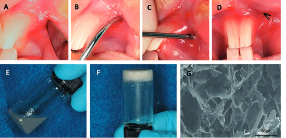
In total, sixteen male New Zealand White rabbits, weighing ~2.5 kg, were allowed to acclimatize for 1 week in the laboratory animal housing prior to the operation. All experiment protocols were approved by the Biomedical Ethics Committees. Under general anaesthesia, through the otomarginal vein with pentobarbital sodium (30 mg/kg), an incision of ~1 mm was made in the mucoperiosteum on the labial side of the maxillary anterior alveolar bone. A subperiosteal space with dimensions of 10×5×4 mm was created with a periosteal elevator. Afterwards, the material was injected through a syringe needle of 1.2 mm in diameter into the tunnel to occupy the space created and to augment the alveolar ridge. The incision was then closed with a
suture (Figure 1A-D).
A total of 32 alveolar augmentation implants were injected bilaterally in 16 rabbits, divided randomly into the following four groups: A- TSAH
(n=12, 4 for each time point: weeks 4, 8, 12); B-
TSAH/rhBMP-2 (n=12, 4 for each time point: weeks
4, 8, 12); C- injectable calcium phosphate/BMP-2 as a positive control (n=4, week 12), and D- PBS
as a blank control (n=4, week 12).
All rabbits were given antibiotics via a
subcutaneous injection 3 days after the operation and were observed grossly every day. Animals were euthanised at 4, 8, and 12 weeks after surgery.
Alveolar bone samples were harvested and ixed in
10% neutral buffered formalin. All specimens were analysed by radiography and histologically.
Radiography analyses and micro-CT assessment
The gross samples were examined using a dental X-ray machine (Warning, Finland) at 60 kV, 8 mA for 0.125 s. Then, they were scanned by micro-computed tomography (micro-CT)
(Siemens Inveon, Germany) at 60 kV, 400 μA in
high resolution scanning mode to determine the
bone volume. Bone mineral density (BMD) and
bone volume fraction (bone volume/total volume, BV/TV) were also determined.
Sequential luorescent labelling
To evaluate the distribution and rate of new
bone formation, the rabbits sacriiced at 12 weeks
post-surgery were labelled with three polychrome
sequential luorescent markers. At 3, 6 and 9
weeks after the surgical operation, the rabbits were injected subcutaneously at the implant sites with 60 mg/kg tetracycline hydrochloride (TE) (Sigma, St Louis, MO, USA), 30 mg/kg alizarin red (AL) (Sigma, St Louis, MO, USA), and 10 mg/kg calcein (CA) (Sigma, St Louis, MO, USA), respectively.
Histological and histomorphometric observations
The samples harvested at 4 and 8 weeks were
decalciied completely in 20% EDTA (pH=7.2), embedded in parafin wax, and sectioned with
a microtome (Leica RM2235, Germany) to a
thickness of 4 μm. Then, haematoxylin and eosin
(HE) staining was performed and the expression of two osteoblast differentiation markers, osteopontin (OPN) and osteocalcin (OCN), was examined by
immunohistochemical staining. Those harvested at 12 weeks post-surgery were bisected. Half of
the gross samples was decalciied and stained
as above. The remaining half was dehydrated, embedded in polymethylmethacrylate and cut into
150-μm-thick sections with a microtome (EXAKT
300CP, Germany). Finally the sections were ground
and polished to a thickness of 40 μm and observed under a luorescence microscope.
Statistical analysis
All data are presented as means±standard
deviations. Signiicant differences in in vitro bone formation analyses were analysed by one-way ANOVA. The differences between groups and time
points were considered statistically signiicant if p<0.05.
RESULTS
Preparation of the TSAH
The TSAH was lowable and could be injected
smoothly through a needle of 1.2 mm in diameter at room temperature. Above the LCST (37°C), it gelled and gained enough strength to support the periosteum (Figure 1E-F).
Morphological observation
The microstructure of the lyophilised TSAH was
observed by scanning electron microscopy (SEM);
the image is shown in Figure 1G. The hydrogel presented an interconnected porous microstructure, like a honeycomb. Interconnected pores with sizes in the range of 30-50 nm could be found in the
TSAH.
Bioactivity of rhBMP-2 released from the hydrogel in vitro
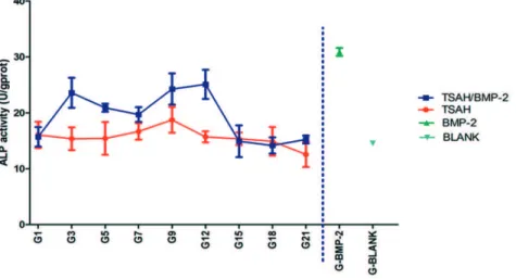
The release proile of rhBMP-2 loaded in hydrogel
is shown in Figure 2. ALP activity increased greatly at day 3, indicating that osteoblast differentiation was induced by the initial exposure to BMP-2, and
then maintained until day 15. It was signiicantly
higher than that in the negative control group
(p<0.05). From day 18 to day 21, there was no
obvious difference between the test group and control group. These results indicated that sustained delivery of BMP-2 from the gel stimulated ALP
activity in hBMSCs from day 3 to day 15; however,
the dose of BMP-2 released might be too low to affect ALP activity before and after this period.
In vivo bone formation
Radiographic examination
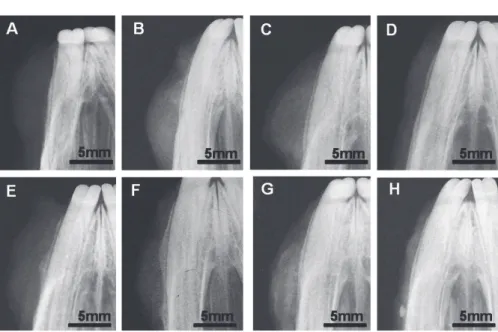
Radiographs were taken at 4, 8, and 12 weeks post-surgery for qualitative assessments of bone formation (Figure 3). There was no visible new bone formation in the samples at 4 and 8 weeks post-surgery in the TSAH and TSAH/rhBMP-2 groups but augmented images above the surface of alveolar ridge can be seen. At 12 weeks, some distributed high density areas were seen within the material. Furthermore, the radiopaque areas in the TSAH/rhBMP-2 group were larger than those
of the TSAH group. The group illed with calcium
phosphate cement (CPC)/rhBMP-2 showed no new bone formation. Interestingly, high-density spots were distinguishable on the buccal side of the
Figure 3- Radiographs of samples in the thermo-sensitive alginate hydrogel (TSAH) group at weeks 4, 8, and 12 (A, B, C, respectively), phosphate buffer solution (PBS) negative control group at week 12 (D), TSAH/bone morphogenetic protein-2 (BMP-2) group at weeks 4, 8, and 12 (E, F, G, respectively), and the calcium phosphate cement (CPC)/BMP-2 control group at week 12 (H)
alveolar ridge, 10 mm from the injecting area in two samples. There was no bone augmentation in the PBS group.
Micro-CT examination
All of the harvested samples were scanned and reconstructed with a micro-CT system for quantitative assessment of bone formation (Figure 4A). The images showed successful bone augmentation on bone surfaces in the TSAH groups with or without rhBMP-2. At the periphery of the protuberant area, the amount of new bone formation was higher than that in the centre. The analysis of regenerated bone volumes also indicated that alveolar ridges were augmented (~10×5×4 mm) in both the TSAH and the TSAH/ rhBMP-2 groups, whereas there were only high density spots (~1×2×2 mm) in the CPC/rhBMP-2 group, and nothing in the PBS group at week 12. The percentage of mineralised tissue in the TSAH/
rhBMP-2 group (41.6±3.79%) was signiicantly higher than that in the TSAH group (31.3±7.21%; p<0.05; Figure 4B). The density of the regenerated
tissue was higher in the TSAH/rhBMP-2 group than
in the other groups (p<0.05; Figure 4C).
S e q u e n t i a l f l u o r e s c e n c e l a b e l l i n g histomorphometric analysis
The regions above the labial cortical bone
were chosen to analyse the luorescence labelling
and assess the mineralisation process (Figure 5). The results showed the shape of tetracycline hydrochloride (TE) labelling (yellow) matched the
linear ibre tissue in both the TSAH and TSAH/
rhBMP-2 groups at week 3. Next, alizarin red (AL) labelling (red) was distributed around the TE labelling at week 6. Finally, calcein (CA) labelling (green) was detected clearly in both groups until week 9, indicating that mineralisation was occurring. There was a trend for areas of TE and AL
labelling in the TSAH group to be weaker than those
in the TSAH/rhBMP-2 group in the irst 6 weeks.
However, at week 9, the areas of CA labelling were comparable. These results demonstrated that TSAH alone can induce calcium deposition.
Histological evaluation
No inlammatory cells were found at 4, 8, or 12 weeks in any group from decalciied sections stained with HE (Figure 6A-D). There was no evidence of
an immune response in any group. Multinucleated giant cells were found adjacent to the remnant hydrogel, which may participate in the degradation of TSAH. The degradation rates of TSAH in groups TSAH and TSAH/rhBMP-2 increased gradually from weeks 4 to 12. However, at 12 weeks, some TSAH was not degraded and remained at the centre of the implantation areas. Nevertheless, some cells, such as osteoblasts and chondrocytes, were found around the remnant hydrogel at 4 weeks post-surgery. Moreover, in some areas, osteoid structures were observed at 12 weeks post-surgery in the peripheral regions of the material in the TSAH and TSAH/rhBMP-2 groups. In the CPC/rhBMP-2 control group very little mineralised tissue was observed in the transplant areas at 12 weeks, consistent with the radiographic and micro-CT results. The normal anatomical structure of intact bone and the periosteum were seen in the PBS group.
Immunohistochemical staining of osteoblast differentiation markers is shown in Figures 6E-F (OPN) and 6G-H (OCN). Newly mineralised tissue showed high expression of OPN in weeks 4-12 and weak immunostaining for OCN in the TSAH and TSAH/rhBMP-2 groups.
DISCUSSION
Bone augmentation techniques have recently developed in a noticeable manner. Subperiosteal tunnelling injection is a minimally invasive technique for bone regeneration. Stevens, et al.21
(2005) reported the use of such technique to obtain new bone tissue21. However, due to the poor
plasticity of most of the injectable bone substitute materials10,14 and their diffusion after injection in
vivo6, the technique has not been used widely.
The sol-gel transformation of the TSAH prepared
here occurred at 37°C. When the lowable material
was injected through a syringe needle into tissue, under the effect of body temperature, in situ it turned into a gel form and was stable on the bone surface. It was strong enough to support the mucoperiosteum effectively without leakage into the surrounding tissues like the CPC. CPC can be washed out by surgical bleeding after injection into the subperiosteum. This highlights the importance of choosing an appropriate protein factor carrier. It also showed that the TSAH material could meet the operating requirements.
In bone tissue engineering, the degradation rate of the scaffold should be consistent with the new
bone formation rate17. Previous research showed
that the degradation time of TSAH can be controlled. With 29% PNIPAAm grafting, 40% of the TSAH was degraded in 28 days, matching the bone formation rate23.
RhBMP-2 can effectively improve the bone formation and many materials have been reported as carriers for it in animal and clinical studies4,11.
Alginate hydrogel can retain rhBMP-2 through an electrostatic interaction with the positively charged protein19. As a scaffold for protein growth factors,
TSAH can provide sustained release of rhBMP-2 and maintain its bioactivity. The rhBMP-2 released from the TSAH into the medium increased the ALP activity of hBMSCs from day 3 to day 15.
Regarding the osteogenic capabilities of TSAH
in vivo, micro-CT showed that both the TSAH and TSAH/rhBMP-2 groups had high-density masses in the augmented area. This suggests that, with or without rhBMP-2, the TSAH material itself can induce mineralisation. When combined with rhBMP-2, the density and volume of mineralised tissue increased and osteogenesis could be enhanced.
Fluorescence labelling revealed that calcium deposition began at week 3 in the TSAH and TSAH/rhBMP-2 groups and was deposited continuously throughout the observation period. The
luorescence intensity in both groups at week 9 was
enhanced versus that at weeks 3 and 6, suggesting that the mineralisation activity increased with time. The deposition expanded beyond the immediate mineralisation centre. Closer to the bone surface, more calcium was deposited, indicating that attachment to the bone surface improved
mineralisation. In the third month, the luorescence
intensity increased, indicating that mineralisation was increasingly active. Prolonging the observation time may lead to detection of more bone tissue formation.
Osteopontin is a non-collagenous bone matrix glycoprotein produced by preosteoblasts and osteoblasts in the early stages of mesenchymal proliferation, pro-mineralization, and mineralization. It is a marker of mature osteoblasts9. Osteocalcin is
expressed by mature osteoblasts in the late stages of osteogenic differentiation. It is considered the
most speciic marker of osteogenesis5. In this study,
immunohistochemistry revealed OPN expression at 4, 8, and 12 weeks post-surgery in both the TSAH and TSAH/rhBMP-2 groups, further indicating that the TSAH material itself can induce mineralisation after contact with the periosteum. There were a few coloured cells after OCN immunohistochemical
staining, in which the scattered dot-like speciic
staining matched the dot-like colouring in bone
luorescence labelling, suggesting that calcium
deposition activity in some areas had resulted in the late stage of mineralization. However, there were
no large areas of OCN-speciic staining after 12
weeks, showing that the osteogenic differentiation of cells in vivo was mostly restricted to the early stage. From specimens at 12 weeks, alginate remained in the centre of the implants. Calcium deposition was located mainly at the edge of the implants. This suggests that the material needs further improvement. Methods could include adding inorganic filler to increase bone conductivity, enhancing nutritional metabolism inside the material, increasing blood supply, and adding further growth factor(s).
CONCLUSIONS
In this study, an injectable, sol-gel reversible thermo-sensitive alginate hydrogel was developed. With a subperiosteal tunnelling injection technique, this bone substitute was injected under the maxillary labial periosteum, gelled in situ, and inally induced
hard tissue formation, to some extent. However, the hard tissue formed was mainly at the early stages of mineralization and predominantly at the periphery of the material. No mature bone tissue was detected. The results suggest the material needs further improvement.
REFERENCES
1- Boix D, Weiss P, Gauthier O, Guicheux J, Bouler JM, Pilet P, et
al. Injectable bone substitute to preserve alveolar ridge resorption after tooth extraction: a study in a dog. J Mater Sci Mater Med.
2006;17(11):1145-52.
2- Boyne PJ, Salina S, Nakamura A, Audia F, Shabahang S. Bone regeneration using rhBMP-2 induction in hemimandibulectomy type defects of elderly sub-human primates. Cell Tissue Bank.
2005;7(1):1-10.
3- Buck BE, Malinin TI. Human bone and tissue allografts. Preparation and safety. Clin Orthop Relat Res. 1994(303):8-17.
4- Cicciù M, Herford AS, Cicciù D, Tandon R, Maiorana C.
Recombinant human bone morphogenetic protein-2 promote and stabilize hard and soft tissue healing for large mandibular new
bone reconstruction defects. J Craniofac Surg. 2014;25(3):860-2. 5- Cowles EA, DeRome ME, Pastizzo G, Brailey LL, Gronowicz GA.
Mineralization and the expression of matrix proteins during in vivo
bone development. Calcif Tissue Int. 1998;62(1):74-82. 6- Driessens FC, Boltong MG, Bermudes O, Planell JA, Ginebra MP,
Fernández E. Effective formulation for the preparation of calcium
phosphate bone cements. J Mater Sci Mater Med.
1994;5(3):164-70.
7- Gauthier O, Bouler JM, Weiss P, Bosco J, Daculsi G, Aguado E.
Kinetic study of bone ingrowth and ceramic resorption associated with the implantation of different injectable calcium-phosphate
bone substitutes. J Biomed Mater Res, 1999;47(1):28-35. 8- Gazdag AR, Lane JM, Glaser D, Forster RA. Alternatives to autogenous bone graft: eficacy and indications. J Am Acad Orthop Surg. 1995;3(1):1-8.
9- Giachelli CM, Steitz S. Osteopontin: a versatile regulator of
inlammation and biomineralization. Matrix Biol.
2000;19(7):615-22.
10- Hjorting-Hansen E, Worsaae N, Lemons JE. Histologic response after implantation of porous hydroxylapatite ceramic in humans.
11- Jiang X, Zhao J, Wang S, Sun X, Zhang X, Chen J, et al. Mandibular repair in rats with premineralized silk scaffolds and
BMP-2-modiied bMSCs. Biomaterials. 2009;30(27):4522-32.
12- Kent JN, Quinn JH, Zide MF, Finger IM, Jarcho M, Rothstein
SS. Correction of alveolar ridge deiciencies with nonresorbable hydroxylapatite. J Am Dent Assoc. 1982;105(6):993-1001.
13- Kent JN, Quinn JH, Zide MF, Guerra LR, Boyne PJ. Alveolar ridge augmentation using nonresorbable hydroxylapatite with or without autogenous cancellous bone. J Oral Maxillofac Surg.
1983;41(10):629-42.
14- Kim J, Kim IS, Cho TH, Lee KB, Hwang SJ, Tae G, et al. Bone regeneration using hyaluronic acid-based hydrogel with bone morphogenic protein-2 and human mesenchymal stem cells.
Biomaterials. 2007;28(10):1830-7.
15- Kolambkar YM, Dupont KM, Boerckel JD, Huebsch N, Mooney DJ, Hutmacher DW, et al. An alginate-based hybrid system for
growth factor delivery in the functional repair of large bone defects.
Biomaterials. 2011;32(1):65-74.
16- Kretlow JD, Young S, Klouda L, Wong M, Mikos AG. Injectable
biomaterials for regenerating complex craniofacial tissues. Adv
Mater. 2009;21(32-33):3368-93.
17- Larsson S, Hannink G. Injectable bone-graft substitutes: current products, their characteristics and indications, and new
developments. Injury. 2011;42:S30-4.
18- Lisignoli G, Zini N, Remiddi G, Piacentini A, Puggioli A,
Trimarchi C, et al. Basic ibroblast growth factor enhances in vitro mineralization of rat bone marrow stromal cells grown on non-woven hyaluronic acid based polymer scaffold. Biomaterials.
2001;22(15):2095-105.
19- Luca L, Rougemont AL, Walpoth BH, Gurny R, Jordan O. The effects of carrier nature and pH on rhBMP-2-induced ectopic bone
formation. J Control Release. 2010;147(1):38-44.
20- Marijnissen WJ, van Osch GJ, Aigner J, van der Veen SW, Hollander AP, Verwoerd-Verhoef HL, et al. Alginate as a chondrocyte-delivery substance in combination with a non-woven scaffold for cartilage tissue engineering. Biomaterials,
2002;23(6):1511-7.
21- Stevens MM, Marini RP, Schaefer D, Aronson J, Langer R,
Shastri VP. In vivo engineering of organs: the bone bioreactor.
Proc Natl Acad Sci U S A. 2005;102(32):11450-5.
22- Tan HP, Ramirez CM, Miljkovic N, Li H, Rubin JP, Marra KG. Thermosensitive injectable hyaluronic acid hydrogel for adipose
tissue engineering. Biomaterials. 2009;30(36):6844-53. 23- Tan RW, She ZD, Wang MB, Fang Z, Liu YS, Feng QL.
Thermo-sensitive alginate-based injectable hydrogel for tissue engineering.
Carbohydr Polym. 2012;87(2):1515-21.
24- Von Arx T, Hardt N, Wallkamm B. The TIME technique: a new method for localized alveolar ridge augmentation prior to placement of dental implants. Int J Oral Maxillofac Implants.
1996;11(3):387-94.
25- Xiao YT, Xiang LX, Shao JZ. Bone morphogenetic protein.
Biochem Biophys Res Commun. 2007;362(3):550-3.
26- Zernik J, Twarog K, Upholt WB. Regulation of alkaline phosphatase and alpha 2 (I) procollagen synthesis during early intramembranous bone formation in the rat mandible.