Fever-Range Hyperthermia vs. Hypothermia
Effect on Cancer Cell Viability, Proliferation
and HSP90 Expression
Dimitra Kalamida1, Ilias V. Karagounis1, Achilleas Mitrakas1, Sofia Kalamida1, Alexandra Giatromanolaki2, Michael I. Koukourakis1*
1Department of Radiotherapy/Oncology, Democritus University of Thrace, Alexandroupolis, 68100, Greece,
2Department of Pathology, Democritus University of Thrace, Alexandroupolis, 68100, Greece
Abstract
Purpose
The current study examines the effect of fever-range hyperthermia and mild hypothermia on human cancer cells focusing on cell viability, proliferation and HSP90 expression.
Materials and Methods
A549 and H1299 lung carcinoma, MCF7 breast adenocarcinoma, U87MG and T98G glio-blastoma, DU145 and PC3 prostate carcinoma and MRC5 normal fetal lung fibroblasts cell lines were studied. After 3-day exposure to 34°C, 37°C and 40°C, cell viability was deter-mined. Cell proliferation (ki67 index), apoptosis (Caspase 9) and HSP90 expression was studied by confocal microscopy.
Results
Viability/proliferation experiments demonstrated that MRC5 fibroblasts were extremely sen-sitive to hyperthermia, while they were the most resistant to hypothermia. T98G and A549 were thermo-tolerant, the remaining being thermo-sensitive to a varying degree. Nonethe-less, as a universal effect, hypothermia reduced viability/proliferation in all cell lines. Hyper-thermia sharply induced Caspase 9 in the U87MG most thermo-sensitive cell line. In T98G and A549 thermo-tolerant cell lines, the levels of Caspase 9 declined. Moreover, hyperther-mia strongly induced the HSP90 levels in T98G, whilst a sharp decrease was recorded in the thermo-sensitive PC3 and U87MG cell lines. Hyperthermia sensitized thermo-sensitive cancer cell lines to cisplatin and temozolomide, whilst its sensitizing effect was diminished in thermo-tolerant cell lines.
Conclusions
The existence of thermo-tolerant and thermo-sensitive cancer cell lines was confirmed, which further encourages research to classify human tumor thermic predilection for patient stratification in clinical trials. Of interest, mild hypothermia had a universal suppressing
OPEN ACCESS
Citation:Kalamida D, Karagounis IV, Mitrakas A, Kalamida S, Giatromanolaki A, Koukourakis MI (2015) Fever-Range Hyperthermia vs. Hypothermia Effect on Cancer Cell Viability, Proliferation and HSP90 Expression. PLoS ONE 10(1): e0116021. doi:10.1371/journal.pone.0116021
Academic Editor:Olivier Gires, Ludwig-Maximilians University, GERMANY
Received:August 12, 2014
Accepted:December 2, 2014
Published:January 30, 2015
Copyright:© 2015 Kalamida et al. This is an open access article distributed under the terms of the
Creative Commons Attribution License, which permits unrestricted use, distribution, and reproduction in any medium, provided the original author and source are credited.
Data Availability Statement:All relevant data are within the paper.
Funding:The study has been funded by the TRAIN-ING AND LIFELONG LEARNTRAIN-ING - ARISTEIA project, code no 520, ESPA 2007–2013, GGET decision number 12605/26.09.2012. The funders had no role in study design, data collection and analysis, decision to publish, or preparation of the manuscript.
effect on cancer cell proliferation, further supporting the radio-sensitization hypothesis through reduction of oxygen and metabolic demands.
Introduction
Cancer cells, similarly to any other cell and living system, respond to mild changes of external temperature by activating homeostatic biological mechanisms, in an attempt to sustain a toler-ant intracellular environment and prevent death. Temperatures above 41°C are toxic both to the tumor vasculature and to the cancer cells themselves, and are used to heat tumors (Oncothermic Hyperthermia) aiming to suppress their growth, achieve regression or to sensi-tize them to radiotherapy and chemotherapy [1]. Randomized trials have demonstrated signifi-cant improvement of local control rates in sarcomas applying regional hyperthermia combined with chemotherapy [2].
Nevertheless, temperatures below 41°C, at the so called fever-range hyperthermia, have also a direct effect on cancer cell and tissue biology, sensitizing tumors to radiotherapy and chemo-therapy. Whole body hyperthermia between 38–40°C has been used in the treatment of wide-spread metastatic tumors in combination with chemotherapy [3,4]. Increased blood flow that allows increased tumor oxygenation and chemotherapy availability is probably one of the main mechanisms of synergism [5,6]. Protein damage is also a critical effect of temperatures above 39°C, but the exact pathways of cell killing remain elusive [7]. Inhibition of homologous re-combination has been also proposed as a mechanism of tumor chemosensitization [8].
In addition, mild hyperthermia as a complementary therapy to cancer immune-therapy has been presented by several preclinical and clinical studies, by improving antitumor immune re-sponses. The hyperthermia- induced improved immune response includes HSPs generation, antigen presenting cells activation and lymphocytes trafficking changes [9]. Furthermore, addi-tional evidence indicates that physiological responses to induced hyperthermia affects the mi-croenvironment of the tumor most likely by a mechanism involving temperature-sensitive check-points regulating tumor vascular perfusion, lymphocyte trafficking, inflammatory cyto-kine expression, tumor metabolism, and last but not least, both adaptive and innate immune action [10]. Increased activity of natural killer cells against colon tumor cells has been verified in mice when temperature was increased at 39.5°C [11].
In any case, cancer cell response to hyperthermia may depend on both temperature levels and exposure times, additional ambient conditions and, certainly, on the cell type under inves-tigation. In the current study, we examined the effect of fever-range hyperthermia on a wide range of cancer cells, providing evidence that its effect on viability and proliferation is cell-dependent. Hypothermia, a far less studied condition in cancer cell systems, was also investi-gated. The effect of hyper- and hypothermia on HSP90 expression was further examined. Fi-nally, hyperthermic chemosensitization was examined in two glioblastoma cell lines (T98G and U87MG) and two lung cancer cell lines (A549 and H1299). T98G and A549, the only tolerant cell lines were examined in comparison with two representative, thermo-sensitive cell lines of the same tumor type, U87MG and H1299, respectively, with the appropri-ate chemotherapy drugs for each type of cancer, together with mild hyperthermia, in order to examine a possible sensitisation or resistance.
Materials and Methods
Cell cultures
A549 (human lung adenocarcinoma, CLS GmbH, Germany), H1299 (human non-small cell lung carcinoma, ATCC), MCF7 (human breast adenocarcinoma, CLS GmbH, Germany), U87MG (human glioblastoma-astrocytoma, CLS GmbH, Germany), DU145 (human prostate carcinoma, CLS GmbH, Germany), PC3 (human prostate adenocarcinoma, CLS GmbH, Ger-many) and MRC5 (human fetal lung fibroblasts, CLS GmbH, GerGer-many) cell lines were cultured using DMEM basal medium (31885-023, Gibco) and T98G (human glioblastoma multiforme, ATCC) cell line was grown in MEM basal medium (10370-047, Gibco). Both basal culture me-diums were supplemented with 10% FBS (FB-1000/500, Biosera), 100 units/ml Penicillin and 100mg/ml Streptomycin (15140-122, Gibco) and 2mM L-Glutamine (25030, Gibco). Cells were maintained at standard conditions, 37°C, 5% CO2 in humidified atmosphere and were used upon reaching 70–90% confluency.
Proliferation Assay
The ability of cell proliferation under different incubation temperatures was tested by a prolif-eration assay. Cells were seeded in 96-well plates at a density of 1000 cells/well, the plates were incubated at 37°C for 3h to facilitate adherence and normal growth and then were placed at 34°C, 37°C and 40°C, respectively, for three consecutive days. Proliferations assays for all the examined cell lines and growth temperatures were performed simultaneously. Cell prolifera-tion measurements were performed in a 24h interval using the AlamarBlue Cell Viability Re-agent (DAL1100, Invitrogen) measured at 540nm excitation and 590nm emission wavelengths, in a FLUOstar Omega microplate reader (BMG LABTECH). The AlamarBlue assay
(DAL1100, Invitrogen) is a reliable method for cell viability [14]. This assay, by using the metabolic activity of cells to reduce resazurin (oxidized form; 7-hydroxy-3H-phenoxazin-3-1-10-oxide) to resorufin, quantifies the number of cells with active mitochondria, since resazurin reduction is performed by mitochondrial enzymes [15]. The experiments were performed three times in order to confirm the significance of the results.
Western Blot analysis
assay kit (Thermo Scientific Pierce, USA). Specific rabbit polyclonal primary antibodies were used for western blot analysis, anti-HSP90 (1:1000; ab13495, Abcam) and anti-Caspase9 (1:1000; ab47537, Abcam).
Whole fraction samples were separated on discontinuous SDS gels using 10% separating and 5% stacking gels. Forty micrograms of the cell extracts were loaded on the gel. Immuno-blotting was performed utilizing PVDF-PSQ membranes (Millipore Corp.). Following a block-ing step with 5% non-fat dry milk in 150 mM NaCl, 10 mM Tris, pH 7.5 containblock-ing 0.1% (v/v) Tween 20 (TBS-T) at room temperature (RT) for 2h, the membranes were hybridized over-night at 4°C with the primary antibodies. The membranes were then hybridized for 2h at 37°C with the secondary antibody, goat polyclonal to rabbit IgG (H+L)-HRP (1:3.000, Biorad, 1706515, USA) and finally developed in Amersham ECL Western blotting detection reagents and analysis system (RPN2209, GE Healthcare) utilizing Chemidoc MP Imaging System (Biorad, USA).
Hyperthermic chemosensitization experiments
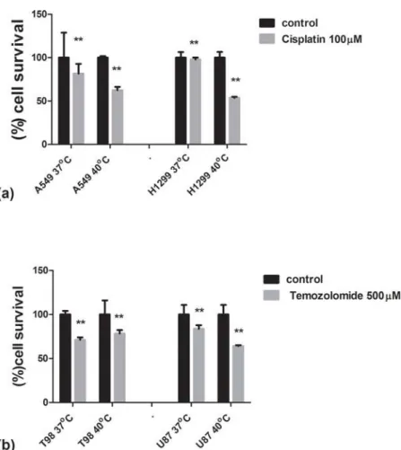
In chemosensitization experiments, 1000 cells/well were plated in a 96-well plate in the appro-priate culture medium. Cells were incubated with clinically established drugs; lung cancer cell lines: A549 and H1299 with 100mΜof Cisplatin and glioblastoma cell lines: T98G and U87MG with 500mΜof temozolomide. The cells, simultaneously with the drug treatment, were incubated for 24h at 37ºC and 40ºC, respectively, while cells’viability was assessed after 24h incubation period by AlamarBlue assay and the cells survival percentages (%) were calculated. Statistical analysis by the 2way ANOVA test (n10 measurements for each group, p = 0.0037 for 5a p = 0.0097 for 5b, which is statistically significant in both cases) and graph presentation has been performed using the GraphPad Prism Version 5.01 statistical package (GraphPad Software Inc., USA). The experiments were performed three times in order to con-firm the significance of the results.
Confocal immunofluorescence and Image analysis
For immunofluorescence staining, cells were grown on No. 1.5 glass coverslips, fixed in 3.7% paraformaldehyde/PBS pH 7.4 for 20 min at 37°C and then permeabilized in PBS/0.1% v/v Triton X-100 pH 7.4 for 5 min at room temperature. In addition, cells were blocked in PBS/5% w/v BSA pH 7.4 for 20 min and stained with various primary antibodies: anti-ki67 mouse monoclonal (1:150; DAKO) Caspase9 rabbit polyclonal (1:100; Abcam), anti-HSP90 rabbit polyclonal (1:100; Abcam), for 1 h at RT. Cells were washed in PBS pH 7.4, incu-bated with appropriate CF 488 and 564 secondary antibodies at RT and DNA was counter-stained with Hoechst 33342 (1 µg/ml; Sigma-Aldrich). After final washes coverslips were mounted in homemade Mowiol mounting medium. Imaging was performed on a customized Andor Revolution Spinning Disk Confocal System built around a stand (IX81; Olympus) with a 60x lens and a digital camera (Andor Ixon+885) (CIBIT Facility, MBG-DUTH). Image acqui-sition was performed in Andor IQ 2 software. Optical sections were recorded every 0.3 µm. All confocal microscopy images presented in this work are 2D maximum intensity projections of z-stack images (ImageJ 1.47v National Institute of Health, USA).
Image intensity analysis for the obtained data sets has been performed using ImageJ 1.47v (National Institute of Health, USA) software. Image processing macros have been custom de-veloped in order to quantify the levels of the examined proteins (% Fluorescence Intensity) for Caspase 9 and HSP90 in the area of interest.
A population of n cells (n20) has been analyzed, for ki67 quantification. The cells were cate-gorized in three classes, according to their pixel units, representing ki67 expression levels. Class I was the lower class including cells with 1000–5000 pixels in the green channel (ki67 imaging) and Class III was the higher class including cells with more than 7501 pixels, while Class II in-cluded 5001–7500 values, respectively.
The two-dimensional (2D) average projection of z-stack images were quantified using a standard size square area where integrated intensity values have been measured. Statistical analysis by the 2way ANOVA test (n20 cells for each group,p<0.0001) and graph
presen-tation has been performed using the GraphPad Prism Version 5.01a statistical package (Graph-Pad Software Inc., USA).
Results
Effect of hyper- and hypothermia on cell growth
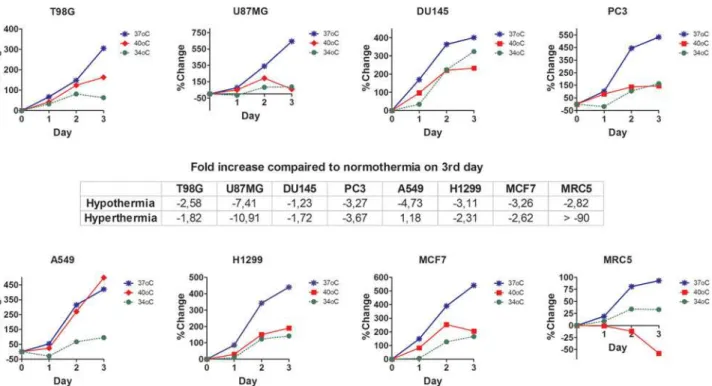
Following a 3-day incubation period under fever range hyperthermia, changes of cell prolifera-tion/viability were strongly dependent upon each cell line (Fig. 1). The normal fibroblast cell line, MRC5, was extremely sensitive to hyperthermia, showing a prevalent cell death effect. U87MG glioblastoma cell line was also very sensitive to hyperthermia, which induced a 10-fold reduction in cell growth. The prostate DU147 and PC3, as well as the lung H1299 and breast MCF7 cancer cell lines were also thermo-sensitive. On the contrary, T98G glioblastoma and A549 lung cancer cell lines were thermo-tolerant, demonstrating a growth rate increase by 1.87 and 1.18 fold, respectively.
Hypothermia at 34°C, on the other hand, had a rather homogeneous effect, resulting in re-duction of both the normal MRC5 fibroblasts and of all cancer cell lines viability (Fig. 1). This
Figure 1. Cell proliferation studies with AlamrBlue after 3days in various incubation temperatures.% change of relative fluorescent units (RFUs) as recorded with the AlamarBlue assay, after 3 days of exposure of cells to hypothermia (34°C) or hyperthermia (40°C) compared to normothermia (37°C).
reduction ranged between 1.23 and 7.40 fold, in DU145 and U87MG cell lines, respectively, compared to control (37°C) cells, as calculated on the third day of incubation.
Effect of hyper- and hypothermia on Ki67 proliferation index
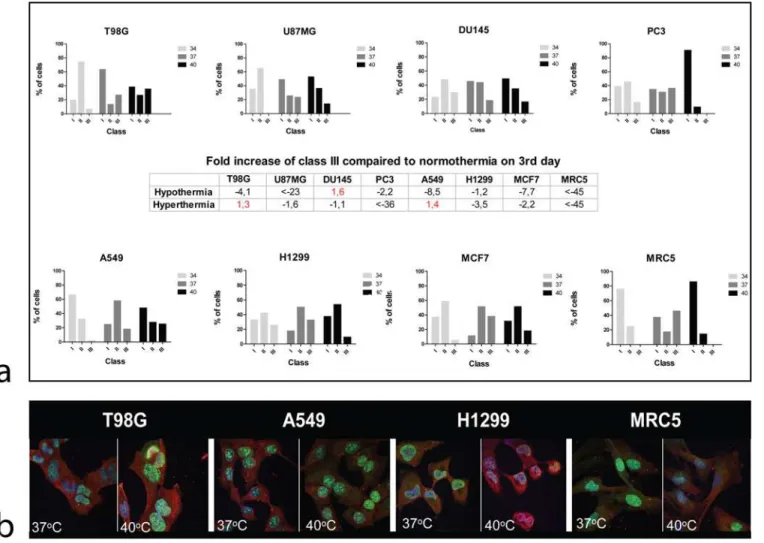
Given that the cell proliferation/viability assessed by the AlamarBlue assay is a result of com-bined proliferation and death events, we further examined the cell proliferation using the ki67 proliferation index. After 3 days of cell incubation, hyperthermia resulted in an increased frac-tion of cells in class III, inT98G and A549 (1.3 and 1.4 fold respectively) cell lines compared to normothermia. This dropped by 1.1 to more than 45 fold in the rest of cell lines (Fig. 2a). Char-acteristic and representative confocal images of Ki67 nuclear staining and changes after expo-sure to hyperthermia are shown inFig. 2b.
Hypothermia resulted in decrease of cell accumulation in the class III group by 1.2 up to more than 45 fold in all cell lines with the exception of DU145 prostate cancer cell line, where this was increased by 1.6 fold.
Figure 2. Confocal immunofluorescent microscopy images and automated quantification of ki67 proliferation marker in various cell lines.2a: changes of Ki67 proliferation index class after 3-day exposure of cells to hypothermia (34°C) or hyperthermia (40°C) compared to normothermia (37°C). 2b: Representative confocal microcopy images showing nuclear Ki67 immunostaining changing intensity after exposure to hyperthermia (40°C).
Effect of hyper- and hypothermia on Caspase 9 levels
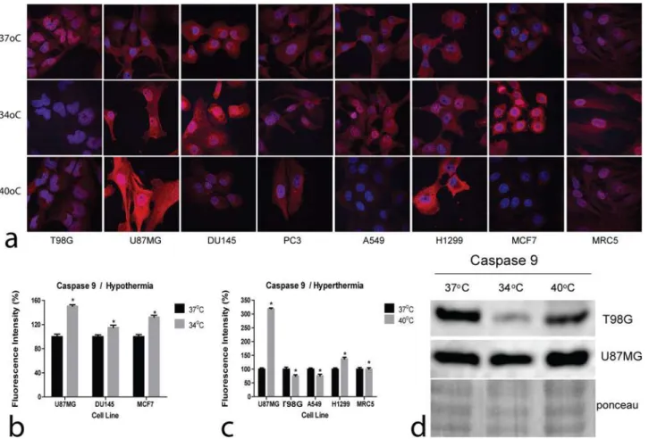
Confocal immunofluorescence images of Caspase 9 expression in cell lines following exposure to hyperthermia and hypothermia are presented inFig. 3a. Plots of the fluorescence intensity changes following exposure to hyperthermia and hypothermia are presented inFig. 3b and 3c, respectively.
Hyperthermia sharply induced Caspase 9 in the U87MG thermo-sensitive cell line, which also exhibited the most profound reduction of cell viability in Alamarblue experiments. Of in-terest, both T98G and A549 thermo-tolerant cell lines, Caspase 9 levels were reduced by hyper-thermia, suggesting an apoptosis suppressing effect of hyperthermia in these cell lines. The most thermo-sensitive of all, MRC5 cell line, however, did not show increased Caspase 9 levels suggesting death by independent of Caspase 9 pathways.
Hypothermia induced Caspase 9 in U87MG, DU145 and MCF7 cells, in agreement with the reduced viability, demonstrated in Alamarblue experiments. In the rest of cell lines, Caspase 9 expression remained stable or it was reduced in the case of T98G cells, suggesting that if apo-ptosis pathways are activated by hypothermia these are Caspase 9 independent.
Western blot analysis performed in the thermo-tolerant T98G and the thermo-sensitive U87MG glioblastoma cell lines are presented inFig. 3d, supporting the previously presented
Figure 3. Confocal immunofluorescent microscopy and western blot images of Caspase9.3a: Representative confocal microcopy images showing cytoplasmic Caspase 9 expression changing intensity after exposure to hypothermia (34°C) or hyperthermia (40°C) compared to normothermia (37°C) (magnification x60). 3b,c: Densitometry performed on confocal microcopy images of Caspase 9 immunostaining after exposure to hypothermia (34°C) or hyperthermia (40°C) compared to normothermia (37°C).
results. Hyperthermia induced Caspase 9 in the thermo-sensitive U87MG cell line, while this was reduced in the thermo-tolerant one, T98G. Hypothermia reduced Caspase 9 levels in the T98G cell line, while no evident change was noted in the U87MG one.
Effect of hyper- and hypothermia HSP90 levels
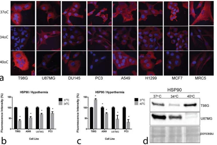
Representative confocal microscopy and immunofluorescence images of the effect of hyper-thermia and of hypohyper-thermia on HSP90 on cell lines are shown inFig. 4a. Plots of fluorescence intensity changes are presented inFig. 4b and c.
Hyperthermia strongly increased the HSP90 levels in the T98G thermo-tolerant cell line, whilst a sharp drop was recorded in the thermo-sensitive PC3 and U87MG cell lines. On the other hand, the HSP90 levels were clearly reduced in all cell lines examined under hypother-mia. Western blot analysis performed in the thermo-tolerant T98G and the thermo-sensitive U87MG glioblastoma cell lines are shown inFig. 4d, confirming the results of confocal microscopy.
Hyperthermic chemosensitization
Exposure of the thermo-tolerant A549 and the thermo-sensitive H1299 lung cancer cell lines to cisplatin, the key drug used in the clinical practice for the treatment of lung cancer, showed
Figure 4. Confocal immunofluorescent microscopy and western blot images of HSP90.4a: Representative confocal microcopy images showing cytoplasmic HSP90 expression changing intensity after exposure to hypothermia (34°C) or hyperthermia (40°C) compared to normothermia (37°C) (magnification x60). 4b,c: Densitometry performed on confocal microcopy images of HSP90 immunostaining. 4d: Western blot images of HSP90 expression in the thermo-tolerant T98G and the thermo-sensitive U87MG cell lines.
that fever range hyperthermia strongly sensitized H1299 to the drug but its effect on the A549 cell line was minimal (Fig. 5a).
Exposure of the thermo-tolerant T98G and the U87MG thermo-sensitive glioblastoma cell lines to temozolomide, the only approved drug for the treatment of human glioblastoma, showed that hyperthermia at 40 ºC strongly sensitized the U87MG cell line to the drug, whilst no sensitizing effect was noted for the T98G one (Fig. 5b).
Discussion
The effect of fever-range hyperthermia on normal and cancer cell biology and its eventual role and influence in cell sensitivity to chemotherapy and radiotherapy remains poorly understood, requiring further investigation. Mild hyperthermia has been reported to have an inhibitory ef-fect on cell proliferation. In a previous study, however, Morriseyet alalso reported a
stimulato-ry effect of mild hyperthermia at 38°C on U87MG cell line that was sharply reversed at 40°C [16]. The conclusion made by these researchers regarding the differential response among cell lines to small temperature elevations is certainly important. The current study has lead as to the identification of two cell lines (T98G and A549) which seem to be resistant to the effect of hyperthermia at 40°C. The human A549 cell line has been previously reported to be resistant to
Figure 5. Hyperthermic chemosensitization experiments with Cisplatin and Temozolomide using the AlamarBlue assay.5a: Viability of lung cancer cell lines A549 and H1299 after a 24h exposure to cisplatin under normothermic and fever range hyperthermic conditions. 5b: Viability of glioblastoma cell lines T98G and U87MG after a 24h exposure to temozolomide under normothermic and fever range hyperthermic conditions.
thermal killing at 43–45°C compared to the U87MG cell line [17], but a differential response ranging from proliferation to cell killing at the well tolerated by the human body 40°C is new. Combinations of fever-range hyperthermia may, therefore, delay progression of metastatic dis-ease in thermo-sensitive tumors with U87MG-like behaviour, while G2-M phase targeting drugs may prove critical to treat thermo-tolerant T98G-like tumors in combination with total body fever induction or local non-toxic heating.
On the other hand, the therapeutic role of hypothermia should not be underestimated and should be thoroughly examined in animal models, as about half of the examined cell lines dem-onstrated a 3–7 fold reduction of viability at 34°C. The current knowledge on its effect on can-cer cell is limited. Hypothermia at 28°C seems to protect preferentially normal fibroblasts compared to cancer cells against 5-fluorouracil [18]. In our study, at 34°C, normal human fi-broblasts suffered a reduced proliferation of an extent, however, quite limited compared to the majority of cancer cell lines. The reduced metabolism and oxygen consumption of tumors ex-posed to hypothermia may also be important in tumor radiosensitization [19], a hypothesis that has been also tested in the clinical practice [20]. The role of hypothermia in inhibiting can-cer cell adhesion to endothelial cells and thus migration as shown by Zhanget al[21], provides
an additional basis for further studies on the usage of hypothermia as a cancer therapy option. We further examined whether the death effect induced by mild temperature changes in sev-eral cell lines is Caspase-9-mediated. The aspartic acid specific protease Caspase-9 is involved in the mitochondrial death pathway. Release of cytochrome c from mitochondria activates apaf-1 (apoptosome), which in its turn cleaves the pro-enzyme of Caspase-9 into its most active form. Nevertheless, cleavage is not essential for Caspase-9 apoptotic activity [22]. In our study, hypothermia and hyperthermia induced Caspase-9 in several sensitive cell lines like U87MG, DU145 and MCF7, in accordance with the reduced viability noted. In the rest of the cases, Caspase-9 expression remained stable, suggesting that if apoptotic pathways are activated by hypothermia these are Caspase 9 independent. For instance, AIF (apoptosis inducing factor) or Caspase-8 and 12 are alternative apoptotic Caspase-9 independent apoptotic pathways [23]. Of interest, in the T98G and A549 thermo-tolerant cell lines, Caspase-9 levels were reduced under fever-ranged hyperthermia, providing evidence for a pathway exploited by some cell lines to escape mitochondrial related apoptosis.
HSP90, on the other hand, is a heavy member of the HSP family, with an important role in tumor growth, being also linked with poor prognosis in breast cancer and other malignancies [24]. The HSPs are up-regulated under hyperthermic conditions [11], to protect cells against heat-induced protein damage by their chaperon activity. Several HSP90 inhibitors have been developed and have been demonstrated invivoto be tumoristatic and to have a synergistic
ef-fect with chemotherapy and other target therapies [25]. The finding that hypothermia reduced the expression of HSP90 in all cell lines examined is interesting. Acting as a physical agent in-hibitor of HSP90, hypothermia could have synergistic effect with chemotherapy, similarly to the chemical inhibitors. Of interest, hyperthermia enhanced HSP90 expression in the thermo-tolerant T98G cell line, which suggests a protective role of HSP90 in such cell lines flourishing under warm conditions.
Therefore, it was further suggested that the trimodal therapy deserves further assessment as a way to enhance the efficacy of irradiation in cases of nodal metastases from head and neck tu-mors [27]. In our study, experiments of simultaneous exposure of lung cancer and glioblastoma cell lines to fever range hyperthermia and clinically established chemotherapeutic drugs showed that sensitization conferred by hyperthermia mainly concerned the thermo-sensitive cell lines, whilst its sensitizing effect was drastically inferior in thermo-tolerant cell lines. This finding further supports the necessity to develop clinical methods able to identify thermo-sensitive tumors that would benefit the most if treated with combined hyperthermia and che-motherapy protocols. Whether thermo tolerant tumors might become sensitive to hyperther-mic chemotherapy by blocking the HSP90 or relevant biological pathways remains an hypothesis for further experimentation.
It is concluded that cancer cells respond differentially to mild temperature changes, whether these are towards mild hypothermia or fever-range hyperthermia. Thermo-tolerant and thermo-sensitive cells lines have been identified to fever range hyperthermia, which encourages research to identify suitable methods for clinical grouping of human tumors according to their thermal predilection. Such characterization would allow clinical trials with non-toxic localized or total body hyperthermia in patients predicted to be sensitive to such therapies. Of interest, mild hypothermia had a suppressing effect on cell proliferation in all cells examined, suggesting that hypothermia would suppress tumor replication and metabolism in most human tumors, further supporting the radio-sensitization hypothesis through increased oxygenation by reduc-ing oxygen and metabolic demands.
Author Contributions
Conceived and designed the experiments: DK AG MIK. Performed the experiments: DK IVG AM SK AG. Analyzed the data: MIK SK. Contributed reagents/materials/analysis tools: SK. Wrote the paper: DK AG MIK.
References
1. Fiorentini G, Szasz A (2006) Hyperthermia today: electric energy, a new opportunity in cancer treat-ment. J Cancer Res Ther 2: 41–46. doi:10.4103/0973-1482.25848PMID:17998673
2. Issels RD, Lindner LH, Verweij J, Wust P, Reichardt P, et al. (2010) European Organisation for Re-search and Treatment of Cancer Soft Tissue and Bone Sarcoma Group (EORTC-STBSG); European Society for Hyperthermic Oncology (ESHO), Neo-adjuvant chemotherapy alone or with regional hyper-thermia for localised high-risk soft-tissue sarcoma: a randomised phase 3 multicentre study. Lancet Oncol 11: 561–570. doi:10.1016/S1470-2045(10)70071-1PMID:20434400
3. Bull JM, Scott GL, Strebel FR, Nagle VL, Oliver D, et al. (2008) Fever-range whole-body thermal thera-py combined with cisplatin, gemcitabine, and daily interferon-alpha: a description of a phase I-II proto-col. Int J Hyperthermia 24: 649–662. doi:10.1080/02656730802104740PMID:18608594
4. Rowe RW, Strebel FR, Proett JM, Deng W, Chan D, et al. (2010) Fever-range whole body thermother-apy combined with oxaliplatin: a curative regimen in a pre-clinical breast cancer model. Int J Hyperther-mia. 26:565–576. doi:10.3109/02656736.2010.483635PMID:20707651
5. Iwata K, Shakil A, Hur WJ, Makepeace CM, Griffin RJ, et al. (1996) Tumour pO2 can be increased markedly by mild hyperthermia. Br J Cancer Suppl 27: S217–221. PMID:8763884
6. Vidair CA, Dewey WC (1988) Two distinct modes of hyperthermic cell death. Radiat Res 116: 157–171. doi:10.2307/3577486PMID:2460896
7. Lepock JR, Frey HE, Ritchie KP (1993) Protein denaturation in intact hepatocytes and isolated cellular organelles during heat shock. J Cell Biol 122: 1267–1276. doi:10.1083/jcb.122.6.1267PMID: 8376462
9. Skitzki JJ, Repasky EA, Evans SS (2009) Hyperthermia as an immunotherapy strategy for cancer. Curr Opin Investig Drugs. 10:550–558. PMID:19513944
10. Repasky EA, Evans SS, Dewhirst MW (2013) Temperature matters! And why it should matter to tumor immunologists. Cancer Immunol Res 1:210–216. doi:10.1158/2326-6066.CIR-13-0118PMID: 24490177
11. Morimoto RI (1998) Regulation of the heat shock transcriptional response: cross talk between a family of heat shock factors, molecular chaperones, and negative regulator. Genes Dev 12: 3788–3796. doi: 10.1101/gad.12.24.3788PMID:9869631
12. Lindquist S (1986) The heat-shock response. Annu Rev Biochem 55: 1151–1191. doi:10.1146/ annurev.bi.55.070186.005443PMID:2427013
13. Pratt WB, Morishima Y, Peng HM, Osawa Y (2010) Proposal for a role of the Hsp90/Hsp70-based chaperone machinery in making triage decisions when proteins undergo oxidative and toxic damage. Exp Biol Med 235:278–289. doi:10.1258/ebm.2009.009250
14. Zachari MA, Chondrou PS, Pouliliou SE, Mitrakas AG, Abatzoglou I, et al. (2014) Evaluation of the Ala-maBlue assay for adherent cells irradiation experiments. Dose-Response. 12:246–58. doi:10.2203/ dose-response.13-024.KoukourakisPMID:24910583
15. Page B, Page M, Noel C (1993) A new fluorometric assay for cytotoxicity measurements in-vitro. Int J Oncol 3:473–476. PMID:21573387
16. Morrissey JJ, Higashikubo R, Goswami PC, Dixon P (2009) Mild hyperthermia as a potential mecha-nism to locally enhance cell growth kinetics. J Drug Target 17: 719–723. doi:10.3109/
10611860903074968PMID:19845488
17. Armour EP, McEachern D, Wang Z, Corry PM, Martinez A, et al. (1993) Sensitivity of human cells to mild hyperthermia. Cancer Res 53: 2740–2744. PMID:8504414
18. Matijasevic Z (2002) Selective protection of non-cancer cells by hypothermia. Anticancer Res 22: 3267–3272. PMID:12530074
19. Nias AH, Perry PM, Photiou AR (1988) Modulating the oxygen tension in tumours by hypothermia and hyperbaric oxygen. J R Soc Med 81: 633–636. PMID:3210193
20. Sealy R, Harrison GG, Morrell D, Korrubel J, Gregory A, et al. (1986) A feasibility study of a new ap-proach to clinical radiosensitisation: hypothermia and hyperbaric oxygen in combination with pharma-cological vasodilatation. Br J Radiol 59: 1093–1098. doi:10.1259/0007-1285-59-707-1093PMID: 3790896
21. Zhang XM, Lv YG, Chen GB, Zou Y, Lin CW, et al. (2012) Effect of mild hypothermia on breast cancer cells adhesion and migration. Biosci Trends 6: 313–324. PMID:23337791
22. Twiddy D, Cain K (2007) Caspase-9 cleavage, do you need it? Biochem J 405(Pt 1): e1. doi:10.1042/ BJ20070617PMID:17555401
23. Susin SA, Lorenzo HK, Zamzami N (1999) Molecular characterization of mitochondrial apoptosis-inducing factor. Nature 397: 441–446. doi:10.1038/17135PMID:9989411
24. Pick E, Kluger Y, Giltnane JM, Moeder C, Camp RL, et al. (2007) High HSP90 expression is associated with decreased survival in breast cancer. Cancer Res 67:2932–2937. doi: 10.1158/0008-5472.CAN-06-4511PMID:17409397
25. Barrott JJ, Haystead TAJ (2013) Hsp90, an unlikely ally in the war on cancer. FEBS Journal 280: 1381–1396. doi:10.1111/febs.12147PMID:23356585
26. Mauz-Körholz C, Dietzsch S, Schippel P, Banning U, Körholz D (2003) Molecular mechanisms of hyperthermia- and cisplatin-induced cytotoxicity in T cell leukemia. Anticancer Res. 23(3B):2643–2647. PMID:12894552