A
FOUR-YEAR
FOLLOW-UP SURVEY OF CHAGASIC
CARDIOPATHY IN CHILE1
Arturo Arribada C., 2 Werner Apt B.,3 and J. MantieZ Ugartti
I
NTRODUCTION
The natural history of Chaga- sic cardiopathy is still not well-known, mainly because innumerable clinical pic- tures develop without symptoms, and the cases seen at hospitals are merely the most overtly serious. This circumstance may tend to create a false impression that this cardiopathy is relatively benign, be- cause of the small demand it generates for hospital beds in comparison with other pathologies. Hence, if it were pos- sible to elucidate what happens to the great majority of asymptomatic cases, that would constitute an important for- ward step in understanding a disease that afflicts millions of people in the Ameri- C&S.
In 1977 we launched a long- term project in Chile that was directed at investigating the epidemiologic picture
’ This article will also be published in Spanish in the Bo- /ehi de la Ojcina Sanitaria Panamen~ana, Volume 101. 1986. The work reported was financed by a grant from the University of Chile and by Grant No. 820599
from the UNDPlWorld Bank/WHO Tropical Diseases Research Program.
* Associate Professor of Medicine, Paula Jaraquemada Hospital, Faculty of Medicine, University of Chile, Southern Division, Santiago, Chile.
3 Professor of Parasitology, Department of Experimental Medicine, Faculty of Medicine, University of Chile, Southern Division, Santiago, Chile.
’ Professor of Biostatistics, Faculty of Medicine, Univer- sity of Chile, Northern Division, Santiago, Chile.
of Chagas’ disease in endemic areas and its cardiac implications. This study re- vealed that all previous assessments had underestimated both the incidence of the disease and its socioeconomic reper- cussions for the country (1-G). Respond- ing to these findings, health authorities took up the problem and adopted a number of health measures-including periodic disinsection of accessible areas and health education at community cen- ters and primary schools. (A simple ex- planatory leaflet was produced for these education campaigns that informed readers about the dangers of the disease and about ways to prevent household in- festations of triatomid vectors.)
We also promoted parallel studies seeking to elucidate epidemio- logic relationships between humans, as- sociated animals, and vector insects (7- 8); to detect different types of vectors
and examine their interrelationships; %i and to define the structures of zymo- t\ demes in various Tgpanosoma crzzi
1 3 strains found in different endemic areas %
(9-10). x
This article reports our experi- ence with long-term field followup of
3
cardiac pathologies observed during the 2 first survey, specifically regarding 2 a,
changes in the subjects’ clinical, sero- logic, and electrocardiographic pictures after a lapse of four years. It also reports findings concerning the risk that people exposed to chagasic infection but show- ing a normal ECG in the original survey would subsequently develop cardiopa- thy.
M
ATERIALS
AND METHODS
Our original survey begun in 1977, whose data were employed in the work reported here, involved 2,938 sub- jects who were given serologic tests and ECGs. Nearly two-thirds of these sub- jects (1,932) yielded serologic results negative for chagasic infection. Four hundred and two (22%) of these 1,932
showed significant ECG alterations, and these 402 were considered to constitute a control group hereafter referred to as “Group A.” The other 1,006 subjects yielded serologic results positive for cha- gasic infection. Three hundred and ninety-one (39 % ) of these 1,006 showed significant ECG alterations, and 298 of the latter yielded findings suggesting chagasic cardiopathy (G,ll). These 298 constituted a group designated “Group B.” As the foregoing implies, 615 (61%) of the 1,006 subjects with positive sero- % logic findings had normal ECGs; these 2 615 were considered to be at risk of con- -. tracting chagasic cardiopathy at any 3 time. For purposes of our study, 67 of z these 615 were selected at random, des- .$ ignated “Group C,” and subjected to
u
B further examination. These three groups 2 were selected in 1981 when we began the followup work reported here.
246
All members of these three groups were given serologic tests for 1: crzlzi antibodies at the beginning of the original study and again after four years (12-15). On both occasions each serum sample was tested by two of three sero- logic methods, the three being indirect hemagglutination (IHA), complement futation (CF), and indirect immuno- fluorescence (IIF). IHA titers above 1: 16,
CF titers above 1: 5, and IFF titers above
1: 20 were considered positive.
During the fourth year after the initial survey, the year depending upon when the first survey had been car- ried out, a clinical study was conducted of 481 people in the three groups; this study was similar to that conducted dur- ing the original survey. In addition, these subjects were given a twelve-lead ECG with an extended II lead for the study of arrhythmias. These ECGs were analyzed independently of the serologic results ac- cording to the following parameters: spa- tial P, QRS, and T axes; PR, RR, RS, and QT intervals; amplitude of R in Vi and V,; intrinsecoid deflection in Vi and V6; ST-T segment; and T-wave items (Soko- low index). The electrocardiographic di- agnoses developed in this manner were grouped according to the guidelines pro- posed by Maguire and colleagues (IT), and a statistical analysis was made of the differences between the groups. In addi- tion, the diagnoses were analyzed ac- cording to the age and sex of the persons surveyed in the three groups during the initial ECG. When no significant differ- ences were found, we used the method of diagnosis employed in the original work (5) to which Student’s t test was applied.
and B, and normalization of an abnor- mality that had been recorded on the ini- tial ECG was regarded as a change for purposes of statistical analysis. However, because we were dealing with multiple abnormalities in many ECGs, normaliza- tion of a specific condition did not neces- sarily constitute normalization of the whole tracing.
All of the studies reported here-performed in 1981, 1982, 1983,
and 1984-were carried out in the 36 vil- lages where the original sample survey was conducted. For reasons to be dis- cussed, by the end of four years Group A had been reduced to 216 subjects and Group B to 198 subjects, and so it was these subjects who provided the basis for the comparison reported here.
The members of all three groups lived in rural, mostly semi-desert areas of northern Chile. All were en- gaged in farming arable land found along the courses of small rivers flowing down from the Andes Mountains, and in general all had very similar working con- ditions. Apart from a few people over 80 years of age who were excluded from Group B in order to eliminate arterioscle- rotic pathologies (11), the age distribu- tion of subjects in Group A and Group B was similar. The members of Group C shared essentially the same socioeco- nomic conditions as members of the other two groups, but they tended to be somewhat younger. The ages of the dif- ferent subjects correspond to those re- corded in the years mentioned above
(1981-1984), so that the recorded ages of all subjects were four years greater than those recorded at the time of the original study.
RE
SULTS
Followup of Groups A and B
Table 1 shows the reasons for reduction in the numbers of subjects in groups A and B during the study period. Of the 402 original Group A subjects (people with negative serology but ab- normal ECGs), nine (2.2 % ) died from causes indicated below; 168 (41.8%)
moved to other areas in search of better financial prospects; seven (1.7%) de- clined to be surveyed; and two (0.5%)
showed a serologic conversion from nega- tive to positive for 1: C~ZLZZ’ antibodies.
Of the 298 original Group B subjects (people with positive serology, abnormal ECGs, and findings suggesting Chagas’ disease), 22 (7.4%) died of cha- gasic cardiopathy; one (0.3 % ) died of stomach cancer; 48 (16.1% ) moved to other areas; and 29 were eliminated for the following reasons: Four were hospi- talized in other areas at the time of the survey; one was away in the mountains looking after livestock; four underwent serologic conversion from positive to negative; two exhibited symptoms of aortic insuffrciency; one showed symp- toms of obstructive cardiomyopathy; 12 had diastolic pressure readings over 100 mmHg; and five had been over 80 years old when initially surveyed.
Causes of death. All deaths in Chile must be certified by the responsi- ble physician of the main provincial hos- pital. Because of this requirement, we were able to obtain reliable data about all of the study subjects who died during the study period. Four of the nine Group A fatalities (see Table 1) were caused by bronchopneumonial conditions, one by stomach cancer, one by a cerebrovascular accident, and three by refractory cardiac insufficiency. In contrast, nearly all the
TABLE 1. Changes in the sizes of groups A and 6 over the four-year study period. Group A Group 6 No. % No. % Size of original group
Deaths due to chagasic cardiopathy Deaths due to other causes Left the area
other status Size after four years
402 fW 298 fW 0 KY 2; (7.4)a 9 (2.2)a (0.3) 168 (41.8) 48 (16.1) (2.2) 29 (9.7) (53.7) 198 (66.4)
a The difference in overall mortali (2.2% in Group A, 7.7% in Group 8) is highly signifint (P<O.COl).
to chagasic cardiomyopathy. Specifically,
12 Group B subjects suffered “instanta- neous death” (two of these people were wearing pacemakers to counteract total auriculoventricular block), 10 died of congestive cardiac insuff%ziency, and only one died of another cause (stomach can- cer).
These four-year data show that the Group B subjects (with chagasic cardiopathy) had an appreciably higher annual risk of dying than did the control subjects with abnormal ECGs in Group A, the annual risks being 0.6% in Group A and 1.9% in Group B. The differ- ence between these mortality figures was found to be highly significant (P< 0.001).
Age and sex of the decedents.
Regarding age and sex, Table 2 shows
that most of the Group A fatalities oc- curred among women, while most of the Group B fatalities occurred among men. The specific ages of the nine Group A subjects at the time of death were as fol- lows: men-63, 78, and 82 years; women-50, 67, 72, 77, 80, and 80 years. The specific ages of the 22 Group B subjects apparently dying of Chagas’ disease were as follows: men-45, 46, 54, 56, 58, 59, 60(2), 61, 62(2), 63, 65, and 76 years; women-47, 48, 50, 55, 56(2), 58, and 65 years. As these figures show, most of the Group A fatalities oc- curred after age 60, while most of the Chagas’-related Group B fatalities oc- curred in younger subjects.
TABLE 2. Sex and age of the nine Group A subjects dying of all causes and the 22 Group B subjjcts apparentty dytng of Chagas’ disease during the four-year study period.
Group A Group B Age at death
(in years) Women Men Total Women Men Total 41-50 51-60 - 1 - - i!m 3 4 2 6 lo” 61-70 1 1 2 1 5 6 71-80 4 1 5 - 1 1 81-90 - 1 1 - - -
Electrocardiographic findings (decedents). As Table 3 indicates, the Group B decedents with chagasic cardi- opathy showed more auriculoventricular and intraventricular conduction disor- ders than did the Group A (control group) decedents.
Specifically, the nine Group A decedents exhibited no A-V or bifascicu- lar block. Two Group A women (72 and 77 years old) exhibited arrhythmias (1 auricular extrasystole and 1 ventricular extrasystole). Two Group A men (63 and
78 years old) exhibited left anterior hemiblock, and a Group A woman (80
years old) exhibited complete left branch block. Left ventricular hypertrophy was observed in two Group A women 50 and
67 years old. An image indicating necro- sis of the diaphragmatic wall of the myocardium was found in one Group A woman 80 years old, and another indi- cating necrosis of the anterior wall was found in one Group A man 82 years old.
In Group B, the four cases of arrhythmia included two cases of polyfo- cal ventricular extrasystole (one in a man of 56 and the other in a woman of 47)
and two of auricular extrasystole (in men of 54 and 58). First degree A-V block was
TABLE 3. ECG abnormalities obsenred in the nine Group A and 22 Group B decedents.
Conditions Group A Group B Arrhythmias 2 4
A-V block, first degree 0 1
A-V block, third degree 0 2 Unifascicular block 3 8
Biiascicular block 0 4
lschemia 0 1 QS image 2 2
Cavity hypertrophy 2 0
Total 9 22
found in a woman of 50, and two cases of total A-V block corrected by fured-rate pacemakers were observed in men of 60
and 76.
The eight cases of unifascicu- lar block included four complete right branch blocks (in two women of 48 and 65 and two men of 61 and 63). There were also three cases of left anterior hemiblock (in three men of 62, 62, and 65) and one case of left posterior hemiblock (in a man of 46).
The four cases of bifascicular block involved left anterior hemiblock associated with complete right branch block (in two men of 59 and 60 and two women of 56 and 58).
One case of anterior-face is- chemia occurred in a man of 45, and two cases with an inactivation or QS image of the anterior wall were observed in two women of 55 and 56.
Analysis of Followup Data on
Groups A, B, and C
2
The distribution by age and sex of all subjects remaining in the three
$
study groups after four years is shown in s Table 4. As may be seen, women pre-
dominated in all three groups, presum- E t ably because the survey was conducted
on week-days during hours when the % male head of the household was gener- 2 ally at work. The age distribution of 3 Group A and Group B subjects was gen- 2 erally similar; it should be noted, how- 2 ever, that five Group B subjects over 81
years old were excluded from the study in
%
order to reduce arteriosclerosis as a factor5
in assessing chagasic cardiopathy. < .
Clinical Cardiopathic Findings
’
In the original survey it was
2
noted that both the Group A and GroupB subjects showed few clinical symp-
3
toms, and the same circumstance pre-
TABLE 4. Distribution by age and sex of all Group A, B, and C subjects remaining in their respective study groups four years after the initial survey.
Group A Group B Group C Age group
(in years) Men Women Total Men Women Total Men Women Total 11-20 10 9 19 2 3 5 4 1 5 21-30 14 23 37 9 11 20 4 6 10 31-40 13 26 39 9 28 37 4 16 20 41-50 15 24 39 19 31 50 7 24 31 51-60 13 16 29 10 24 34 - 1 1 61-70 IO 18 28 14 20 34 - - - 71-80 9 9 18 9 9 18 - - - 81-90 2 5 7 0 oo---
Total 86 130 216 72 126 198 19 48 67
Group A clinical histories re- vealed symptoms of congestive cardiac insufficiency in 10 subjects (4.6%). These included six men 5 2 to 89 years old and four women 52 to 80 years old. (The ECGs showed previous necrosis in five of the 10 cases-in three men of 52, 62, and 89 and two women of 55 and 63.)
Group B clinical histories re- vealed symptoms of chagasic cardiopathy in 21 subjects (10.8%), seven of these having multiple complaints. Nine sub- jects presented ;vith irregular precordial palpitations and sharp precordial pain, the latter most often located on the left side. (The subjects involved were four men of 23 to 40 and five women of 19 to 50; three of these also exhibited cases of Adams-Stokes crisis.)
In all, there were 10 Group B subjects with Adams-Stokes crisis, but only one of these (a man of 60) had third-degree A-V block. Of the other nine, three women of 38, 39, and 42 showed wandering of the auricular pace- maker; one man of 38 had auricular tachycardia; three men of 49, 53, and 65 and a woman of 5 2 exhibited bifascicular block (left anterior hemiblock plus com- plete right branch block), while one man of 33 showed another type of bifascicular
block (left posterior hemiblock plus com- plete right branch block).
Also, 10 Group B subjects ex- hibited symptoms of congestive cardiac insufficiency. (Four were among those with bifascicular block and Adams- Stokes syndrome.) The ECGs of three women of 45,48, and 50 showed altera- tion of ventricular repolarization, while those of the other three subjects (two women of 60 and 62 and a man of 63) showed ischemia images. Neither acute- phase antecedents nor portal of entry (Romaiia’s sign) antecedents were ob- served in any members of the group.
Electrocardiogram Results
Table 5 shows the changes found in the control group (A) and the chagasic group (B) when the first ECGs were compared with those obtained four years later. As may be seen, there was a higher proportion of ECG abnormalities per tracing among the chagasic group on both occasions. Initially, by definition, there were no normal ECGs in either group. A considerable number of these abnormalities (28.4% in the control group, 3 1.7 % in the chagasic group) were found to have normalized them- selves as of the final tracing. However, because some subjects had multiple ab- normalities, the number of abnormali- ties normalized was greater than the
number of ECGs normalized. Overall, 5 1 Group A final tracings (23 % ) were found to be normal, as were 16 Group B final tracings (8%). The number of ab- normalities that became normalized were 99 in Group A and 137 in Group B.
Table 5 also shows that the prevalences of observed abnormalities in the two groups were fairly similar in most cases, and that there were only a few in- stances when the Group A and Group B data showed differences that were statis- tically very significant (P < 0.01). More specifically, initial abnormalities very sig- nificantly more prevalent in Group B than Group A (P < 0.01) included bifas- cicular block, subepicardial damage (which was not found in Group A), and the presence of a prolonged QT interval on the initial tracing. The greater fre- quency of QS images in Group A related to coronary cardiopathy (a history of in-
TABLE 5. ECG abnormaliies found in the 216 Gmup A and 198 Group 8 study subjects.
Group A Group B
Initial ECG Final ECG Initial ECG Final ECG Abnormal ECG findings No. (%) No. VW No. (%) No. (%I 1. Normalized 99 (28.4) (0) 137 (31.7) 2. Auricular arrhythmia 6: $3) 47 (13.5) 4: (14.3) 55 (12.7) 3. Ventricular arrhythmia
4. A-V block, 1st degree 5. A-V block, 2nd degree
6. A-V block, 3rd degree (0.7) 7. Unifascicular block (21.7) 62 (14.4) 8. Bifascicular block (11.81” 47 (10.9)” 9. lschemia image (2.3) 10. Subepicardial damage (0.9)b Il. Subendocardial damage (4.4)a 12. QS image
13. Repolartzation change
14. Cavity hyper-trophy 27 (10.7) 32 (4.9) 15. Prolonged QT 1 (0.4) 29 (8.3) 45 (4.4) Total No. of abnormalities 253 (100) 349 (100) 322 (100) 432 (100) Total No. of ECGs 216 216 198 198
farct) in seven Group A subjects. In con- trast, the three Group B subjects initially showing QS images had no history of an- gina. Regarding the final ECGs, the only abnormalities very significantly more fre- quent in Group B than Group A
(P<O.Ol) were bifascicular block and subendocardial damage.
Aticular arrhythmias. Table 6 provides more information about the ob- served cases of auricular arrhythmia in the two groups. In general, it shows a higher proportion of functional abnor- malities (especially short PR syndrome and bradycardia associated with simple A-V block among miners working at high altitudes) in the Group A subjects. Although auricular fibrillation was ini- tially more common in the Group B (chagasic) subjects, the final ECGs showed it in about equal proportions of both groups. Initial abnormalities ob-
served much more frequently (P < 0.01) in the Group B subjects included wan- dering of the auricular pacemaker, auric- ular tachycardia, and auricular fibrilla- tion. Of these, the only one sufficiently more frequent in Group B than Group A on both the initial and final ECGs to reg- ister a highly significant difference (P < 0.01) was auricular tachycardia. Many of the Group B abnormalities were consistent with possible sick sinus syn- drome. The difference between the numbers of auricular arrhythmias ob- served in the two groups was not very sig- nificant (P < 0.01).
Sex and age data. Table 7 pro- vides data on the sex and age of Group A and Group B subjects whose initial ECGs yielded abnormal findings. While most
TABLE 6. ECG abnormalities associated with arrhythmias observed in the 216 Group A and 198 Group B study subjects. Group A Group B
Initial ECG Final ECG Initial ECG Final ECG Abnormalities No. (%I No. (%I No. (%I No. (%I Short PR syndrome 28 (40.6) 33 (70.2) 13 (28.3) 14 Wolff-Parkinson-White syndrome 4 (0) 0 ‘S5’ Wandering auricular pacemaker 2 ;;q 8
(24:6) 6
1:; i (19.6)” 1 (1.8)b Auricular extrasystole 17 (12.8) 8 (17.4) 13 (23.6)b Auricular tachycardia 1
Auricular bradycardia 8 (1.4) y (0) 6
(13.0)” 10 (18.2)” (11.6) (2.1) ; (0) 5
Auricular fibrillation 1 (1.4) 5 ‘l?’
(13.0)” 5 1i.i; b
Pulmonary P wave 3 (4.3) 0 0 (4 Nodal rhythm 1 (0) 2 (0) 2” (3.6) lsorhythmic A-V dissociation 1 (1.4) y lt3’ 2 (3.6) Nodal tachycardia 1 1i.i; 0 (0) (2.1) ; (0) y (0) Sick sinus syndrome 0 (0) 0 (0) 1 (2.2) (1.8) Nodal extrasystole 1 (0) 0 (0) Sinus arrest 0 (0) (1.4) y (0) ‘: (1.8) 2/l flutter
il
(1.4) 0 (2.1) (0) ; 1:; 0 (0) Left auricular rhythm 04 0 (0) y (0)
(2.2) :, (1.8) Pacemaker defect with arrhythmia 0 (0) cl 03 (0)
Total 69 (100) 47 (100) 46 (100) 55 (100)
categories of findings reflected the fe- male predominance seen previously in the two study groups, there were more Group A men than women with first de- gree A-V block, and about equal num- bers of Group A men and women with unifascicular block and bifascicular block, as well as more Group B men than women with cavity hypertrophy and about equal numbers of Group B men and women with ventricular arrhythmia, first degree A-V block, bifascicular block, and ischemia image. Aside from the fact that the Group B (chagasic) subjects with repolarization disorders tended to be younger, no highly significant age differ- ences were found. More specifically, no highly significant age differences were observed between Group A and Group B subjects with auriculoventricular block.
Unifascicular blocks. Table 8 provides information about the unifasci- cular intraventricular blocks observed in Group A and Group B subjects. As may be seen, the initial ECG showed com- plete right branch block to be three times as prevalent in the Group B (chagasic) subjects (P<O.Ol), but this difference diminished markedly in data from the fi- nal ECG. Incomplete right branch block was far more prevalent among Group A subjects than among those in Group B.
Regarding the sex and age ranges of affected subjects, the data in Table 9 indicate that about equal num- bers of Group A men and women ini- tially exhibited the various types of un”i- fascicular blocks encountered, while in Group B there was a greater (though not statistically very significant) predomi- nance of women with complete right branch block and left anterior hemi-
TABLE 7. Abnormal initial EGG findings among the 216 Group A and 198 Group B subjjcts, showing the numbers and age ranges of the affected subjects, by sex.
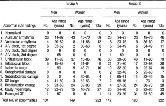
Men
Group A
Women Men
Group B Women Age range Age range Age range Age range Abnormal ECG findings No. (years) No. (years) Total No. (years) No. (years) Total 1. Normalized 0 0 0 0 0 0 0 0 0 0 2. Auricular arrhythmia 26 11-82 43 16-70 69 23 24-73 23 19-73 46 3. Ventricular arrhythmia 4 20-82 8 11-86 12 8 23-76 9 36-80 17 4. A-V block, 1st degree 6 32-59 2 30-63 8 5 24-49 6 34-49 11 5. A-V block, 2nd degree 0 0 0 0 0 0 0 0 6. A-V block, 3rd degree 1 65 0 8 1 1 6: 0 0 1 7. Unifascicular block 39 11-83 37 15-86 76 30 20-30 40 II-80 70 8. Bifascicular block 5 15-83 4 24-64 9 21 21-80 17 22-68 38 9. lschemia image
:,
TABLE 8. Unifascicular blocks observed in the 216 Group A and 198 Group B study subjects.
Initial EGG Final ECG Group A Group B Group A Group B Type of
unifascicular block No. (%) No. WI No. (%I No. WI Incomplete right branch block 30 (39.5) 7 (10.0) 30 (42.9) 10 (16.1) Complete right branch block 7 (9.2) 21 (30.0)” 6 (8.6) 16 (25.8)b Left anterior hemiblock 29 (38.2) 36 (51.4)a 26 (37.1) 32 (51 .6)b Left posterior hemiblock 8 (10.5) 4 (5.7) 4 (5.7) 2
Complete left branch block 2 (2.6) 2 (2.9) 4 (5.7) 2 I?$ . Total 76 (100) 70 (100) 70 (100) 62 (100)
a Difference between data for Group A and Group B (mltial) is highly signkant (PC 0.01). b Difference between data for Group A and Group B (final) IS not highly sigmflcani (P> 0 01).
TbLE 9. Numbers and age ranges of the 216 Group A and 198 Group 6 subjects whose initial ECGs showed unifascicular blocks, by sex.
Type of unifascicular block
Group A Group B Men Women Men Women
Age range Age range Age range Age range No. (years) No. (years) Total No. (years) No. (years) Total Incomplete right branch block 15 11-77 15 2158 30 3 20-47 4 19-65 Complete right branch block 4 34-83 3 31-62 26-57 13 21-81 2: Left anterior hemiblock 16 15-74 13 15-75
2; 1:
23-80 20 29-75 36 Left posterior hemiblock 4 30-47 4 18-43 8 2 42-49 2 II-34 4 Complete lefl branch block 0 0 2 42-54 2 1 65 1 61 2 Tota 39 37 76 30 40 70
block. Regarding the ages of subjects with unifascicular blocks, it is worth not- b ing that in Group B both incomplete 3 and complete right branch block was
. found to occur predominantly in young- 53 er men.
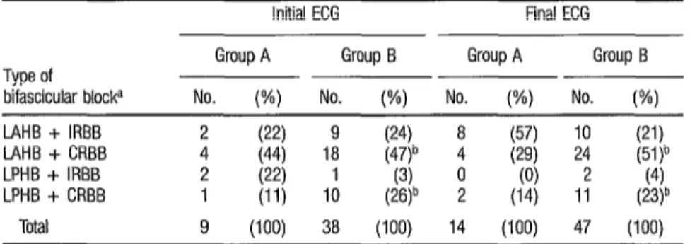
% Bifasciculat blocks. Table 10 .j$ shows data on bifascicular blocks found
9,
a 3 in both initial and final ECGs of Group 2 A and Group B subjects. The quantita-
2
tive differences involved demonstrate that these blocks predominated among Group B (chagasic) subjects. Regarding distribution of the various types of blocks 254 within Group A and Group B, however,
statistical analysis failed to reveal any very significant (P < 0.01) differences in the distribution of particular types of bi- fascicular blocks within the groups. This suggests that a quantitative rather than a qualitative difference is involved.
Regarding distribution of bi- fascicular blocks by sex, the data in Table
TABLE 10. Bifascicular blocks observed in the 216 Group A and 198 Group 6 study subjects.
Type of
bifascicular blocka
Initial ECG Group A Group B No. (%I No. (%I
Final EGG Group A Group B No. w No. (%I L4HB + IRBB
IAHB + CRBB LPHB + IRBB LPHB + CRBB
Total
2 (22)
1: (21)
4 (44)
2 (22) (0)
1 (11) 1: (26)b 2 (14) 1: (23)b
9 (100) 38 (100) 14 (100) 47 WV
a L4HB = left anterior hemiblcck: LPHB = left posterior hemiblock: IRBB = incomplete right branch block, and CRBB = complete right branch black
b Ddferences between the indicated Group A and Group B data are not highly significant (P>O 01)
TABLE 11. Numbers and age ranges of Group A and Group B subjects whose initial ECGs showed bifascicular bloc@, by sex.
Group A Group B
Type of bifascicular blocka
Men Women Men Women Age range Age range Age range Age range No. (years) No. (years) Total No. (years) No. (years) Total LAHB + IRBB 1 15 1 64 2 5 37-62 4 32-63 9
LAHB + CRBB 3 26-83 1 4 10 29-80 8 22-69 18
LPHB + IRBB 0 0 2 2&I 2 1 54 0 0 1 LPHB + CRBB 1 38 0 0 1 5 21-80 5 42-54 IO
Total 5 4 9 21 17 38 a LAHB = left anterior hemibkck; LPHB = left posterior hemlblock; IRBB = incomplete right branch block, and CRBE = complete right branch block
of Group A subjects with such blocks were too small for conclusions to be drawn.
ECG changes. So far we have examined numerical data that establish certain differences between the two groups studied but do not express the variation in ECG abnormalities over time. To assess this variation we have used cross-indexed tables in which the 15 ECG abnormalities diagnosed from the first tracing are listed vertically, and the same abnormalities diagnosed from
QS image (in one case), repolarization change (in one case), and cavity hyper- trophy (in three cases). In addition, look- ing down column three it may be seen that five other subjects (for a total of eight) exhibited ventricular arrhythmia on the final ECG.
Table 12, which lists only data for Group A (the control group), shows that 99 of the 25 3 abnormalities found in the initial 216 ECGs had normalized on the final ECG. Those abnormalities that disappeared were as follows:
(a) 32 auricular arrhythmias-four cases of Wolff- Parkinson-White syndrome (in men given the final ECG when they were 35 to 54 years old), two cases of wandering auricular pacemaker (in women of 23 and 35) one case of auricular tachycardia (in a woman of 25) 11 cases of au- ricular extrasystole (in five men 16 to 54 and six women 17 to 68) seven cases of auricular bradycardia (in four men 28 to 35 and three women 18 to 40), three cases of pulmonary P wave (in women 27 to 49) one case of nodal tachycardia (in a woman of 27) one case of nodal extrasystole (in a woman of 70). one case of nodal rhythm (in a man of 35), and one case of 2:l auricular flutter (in a man of
61);
(b) nine ventricular arrhythmias (all of ventricular extrasystole in three men 20 to 82 years old
(4
(4
d k) 3
and sh women 38 to 61 years old);
five cases of first-degree A-V block (in four men 32 to 59 and one woman of 30);
13 cases of unifascicular block-six cases of in- complete right branch block (in three men 31 to 42 and three women 21 to 39), four cases of left posterior hemiblock (in two men 35 and 56 and two women 30 and 43), and three cases of left anterior hemiblock (in two males 15 and 24 and a girl of 15);
six ischemia images (in women 43 to 71 years old);
all four images indicating subendocardial damage (in women 30 to 50);
19 cases of repolarization alterations (in four men 23 to 55 and 15 women 17 to 68);
z
(h) 11 images of cavity hypertrophy (in three men 345 to 56 and eight women 17 to 76).
The remaining Group A ab- normalities persisted over time. In this 256 regard, it should be noted that all seven
of the initial QS image abnormalities as- sociated with myocardial necrosis ap- peared on the final ECG. In addition,
105 new abnormalities appeared on the final ECG, these being as follows:
(4 10 new auricular arrhythmias-five cases of short P-R syndrome (in men 17 to 54) four :ases of auricular fibrillation (in women 70 to 78) and one case of sinus arrest (in a woman Id 58);
(b) Iive new cases of ventricular arrhythmia (all of ventricular extrasystole in men 54 to 73);
(c) one new case of first-degree A-V block (in a man of 68 whose ECG also showed a picture of left ventricular hypertrophy);
(4 one new case of third-degree A-V block (in a man of 68 with an image indicating subendo- cardial damage);
(4 13 new cases of unifascicular block-three cases in which a bifascicular block had re- verted to a unifascicular form (left posterior hemiblock plus incomplete right branch block becoming left posterior hemiblock in a woman of 38; left anterior hemiblock plus in- complete right branch block becoming in- complete right branch block in a boy of 15; and left anterior hemiblock plus complete right branch block becoming complete right branch block in a woman of 21); five cases of incomplete right branch block (in three men 15 to 56 and two women 36 and 47); three cases of complete left branch block (in two men 54 and 79 and one woman of 70); and two cases of left anterior hemiblock (in men
56 and 75);
(0 eight new cases of bifascicular block, six of which developed in subjects whose initial ECGs showed unifascicular blocks (three cases of incomplete right branch block changed to left anterior hemiblock plus complete right branch block in men of 22, 37, and 57, and three other cases of incomplete right branch block changed to left anterior hemiblock plus incomplete right branch block in women of
29, 39, and 47); in addition, two new cases of left anterior hemiblock plus incomplete right branch block appeared in women of 48 and
74;
TABLE 12. Group A abnormalities found by analysis of the initial and final ECGs of the 216 Group A study subjects. The numbers of abnormalities found initially, by type, are shown in column 2. The numbers of subjects with particular abnormalffes found on the final ECG, by type, are shown at the bottom of the table (e.g., four subjects showed first-degree A-V block on their final ECGs). The numbers of subjects in whom the initially observed abnormality persisted are shown by the dark numbers running diagonally through the table. And the numbers of subjects who initially had a particular abnormality that disappeared on the final ECG or who showed other abnormalities on the final ECG may be seen by reading horizontally across the table (e.g., of eight subjects with first-degree A-V block on the initial ECG, five did not show this abnormali on the final ECG, three did show it, and one showed another abnormality, prolonged GT).
Total No. of
initial Final ECG findings (l-15) abnormalities
Initial ECG findings (I-15) observed 1 23 4 5 6 7 8 9 IO 11 12 13 14 15 I. Abnormality normalized ____---
2. Auricular arrhythmia 69 32 37 1 - - - 2- 3-- I- 2 6 3. Ventricular arrhythmia 12 g-3--- l---- 1 13- 4. A-V block, 1st degree 8 5--3---l
5. A-V block, 2nd degree - _____--- 6. A-V block, 3rd degree 1 ---all---
7. Unifascicular block 76 13 1 1 - - - 49 6 4-- 2 3 5 9 8. Bifascicular block g --- 3 6--- 13 9. lschemia image 10 6-j--- l- 4--- 111 10. Subepicardial damage - _---_---
11. Subendocardial damage 4 4----l-.- 1 - - - 1 - - 12. QS image 7 - 4---- I---- 7 I-- 13. Repolarization change 29 19 - 1 - - - 2 l-- 1 1 10 4 5 14. Cavity hypertrophy 27 11 5 1 1 - - 3 1 l- 1 - - 16 3 15. Prolonged CIT , -~---.---~~~l
Total abnormalities or normalized 99 47 8 4 - 2 62 14 13 - 2 12 17 32 29 events observed on final ECG
s:
04
(9
(j)
(kj
five new images of myocardial necrosis-QS image (in two men of 53 and 82 and three women of 29, 61, and 68, the last two being associated with left anterior hemiblock; the woman of 29 had no history of angina); seven new cases of repolarization alterations (in two men of 31 and 82 and five women of
38 to 62);
16 new images of cavity hypertrophy-three cases of hypertrophy of the left auricle (in a man of 47 and two women of 67 and 74) and 13 cases of left ventricular hypertrophy (in five men of 55 to 82 and eight women of 58 to
78);
28 new cases of prolonged QT (in seven men of 43 to 83 and 21 women of 21 to 66).
Table 13, which presents the data for Group B, shows that 137 of the 322 abnormalities found in the initial
198 ECGs had normalized on the final ECG. Those abnormalities that disap- peared were as follows:
(4
(b)
(4 (4
(4
10 auricular arrhythmias-eight cases of wan- dering auricular pacemaker (in three men of 38, 44, and 73 and five women of 46 to 73), one case of auricular fibrillation (in a woman of 65) and one case of arrhythmia in a man 68 years old with a fLued-rate pacemaker that was complicated by multiple extrasystole; nine ventricular arrhythmias (all of ventricular extrasystole in four men of 23 to 76 and five women of 36 to 63);
five cases of first-degree A-V block (in four men of 26 to 49 and a woman of 34); 11 cases of unifascicular block-four cases of incomplete right branch block (in a man of 47 and three women of 19 to 38), five cases of left anterior hemiblock (in two men of 47 and 49 and three women of 29,’ 35, and 66), one case of left posterior hemiblock (in a man of 49), and one case of complete right branch block (in a man of 30);
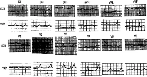
two cases of bifascicular block (both left ante- rior hemiblock plus incomplete right branch block, in a man of 37 and a woman of 38); I ECG tracings obtained from this subject are shown in
Figure 1.
6) (g) (h) (9 (i) (k) (1)
11 cases of ischemia image (in six men of 36 to 71 and five women of 37 to 66);
six images of subepicardial damage (all in the apical region, in two men of 32 and 48 and four women of 25 to 63);
11 images of subendocardial damage (in two men of 65 and 71 and nine women of 33 to
49);
two cases of myocardial necrosis-QS image (in a man of 76 and a woman of 73); 21 cases of repolarization change (in two men of 51 and 56 and 19 women of 30 to 73); 13 images of cavity hypertrophy (in nine men of 29 to 77 and four women of 60 to 80); 36 cases of prolonged QT syndrome (in 12 men of 29 to 80 and 24 women of 20 to 60).
The remaining Group B ab- normalities persisted over the four-year study period. In addition, 130 new ab- normalities appeared on the final ECG, these being as follows:
(4
(b)
(cl
(4
(4
19 new auricular arrhythmias-one case of short P-R syndrome (in a man of 47), five cases of auricular extrasystole (in a man of 47 and four women of 47 to 57), four cases of auricu- lar tachycardia (in women of 30 to 43), one case of left auricular rhythm (in a woman of 46) two cases of isorrhythmic A-V dissociation (in a man of 52 and a woman of 73) one case of sinus arrest (in a woman of 44), and five cases of sinus bradycardia (in four men of 37 to 76 and a woman of 53);
11 new cases of ventricular arrhythmia (all of ventricular extrasystole in six men,of 36 to 60 and five women of 36 to 60); six of these cases were multifocal;
four new cases of first-degree A-V block (in two men of 47 and 77 and two women of 39 and 61); all four were associated with in- traventricular conduction blocks;
two new cases of third-degree A-V block (in a man of 66 whose first ECG had shown a bifas- cicular block and a woman of 5 1 without prior evidence of an intraventricular conduction disorder);
TABLE 13. Group B abnormaliis found by analysis of the initial and final ECGs of the 198 Group B study subjects. The numbers of abnormalities found initially, by type, are shown in column 2. The numbers of subjects with particular abnormalities found on the final ECG, by type, are shown at the bottom of the table (e.g., 10 subjects showed first-degree A-V block on their final ECGs). The numbers of subjects in whom the initially obsenred abnormality persisted are shown by the dark numbers running diagonally through the table. And the numbers of subjects who initially had a particular abnormali that disappeared on the final ECG or who showed other abnormalities on the final ECG may be seen by reading horizontally across the table (e.g., of 11 subjects with first-degree A-V block on the initial ECG, five did not show this abnormality on the final ECG, six did show it, and one showed another abnormalii, unifascicular block).
Initial ECG findings (I-15)
Total No. of
initial Final ECG findings (1-15) abnormalities
observed 12 3 45 6 7 8 9 10 11 12 13 14 15 1. Abnormality normalized - _---
2. Auricular arrhythmia 46 IO 36 i--i 3 - - - l--- 2 3. Ventricular arrhythmia 92 8--- 1 - - I--- I- 4. A-V block, 1st degree i: 5-B 6-A l---.e.m
5. A-V block, 2nd degree - ----_---_--
6. A-V block, 3rd degree 1 -- ~--l---~~-
7. Unifascicular block 70 11 8 3 3 - - 44 15 1 - 4 3 4 2 2 8. Bifascicular block 38 2341-l 4 31 I- 1 2 1 2 2 9. lschemia image 18 11 I- - - 7 - 3- 2 11
10. Subepicardial damage 6 1 1 - - - 2--11-i-i ’ 11. Subendocardial damage 1: 11 1- - - - 2 - - - 4 - 4 - 2 12. QS image 3 2--- 1 - - - 13. Repolarization change 28 21 1 I--- 4- 1- 5- 7 4- 14. Cavity hypertrophy 23 13 1 - - - 1 - - - 10 - 15. Prolonged QT 45 36 1 - - - - 1 1 - I-- 119
Total abnormalities or normalized 137 55 19 10 - 3 62 47 10 4 19 6 20 21 19 events observed on final EGG
h,
FIGURE 1. In 1978 the ECG tracings of a Group B woman 25 years old revealed an evident left anterior hemiblrxk. About four years later the same subject yielded normal tracings with no evidence of any itiraventricular conduction defect.
aVR aVL aVF
becoming complete right branch block in two women of 60 and 68); nine cases of left ante- rior hemiblock (in four men of 37 to 65 and five women of 38 to 68); one case of left poste- rior hemiblock (in a man of 54); two cases of complete right branch block (in two men of 47); and two cases of incomplete right branch block (in a man of 43 and a woman of 33); (f) 16 new cases of bifascicular block, 15 of which
developed in subjects whose initial ECGs showed unifascicular blocks (four cases of left anterior hemiblock changed to left anterior hemiblock plus incomplete right branch block in two men of 55 and 58 and two women of 54 and 68; another two cases of left anterior hemiblock changed to left anterior hemiblock plus complete right branch block in a man of b
5
66 and a woman of 47; three cases of complete right branch block changed to left anterior -
3
hemiblock plus complete right branch block in two men of 57 and 66 and a woman of 5 1; % another three cases of complete right branch
.g block changed to left posterior hemiblock plus $ complete right branch block in one man of 66 and two women of 3 1 and 34; two cases of left 2 posterior hemiblock changed to left posterior 2
hemiblock plus complete right branch block in a man of 42 and a woman of 2 1; and a case of incomplete right branch block changed to left anterior hemiblock plus incomplete right branch block in a man of 20); the sixteenth 260 new case of bifascicular block (left posterior
hemiblock plus incomplete right branch block in a woman of 34) was associated with a pro- longed QT syndrome;
(g) three new images of ischemia (in a man of 49 and two women of 36 and 66);
(h) three new images of subepicardial damage (in two men of 36 and 66 and a woman of 63); (i) 15 new images of subendocardial damage (in
two men of 49 and 67 and 13 women of 26 to 66);
(j) five new images of myocardial necrosis-QS image (in two men of 36 and 54 and three women of 44, 47, and 55); these images ap- peared to involve the interior wall of the left ventricle and were associated in all cases with uni- or bifascicular block;
(k) 13 new cases of repolarization alteration (in three men of 49 and 10 women, six of them under age 50, who were 32 to 67 years old); (1) 11 new images of cavity hypertrophy-four
cases of left ventricle hypertrophy (in a man of 21 and three women of 58, 67, and 73) and seven cases of left auricle hypertrophy (in two men of 23 and 60 and five women of 43 to
67);
A comparison of Tables 12 and 13 indicates that Group B (the cha- gasic group) exhibited more changes in its abnormalities over the four-year pe- riod between ECGs. Also, in comparison with Group A, fewer of the Group B au- ricular arrhythmias disappeared, while more indications of ischemia, subepicar- dial damage, subendocardial damage, and prolonged QT syndrome disap- peared. In addition, it should be noted that in comparison to Group A, Group B exhibited larger numbers of new auric- ular arrhythmias ( 19 versus lo), ventricu- lar arrhythmias (11 versus 5) unifasci- cular blocks (18 versus 13), bifascicular blocks (16 versus 8), images of subendo- cardial damage (15 versus 2), and repo- larization alterations (13 versus 7). Also, the ages of the Group B subjects experi- encing new abnormalities tended to be younger, overall, than the ages of their Group A counterparts.
Another important point is not merely that auricular and ventricular arrhythmias were associated with many Group B diagnoses, but that a relatively high proportion of them were associated with intraventricular conduction blocks. It is also clear that many of the bifascicu- lar blocks developed from previously es- tablished unifascicular blocks.
Development of ECG
Abnormalities in Infected
Subjects
This matter was studied by ex- amining the 67 Group C subjects, whose serologic responses to the initial survey had indicated chagasic infection but whose initial ECGs had shown no abnor- malities. This group, comprised of 19 men and 48 women not over 60 years of age at the time of the initial survey (all
but two were 50 or under), received the same final tests and examinations as groups A and B. Because this group was selected at the time of the final survey, its size was naturally unaffected by deaths or migrations during the study period. However, as Table 14 shows, the final ECGs revealed abnormalities in 26 of the 67 study subjects. These abnormalities, 28 in all, included one case of auricular arrhythmia (corresponding to a nodal rhythm problem in a woman of 48) five cases of incomplete right branch block (in two men of 16 and 26 and three women of 41, 46, and 47) one case of bifascicular block (left anterior hemiblock plus complete right branch block in a man of 37), two images of is- chemia (in women of 27 and 43) three cases of repolarization change (in a man of 35 and two women of 3 1 and 47)) and 16 cases of prolonged QT (in four men of 38 to 48 and 12 women of 37 to 47).
TABLE 14. ECG abnormaliies indicated on the final ECGs of the 67 Group C subjects with chagasic infections, whose initial ECGs had shown no abnormalities.
ECG findings No. % 1. Abnormality normalized
2. Auricular arrhythmia 3. Ventricular arrhythmia 4. A-V block, first degree 5. A-V block, second degree 6. A-V block, third degree 7. Unifascicular block 8. Bifascicular block 9. lschemia image IO. Subepicardial damage 11. Subendocardial damage 12. CS image
13. Repolarization change 14. Cavity hypertrophy 15. Prolonged QT
Total No. of abnormalities Total No. of ECGs with abnormal&s
N/As N/As 1 3.6 - -
- - - - - -
5 17.9 1 3.6 2 7.1 - -
- - - -
3 10.7 - - 16 57.1 28 100.0 26
The number of abnormalities arising in this group of relatively young subjects (all those afflicted were under the age of 50 at the time of their initial ECGs) is quite striking. Except for the two women with ischemia images, who complained of sharp precordial pain without the characteristics of angina, none of the Group C subjects involved showed any overt symptoms.
On the basis of these data, it appears that the risk of contracting cardi- opathy for a population of persons with chagasic infections is on the order of 38.8 % over a period of four years, or about 9.7% per annum.
D
ISCUSSION
AND CONCLUSIONS
This longitudinal study yielded a number of findings about the natural history of chagasic cardiopathy that seem significant. To begin with, the data indicate that mortality was four times higher among infected subjects and that the disease threatens a much younger population than is threatened by nonchagasic cardiopathy. Also, it ap- pears noteworthy that cardiopathy devel- oped among the infected Group C sub- jects at a rate of 9.7 % per year, and that the ECG abnormalities involved were
% similar to those found in Group B, the 2 chagasic group with initial cardiopathy
.
3 This 9.7 % rate is considerably higher s
than that cited by Puigb6 (I 1) in his lon- gitudinal study. In addition, these find- .,g
9, ings point up the fact that cases with car- a 3 diopathologic problems can be detected. : This is significant, because cases in which
3
lesions have only begun to appear lend themselves to timely treatment, and so ECG findings of the sort described could serve as criteria for selecting patients to 262 receive therapeutic treatment (16).
The study also indicates that the Chagas’ disease picture in Chile dif- fers from that found in a number of other countries in the Americas. Specifi- cally, we did not see the acute phase with characteristic myocarditis, nor did we find cases with Romaiia’s sign (16-19), or chronic cases with bradycardia (20). In- deed, the overt symptoms exhibited were scant, disproportionately so considering the highly abnormal ECGs obtained from many of the same subjects and the severe myocardial compromise evidenced by high mortality, including a large pro- portion of sudden deaths. (The fact that many of the fatalities were sudden sug- gests that death was caused by profound changes in myocardial rhythm and elec- trical conduction-21 .)
cumstances, there seems no reason to doubt that if the work described here were repeated, a similar result would be obtained. Another investigation, con- ducted by Schenone and coworkers (26) and carried out on 9,990 subjects in Chile, describes ECG abnormalities in the control group that were similar but less frequent than those seen in the cha- gasic subjects. Unfortunately, no detailed report of these findings is available.
Another point worth noting is that for many Group B (chagasic) sub- jects, the abnormalities seen on the final ECG differed from those seen on the ini- tial ECG. This is in accord with available information indicating that the electrical tracing can “blank out,” i.e., pass through periods of normality, a circum- stance described by Anis Rassi in report- ing on his cases (27). As time goes by, many more changes occur, and occur in a much higher proportion of the subjects, than is true in the control group; the ar- rhythmias are associated with a larger number of other events and are more se- rious; and intraventricular blocks are much more frequent.
In our own study, if the Group B intraventricular (unifascicular and bifascicular) blocks that disappeared are added to those that did not change and to the new ones, the total obtained is 122 intraventricular blocks as compared to 89 in the control group. The number of unifascicular and bifascicular blocks disappearing in Group B was less than the number appearing, so the total ap- parent number increased, with many of the abnormalities appearing in relatively young subjects, In general, these find- ings tend to confirm the classic picture of the disease.
This longitudinal study also demonstrated that both unifascicular
and bifascicular blocks appear together with repolarization alterations, ischemia images, and prolonged QT syndromes as the first manifestations of abnormality in previously normal ECGs. It is possible that the apparent disappearance of some of these abnormalities in our study sub- jects was only temporary, a point that re- quires further investigation.
Regarding our serologic fmd- ings, the low rate of conversion from negative to positive for chagasic infection (in only two of 218 Group A subjects tested in the followup study) is not con- sistent with the situation generally ob- served in endemic areas (11). It seems likely that this finding derives from the effectiveness of government anti-vector efforts that have reduced public exposure to triatomid bugs. The conversion in some subjects from positive to negative is harder to explain. However, the four subjects involved showed minimum pos- itive titers to begin with, and so the results could have represented errors (false positive findings in the initial sur- 2 vey or false negative findings in the fmal
2 b survey), or else could have arisen from s immunologic depression, a phenome-
non observed by Breniere and coworkers s that could explain periodic conversions 2 in subjects with marginally positive or 8 negative sera (28, 29). 5
In conclusion, our study has sought to probe the “great silent group”
described by Laranja (17), and has ob- % tained from it findings that represent no 3 more than a tiny fraction of the informa-
tion needed about the pathology of Cha- z gas’ disease. In the process, by selecting a l
control group with ECG abnormalities, 4; Q we have also dealt with the broader 2 group of pathologies described by Black-
burn (30). As a result of this investiga-
2
tion, we have learned that it is relevant, indeed necessary, to study the problem of
2
chagasic cardiopathy over an extended
heightened awareness of the fact that many matters remain to be resolved and that a great deal of work must still be done.
A
CKNOWLEDGMENTS
The authors wish to thank Dr. Luis Cabrera, Dr. Fernando Arab, and Dr. Ricardo Estela for assisting with the clinical work described herein.
S
UMMARY
In 1977 the authors launched a long-term investigation of the epide- miology of Chagas’ disease and its car- diac implications in Chile. During this work, clinical examinations were per- formed, blood samples were drawn for serologic tests, and electrocardiograms were obtained using a study population of 2,938 subjects residing in rural settle- ments of northern Chile; and this was done again four years later with 481
study subjects remaining in the area. This article reports on the latter four-year followup study.
To begin with, the study sub- jects were divided into three specific groups: those with no evidence of Cha- gas’ disease but with cardiopathy (as in- b co dicated by abnormal ECGs) were desig- a C‘I nated Group A; those with positive
-
G3 chagasic serology and cardiopathy were G designated Group B; and 67 subjects .$ randomly selected from those with posi-
9, %
tive chagasic serology and normal ECGs
2
were designated Group C. At the time of the followup work, 216 subjects were re-
2
tained in Group A and 198 in Group B. Comparison of the initial and final Group A and Group B data yielded
264
a number of results that seem significant. To begin with, they indicated that mor-
tality was four times higher among the infected subjects and that the disease tended to threaten a much younger pop- ulation than was threatened by noncha- gasic cardiopathy. Also, cardiopathy ap- peared to develop among the infected Group C subjects at a rate of about 9.7% per year, considerably faster than indi- cated by a previous longitudinal study (II). In addition, the results point up the ability to detect developing chagasic cardiopathy on ECGs, an ability that suggests ECGs could prove useful in se- lecting patients to receive therapeutic treatment.
The findings also suggest that the clinical picture of Chagas’ disease in Chile differs from that found in certain other parts of the Americas, in that overt symptoms are scant despite the fact that the disease gives every appearance of do- ing severe damage.
Another point worth men- tioning is that Group B subjects exhib- ited higher frequencies of bifascicular blocks, images indicative of subepicar- dial damage, and prolonged QT syn- dromes than did their Group A counter- parts. This finding is at variance with the classical descriptions of chagasic car- diomyopathy. It also appears that unifas- cicular and bifascicular blocks, together with repolarization changes, ischemia images, and prolonged QT syndromes, were the first manifestations of abnor- mality in previously normal ECGs. The apparent appearance and disappearance of some of these abnormalities in our study suggests some could be only tem- porarY*
or erroneous (false positive or false nega- tive) serologic findings. The seroconver- sion of only a few (two) study subjects from negative to positive during this pe- riod may have been due to the effective- ness of a government disinsection cam- paign designed to cut public exposure to the triatomid bugs that carry the disease.
FL
FERENCES
Arribada, A., W. Apt, J. M. Ugarte, and J. Sandoval. Cardiopatia chagkica en el VaIle de Elqui: Estudio epidemiologico y electrocardio- grifico. Rev Mea’Ch3 107:9-15, 1979. Apt, W., C. A. Arribada, M. A. Arribada, J. Sandoval, and J. M. Ugarte. Cardiopatia cha- g&ica en el Valle de1 Rio Limari: Estudio se- roepidemiolo ice, clinic0 y electrocardiogra- fico. Rev Me c.f Chd 108:203-209, 1980. Arribada, C. A., W. Apt, M. A. Arribada, J. M. Ugarte, and J. Sandoval. Cardiopatia cha- g%sica en la Provincia de Cha&ual. Rev Med 0% 108:1118-1124. 1980.
Apt, W., C. A. Arribada, J. M. Ugarte, M. A. Arribada, and J. Sandoval. Cardiopatia chaga- sica en la IV Regi6n: Estudio clinico, epide- miol6gico y electrocardiografico en la localidad de Salamanca, Combarbald e Illapel. Rev Med C/!d 109:197-205, 1981.
Arribada, C. A., W. Apt, J. M. Ugarte, M. A. Arribada, and J. Sandoval. Epidemiologia de la cardiopatia chagbica en Chile. Rev Med Chi~109:1199-1207, 1981.
Arribada, A., and W. Apt. Cardiopathpara- sitrariar. Editorial Universitaria, Santiago, 1980, pp. 39-119.
Arrau, S. Prevalencia de la enfermedad de Chagas en ca rinos de1 Norte de Chile. Thesis for Doctor o P Veterinary Medicine. Escuela de Medicina Veteriiaria. Universidad de Chile. Santiago, 1980.
8 Rios, A. Enfermedad de Chagas en animales sinantropicos de1 Norte de Chile. Thesis for Master of Veterinary Sciences. Escuela de Me- dicina Veterinaria, Universidad de Chile, San- tiago, 1980.
9 Miles, M., W. Apt, C. Widmer, C. Schofield, M. Povda, and R. Cibulskis. Isozyme hetero- geneity and numerical taxonomy of Tvpano- soma cmzi from Chile. 5an.r R Sot Eop Med Hyg (in press).
10 Schofield, C. J., W. Apt, and M. Miles. The ecoloev of Chaeas’ disease in Chile. EGOS Dis 1:11?129, 19822.
11
12
13 14
15
16
17 18
PuigbB, J. J., J. R. Nava Rhode, M. Garcia Barrios, and C. Gil-Yepez. Cuatro silos de es- tudio longitudinal de una comunidad rural con endemicidad Chagasica. Bol Of Sanit Panam 67:112-119, 1969.
Knierim, I?, and P. Saavedra. TEcnica de la reacci6n de hemoaglutinacion aplicada al diagn6stico serologic0 de las parasitosis. Bd Chil’ Paras&o/ 2 1: 39-44, 1966.
Knierim, F!, J. Sandoval, and E. Mtinoz. Reac- cion de hemoaglutinacion indirecta a la en- fermedad de Chagas cronica. BoL Cbil Para.n- to/ 28154-57, 1973.
Cerisoia, J. A. Immunodiagnosis of Chagas’ disease: Haemoagglutination and immuno- fluorescence tests. J Parasitol 56~409-410, 1970.
Maguire, J., K. Mott, J. A. Souza, E. CarvaIho A., N. Borges R., and A. C. Gurmaraes. Elec- trocardiographic classification and abbreviated lead svstem for nonulation-based studies of Chaga? ~disease. k& Pan Am Health Organ 16:47-58, 1982.
World Health Organization. Report. U%&-
shob on x &idehnes for Standardized Protocob 3
for the Chemotherapy of Chagar’ Disease 11:2-g, 1981.
Laranja, F. S., E. Dias, G. Nobrega, and A. Miranda. Chagas’ disease: A clinical, epidemi- ologic and pathologic study. Circdation 14:1035-1060, 1956.
Apt, W., A. Arribada, L. Cabrera, and J. San- doval. Natural history of chagasic cardiopathy in Chile: Follow-up of 71 cases after 4 years. J ??op MedHyg g&217-222, 1983.
19 Enos, W. E, and N. W. Elton. Fatal acute Cha- gas’ disease associated with myocarditis in the Canal Zone. Am J Trap MedHyg 30:829-833,
20
21
22 23 24
25
PaImero, H. A., T F. Caciro, and D. Iosa. Prevalence of slow heart rates in chronic Cha- gas’ disease. Am J %o/ MedHyg 30(6):1179- 1182, 1981.
Pinto Dias, J. C., and K. Kloetzel. The prog- nostic value of the electrocardiographic fea- tures of chronic Chagas’ disease. Rev Zmt Mea’ ?+op 320 Paul0 10:158-162, 1968.
Brener, Z., and 2. Andrade. Trypanosoma cruzi e a’oengu de Chugas. Guanabara Koogan, Rio de Janeiro, 1979, pp. 299-300.
Rosenbaum, M., and A. J. Alvarez. The elec- trocardiogram in chronic chagasic myocarditis. Am Heart J 50492-527, 1955.
Ducci, H., and T P&i. Miocarditis Chagasica. Rev Mea’ ChiL 77~207-209, 1949.
Schenone, H., and G. Niedmann. Nuevos aportes al estudio de la cardiopatia chag;isica en Chile. Bol’ Chd Parasitoll2:2-7, 1957.
26
27
28 29
30
Schenone, H., J. P. Perez Olea, and M. de1 C. Contreras. Enfermedad de chagas en Chile, sectores w-ales: Frecuencia de alteraciones electrocardiograficas en I .7 14 personas con serologia positiva y 8.276 personas con sero- Iogia negativa. Bol Chil Pamsitol 38:67-68, 1983.
Rassi, A. Cardiopatia chagasica: Pattern elec- trocardiognlfico en el tiempo. Primer Simpo- sio sobre miocardiopatias. Montevideo, Uru- guay, 15-18 Agosto 1984.
Camargo, M. Personal communication, 1982. Breniere, E, G. Poch, H. Selaes, M. Tibay- rene, E L. Lemesre. G. Antezana, and I? H. Desyeuy. Specific humor depression in chronic patients infected by Tvpanosoma crzzi. Rev Inst Med Tro) Srio Pado 26:254-258, 1984. Blackburn, H., A. Keys, S. Simonson, P. Ran- taharvn. and S. Punsar. The electrocardioeram in po’puiation studies: A classification sysyem. Circdation 21:1160-1175, 1960.