Authors
Basil M. Saour1,2,3 Jeffrey H. Wang4 Michael P. Lavelle1 Roy O. Mathew5 Mandeep S. Sidhu2,3 William E. Boden2,3 Joseph D. Sacco2,3 Eric J. Costanzo6 Mohammad A. Hossain6 Tuhsar Vachharanji7 Anas Alrefaee6 Arif Asif6
1 Albany Medical College, Albany,
NY, USA.
2 Stratton VA Medical Center,
Department of Medicine, Division of Cardiology, Albany, NY, USA.
3 Albany Medical College,
Department of Medicine, Division of Cardiology, Albany, NY, USA.
4 Hennepin County Medical
Center, Department of Medicine, Division of Nephrology, Minneapolis, MN, USA.
5 WJB Dorn VA Medical Center,
Department of Medicine, Division of Nephrology, Columbia, SC, USA.
6 Jersey Shore University Medical
College, Seton Hall Hackensack-Meridian School of Medicine, Department of Medicine, Neptune, New Jersey, USA.
7 Salisbury VA Health Care System,
Department of Nephrology, North Carolina, USA.
Submitted on: 12/08/2017. Approved on: 28/05/2018.
Correspondence to:
Arif Asif.
E-mail: arif.asif@hackensackmeridian. org
death in ESRD?
TpTe e TpTe/QT: novos marcadores para prever morte súbita cardíaca
na DRT?
Introdução: Marcadores confiáveis para predizer morte súbita cardíaca (MSC) em pacientes com doença renal terminal (DRT) permanecem elusivos, mas os parâmetros do ecocardiograma (ECG) podem ajudar a es-tratificar os pacientes. Devido a seus papéis como marcadores para a dispersão miocárdi-ca, especialmente em populações de alto risco, como aquelas com síndrome de Brugada, nós hipotetizamos que o intervalo pico da onda T ao final da onda T (TpTe) e TpTe/QT são fatores de risco independentes para MSC na DRT. Métodos: Revisão retrospectiva do prontuário foi realizada em uma coorte de pacientes com DRT iniciando a hemodiálise. Os pacientes eram veteranos de guerra ame-ricanos que utilizavam os centros médicos do Veterans Affairs para atendimento médico. A idade média de todos os participantes foi de 66 anos e a maioria era do sexo masculino, consistente com uma população veterana dos EUA. ECGs que foram realizados dentro de 18 meses após o início da diálise, e foram avaliados manualmente para TpTe e TpTe/ QT. Os desfechos primários foram MSC e mortalidade por todas as causas, e estes foram avaliados até 5 anos após o início da diálise. Resultados: Após o critério de exclusão, foram identificados 205 pacientes, dos quais 94 com TpTe prolongado e 61 com intervalo TpTe/ QT prolongado (não mutuamente exclusivo). A mortalidade geral foi de 70,2% em 5 anos e a MSC foi de 15,2%. Nenhuma diferença significativa foi observada nos desfechos pri-mários ao se avaliar o TpTe (MSC: prolon-gado 16,0% versus normal 14,4%, p = 0,73; mortalidade por todas as causas: prolongado 55,3% vs. normal 47,7%, p = 0,43). Da mes-ma formes-ma, nenhumes-ma diferença significativa foi encontrada para TpTe/QT (MSC: prolongado 15,4% vs. normal 15,0%, p = 0,51; mortali-dade por todas as causas: prolongado 80,7% vs. normal 66,7%, p = 0,39). Conclusões: Em
R
ESUMOIntroduction: Reliable markers to pre-dict sudden cardiac death (SCD) in patients with end stage renal disease (ESRD) remain elusive, but echocardio-gram (ECG) parameters may help strati-fy patients. Given their roles as markers for myocardial dispersion especially in high risk populations such as those with Brugada syndrome, we hypothesized that the Tpeak to Tend (TpTe) interval and TpTe/QT are independent risk fac-tors for SCD in ESRD. Methods: Retro-spective chart review was conducted on a cohort of patients with ESRD starting hemodialysis. Patients were US veterans who utilized the Veterans Affairs medi-cal centers for health care. Average age of all participants was 66 years and the majority were males, consistent with a US veteran population. ECGs that were performed within 18 months of dialysis initiation were manually evaluated for TpTe and TpTe/QT. The primary out-comes were SCD and all-cause mortal-ity, and these were assessed up to 5 years following dialysis initiation. Results: Af-ter exclusion criAf-teria, 205 patients were identified, of whom 94 had a prolonged TpTe, and 61 had a prolonged TpTe/ QT interval (not mutually exclusive). Overall mortality was 70.2% at 5 years and SCD was 15.2%. No significant difference was observed in the primary outcomes when examining TpTe (SCD: prolonged 16.0% vs. normal 14.4%, p=0.73; all-cause mortality: prolonged 55.3% vs. normal 47.7%, p=0.43). Likewise, no significant difference was found for TpTe/QT (SCD: prolonged 15.4% vs. normal 15.0%, p=0.51; all-cause mortality: prolonged 80.7% vs. normal 66.7%, p=0.39). Conclusions:
In ESRD patients on hemodialysis,
pro-A
BSTRACTI
NTRODUCTIONSudden cardiac death (SCD) is the leading cause of mortality in patients with end stage renal disease (ESRD) treated with hemodialysis, accounting for 26.9% of all deaths in this population.1 In the United
States, the incidence of SCD in the general popula-tion is 53/100,000; the primary identifiable risk fac-tors are reduced systolic function with a depressed left ventricular ejection fraction or a history of prior sudden cardiac arrest.2-4 These characteristics do not
have the same predictive ability in ESRD. Bleyer et al. reported that 75% of dialysis patients who died of SCD have a left ventricular ejection fraction > 35%.5
To date, there is no reliable risk stratification marker to identify dialysis patients at high arrhythmic risk for sudden cardiac arrest.5-7
Current efforts aimed at identifying SCD risk stra-tification markers have focused on ECG data. In the general population, ECG findings with validated evi-dence to support primary prevention of SCD with an implantable cardioverter-defibrillator (ICD) are those linked to an underlying cardiomyopathy or impai-red ion channel function, such as Brugada pattern, Arrhythmogenic Right Ventricular Dysplasia with an Epsilon wave, or prolonged QT.8 Electrolyte and fluid
shifts during hemodialysis combined with the increa-sed prevalence of myofibrosis in this population, is thought to predispose individuals to ventricular ar-rhythmias, which may manifest as derangements in ECG parameters.6,9 Recent literature suggests that
va-rious ECG changes, such as prolonged PR, QRS, or QTc intervals, may be independent risk predictors for cardiovascular (CV) death in patients with chronic kidney disease.10-12
Tpeak-Tend (TpTe) and TpTe/QT intervals are ECG markers of arrhythmogenesis, which reflect the degree of heterogeneity of repolarization in the myocardium.13 In the general population, a
prolon-ged TpTe is associated with a 2-fold higher risk of SCD.14 Furthermore, prolonged TpTe or prolonged
TpTe/QT intervals have demonstrated potential utili-ty for prediction of SCD in patients with hypertrophic longed TpTe or TpTe/QT was not associated with a significant increase in SCD or all-cause mortality.
pacientes com insuficiência renal terminal em hemodiálise, TpTe ou TpTe/QT prolongados não foram associados a um aumento significativo da morte súbita ou mortalidade por todas as causas.
Keywords: Death, Sudden; Kidney Failure, Chronic; TpTe.
Palavras-chave: Morte Súbita; Falência Renal Crôni-ca; TpTe.
obstructive cardiomyopathy, long QT syndrome, and those undergoing percutaneous coronary interven-tion.15,16 Although hemodialysis has been shown to
prolong the TpTe interval, no study have examined the predictive ability of a baseline TpTe interval in patients with ESRD.17 We hypothesized that TpTe
and TpTe/QT are independent risk factors for SCD in ESRD. The aim of this study was to assess the prog-nostic value of TpTe and TpTe/QT for SCD in ESRD patients, independent of the mechanism for prolonga-tion of the TpTe interval.
M
ETHODSSTUDY POPULATION
This retrospective cohort study included veterans with ESRD from the 5 upstate New York Veterans Affairs medical centers. All data was obtained from clinical information that was already collected and stored wi-thin the Veterans Affairs corporate data warehouse, no patients were formally interviewed or examined as the study was retrospective in nature. All consecu-tive patients who initiated outpatient in-center hemo-dialysis between January 1, 2000, and December 31, 2007, and dialyzed for at least 90 days were included.
Although it is routine that an ECG be conducted prior to the initiation of hemodialysis, this was un-fortunately not always done in our patient sample. Furthermore, given the dynamic nature of ECGs in patients, especially in those with advanced renal di-sease, the first ECG that was examined was often not interpretable for evaluation of the TpTe segment. Thus, we defined the “baseline” ECG as the first sui-table ECG after dialysis initiation.
The Albay Stratton VA Medical Center Institutional Review board and Research and Development Committee approved this study under expedited review.
ELECTROCARDIOGRAPHICANALYSIS
ECGs were analyzed at 25 mm/s paper speed and 10 mm/mV amplitude. All measurements were perfor-med by a board-certified cardiologist. Baseline para-meters from the ECG included manual measurements of the TpTe segment and QT interval. TpTe was cal-culated from the difference of the QT interval and the QRS complex to Tpeak interval (Figure 1). The QT interval was measured from the beginning of the QRS complex to the end of the T wave. The corrected QT interval (QTc) was obtained using Bazett’s formula (QTc = QT/√RR interval). A prolonged TpTe segment was defined as > 85 ms, while a prolonged TpTe/QT segment was defined as > 0.25.14 The axis,
presen-ce of left ventricular hypertrophy (via Sokolow-Lyon criteria), right bundle branch block, non-specific in-traventricular conduction delay, and left anterior or posterior fascicular block were recorded. QRS dura-tion, heart rate, and PR interval were obtained from the ECG computer measurement. In accordance with previous studies, lead V5 was used for the measure-ments.14-16 If V5 was not interpretable, V4, then V6
was used.
POWERANALYSIS
There is no previous report in the literature on the rate of SCD in dialysis patients with a normal TpTe interval to guide the sample size calculation. Although we acknowledge that ESRD patients are a different substrate than the general population, using data from the general population we estima-ted that a dialysis patient with a normal TpTe has a 19% probability of dying from SCD 5 years after dialysis initiation.14 We hypothesized that having a
prolonged TpTe interval increases the probability of SCD at 5 years by 2.2-fold. Thus, if we had 2 controls per case, we needed 135 patients (45 with prolonged TpTe and 90 with non-prolonged TpTe) to be able to reject the null hypothesis with power of 0.8. The type 1 error probability associated wi-th wi-this test of null hypowi-thesis is 0.05. The sample size calculation was performed with the Power and Sample Size Program 3.0 (Vanderbilt University, Nashville, TN).
ADJUDICATIONOFSUDDENCARDIACDEATH
Mortality status and cause of death were obtained from the Centers for Medicare and Medicaid Services 2746 death notification form through a data re-quest to the United States Renal Data System regis-try. Death from cardiac arrhythmia or cardiac arrest, cause unknown, was considered meeting criteria for SCD. Outcomes were assessed up to a maximum of 5 years following initiation of hemodialysis therapy.
DATAANALYSIS
Statistical analyses were conducted using SigmaPlot 12 (San Jose, CA). Baseline characteristics between subjects with a prolonged TpTe (and TpTe/QT) were compared using the two-tailed unpaired Student’s t--test for continuous variables and the Pearson χ2 test
for categorical data. Time-to-event analysis was per-formed using the Kaplan-Meier method with log-rank test. Statistical significance was defined as p < 0.05.
R
ESULTSPATIENTSELECTION
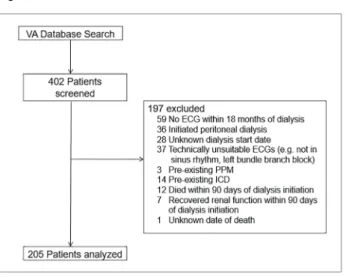
The initial search of the VA database yielded 402 pa-tients. After exclusion criteria were applied, 205 sub-jects remained. The major reasons for exclusion were the absence of an acceptable ECG within 18 months after starting dialysis (N = 59), use of peritoneal dialy-sis (N = 36), and technically unsuitable ECG (N = 37) (Figure 2). Of the 205 that remained, 94 were found to have a prolonged TpTe, while 61 had a prolonged TpTe/QT.
BASELINECHARACTERISTICS
Of the 205 identified patients, 99.5% were male, 66.8% were Caucasian and the mean age was 66.6 +/-12.3 years (Table 1). The mean duration on dialy-sis prior to the first ECG being obtained was 104 +/- 11.7 days.
NORMAL VS. PROLONGED TPTE
Figure 1. Pictorial representation illustrating how TpTe was calculated.
Figure 2. Patient selection.
failure or other comorbidities (Table 1). The QRS du-ration was significantly longer in the prolonged TpTe group compared to normal TpTe (98 ms vs. 90 ms, p = 0.01). There was no statistical difference betwe-en normal or prolonged TpTe groups with regards to any other ECG parameter evaluated.
NORMAL VS. PROLONGED TPTE/QT
There was no statistically significant difference be-tween normal and prolonged TpTe/QT patients in any demographic or co-morbid condition (Table 1). In contrast to the TpTe comparisons, there was no difference in racial category distribution between prolonged TpTe/QT and normal TpTe/QT groups. The QRS duration was significantly longer in the pro-longed TpTe/QT group (98 ms propro-longed vs. 92 ms normal, p = 0.046) but no other ECG parameter was significantly different between the two groups.
OUTCOMES - NORMAL VS. PROLONGED TPTE
Subjects were followed for a mean of 3.5 years. The mean survival times for patients with a normal and
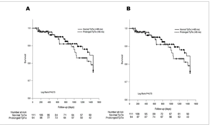
prolonged TpTe interval after dialysis initiation were 2.91 and 2.83 years, respectively (Figure 3A). No sig-nificant difference was observed in the rates of SCD or all-cause mortality between patients with a prolon-ged TpTe compared to a normal interval (Figures 3a and 3b). All-cause mortality in patients with a prolon-ged TpTe vs. normal was 72.3 vs. 68.5% (p = 0.76). SCD in patients with prolonged TpTe vs. normal was 16.0 vs. 14.4% (p = 0.52).
OUTCOMES - NORMAL VS. PROLONGED TPTE/QT
The median survival time for patients with a normal and prolonged TpTe/QT interval was 2.94 and 2.67 years, respectively. Once again, there was no statisti-cally significant difference seen in the rates of SCD or all-cause mortality between the normal and prolon-ged TpTe/QT groups. All-cause mortality was 68.8 vs. 70.8% in patients with prolonged compared to normal TpTe/QT (p = 0.26) (Figure 4A). SCD was present in 13.1 vs. 16% in patients with prolonged TpTe/QT compared to normal TpTe/QT (p = 0.51) (Figure 4B).
CAUSEOFDEATH
No statistical difference was found in cause of death between patients with normal and prolonged TpTe (Table 2). However, there was a trend of increased mortality from infection in patients with normal TpTe (p = 0.09). No significant difference in cause of death was observed when comparing normal and prolonged TpTe/QT groups.
D
ISCUSSIONAll (n = 205)
Normal TpTe (n = 111)
Prolonged TpTe (n = 94)
p value (Normal
vs.
Prolonged TpTe)
Normal TpTe/QT (n = 144)
Prolonged TpTe/QT
(n = 61)
p value (Normal
vs. Prolonged
TpTe/QT)
Age, years 66.6 ± 12.3 65.9 ± 12.4 67.3 ± 12.2 0.40 65.7 + 1.1 67.5 + 1.5 0.36
Gender, males (%) 99.5 100 98.9 0.93 100 98.4 0.66
Time to ECG, days 104 ± 11.7 95 ± 11.9 115 ± 11.4 0.069 94 + 9.6 128 + 15.3 0.055
Race, n (%)
White 137 (66.8) 66 (59.5) 71 (75.5) 0.022 93 (64.6) 44 (72.1) 0.58
African American 61 (29.8) 41 (36.9) 20 (21.3) 0.18 45 (31.3) 15 (24.6) 0.55
Cause of ESRD, n (%)
Diabetic nephropathy 94 (45.9) 51 (45.9) 43 (45.7) 64 (44.4) 30 (49.2)
Hypertension 24 (11.7) 15 (13.5) 9 (9.6) 18 (12.5) 6 (9.8)
Glomerular disease 22 (10.7) 11 (9.9) 11 (11.7) 17 (11.8) 5 (8.2)
Acute kidney injury 15 (7.3) 10 (9) 5 (5.3) 12 (8.3) 3 (5)
Obstruction 8 (3.9) 2 (1.8) 6 (6.4) 4 (2.8) 4 (4.9)
Ischemic nephropathy 5 (2.4) 5 (4.5) 0 (0) 5 (3.5) 0 (0)
Polycystic kidney 5 (2.4) 3 (2.7) 2 (2.1) 3 (2.1) 2 (3.3)
Unknown/Other 32 (15.6) 14 (12.6) 18 (19.2) 21 (14.6) 11 (18)
ECG (mean, 95% CI)
PR interval 170
(152, 194)
168
(151, 194) 0.99
172 (152,194)
166
(148,194) 0.47
QRS duration, ms 90
(84, 102)
98
(89.5, 112) 0.01
92
(84, 102) 98 (90, 114) 0.05
Left
ventricularhypertrophy, n (%)
8 (7.2) 12 (12.8) 0.27 12 (8.3) 8 (13.1) 0.41
Fascicular Block 9 (8.1) 9 (9.6) 0.9 3 (8.1) 5 (8.2) 1
RBBB 12 (10.8) 13 (13.8) 0.66 3 (8.1) 5 (8.2) 1
QTc interval, ms 451
(426, 470)
453
(434, 476) 0.3
447 (426, 473)
457
(434, 475) 0.2
TpTe interval, ms 71.3
(56.3, 80.5)
103.5
(93.8, 120) < 0.001 n/a n/a
TpTe/QTc interval, ms n/a n/a 0.18
(0.15, 0.21)
0.28
(0.26, 0.32) < 0.001
Comorbidities n (%)
Hypertension 182 (88.8) 97 (87.4) 85 (90.4) 0.64 128 (88.9) 54 (88.5) 0.87
Diabetes 135 (65.9) 74 (66.7) 61 (64.9) 0.91 96 (66.7) 39 (63.9) 0.83 CAD 87 (42.4) 46 (41.4) 41 (43.6) 0.86 63 (43.8) 24 (39.3) 0.67
CHF 80 (39.0) 37 (33.3) 43 (45.7) 0.09 54(37.5) 26 (42.6) 0.6
BMI (Mean, 95% CI) 27.4
(24.8, 32.3)
28.9
(24.3, 32.3) 0.54
28.2 (25.1, 32.3)
28.4
(23.9, 32) 0.75 TABLE 1 BASELINEDEMOGRAPHICS. DATAAREREPORTEDASMEAN ± SD UNLESSSTATEDOTHERWISE
Figure 3. Overall survival (A) and survival without sudden cardiac death (B) between hemodialysis patients with normal and prolonged TpTe interval.
Figure 4. Overall survival (A) and survival without sudden cardiac death (B) between hemodialysis patients with normal and prolonged TpTe/QT interval.
dialysis was not associated with a statistically signifi-cant increase in SCD or all-cause mortality.
It is well established that there is a high incidence of cardiovascular morbidity and mortality in the ESRD population.1,18 Unfortunately, traditional risk factors
for cardiovascular (CV) disease, which are ubiquitous among patients with ESRD, provide little predictive discrimination for those at higher risk of developing CV events, especially SCD.5-7 Traditional markers
Cause of Death
All (N = 205)
Normal TpTe (N = 111)
Prolonged
TpTe
(N = 94)
p value (Normal
vs. Prolonged
TpTe)
Normal TpTe/QT (N = 144)
Prolonged TpTe/QT
(N = 61)
p value (Normal
vs. Prolonged
TpTe/QT)
p value (Prolonged
TpTe vs Prolonged
TpTe/QT)
SCD, n (%) 31 (15.1) 16 (14.4) 15 (15.9) 0.52 23 (16) 8 (13.1) 0.51 0.94
Infection, n (%) 22 (10.7) 14 (12.6) 8 (8.5) 0.09 18 (12.5) 4 (6.5) 0.88 0.87
Non-SCD
cardiac, n (%) 17 (8.2) 10 (9.0) 7 (7.4) 0.28 12 (8.3) 5 (8.1) 0.82 0.65
Unknown, n (%) 18 (8.7) 11 (9.9) 7 (7.4) 0.19 12 (8.3) 6 (9.8) 0.54 0.42
Other, n (%) 56 (27.3) 25 (22.5) 31 (32.9) 0.86 37 (25.6) 19 (31.1) 0.13 0.70
All-cause
Mortality, n (%) 144 (70.2) 76 (68.5) 68 (72.3) 0.76 102 (70.8) 42 (68.8) 0.41 0.26 TABLE 2 CAUSESOFDEATH
SCD: sudden cardiac death; TpTe: T peak to T end interval; TpTe/QT: TpTe interval corrected for the QT interval.
may not be specific enough to influence adequate risk reduction measures in regard to SCD in patients with ESRD. The majority of SCD cases are presumed to be related to ventricular arrhythmias, although the pos-sibility of PEA/asystole or bradycardic arrest is not ru-led out in analyses focusing on baseline ECG parame-ters, as ours. Patients with ESRD, especially those on hemodialysis, are chronically exposed to homeostatic changes that could perturb the electrical conductance of the myocardium. Such changes should readily be evident in a resting ECG but investigating the predic-tive ability of ECG findings in ESRD patients is not novel. In a sub analysis of the German diabetic dialy-sis study (4D), 9 ECG parameters were examined for their ability to predict mortality in ESRD patients.19
The authors found that the only ECG parameter pre-dictive of increased mortality was the absence of si-nus rhythm, while signs of MI, heart rate, QRS axis, AV block, complete LBBB or RBBB, and QT interval had no significant association with outcomes. Several possibilities might explain these findings. First, struc-tural defects are perhaps more important than con-duction abnormalities in the genesis of SCD in ESRD. Second, uremia and the subsequent CV changes with the additional stress of the dialysis procedure are the primary determinants of SCD, and thus indifferent to the underlying electrical and structural defects of the heart. Third, routine ECG measurements are not specific enough to the high risk conduction abnorma-lities in the highly altered environment of ESRD. Our investigation sought to examine this last possibility. We examined the predictive ability of TpTe, an ECG marker of arrhythmogenesis that has been shown to be predictive of SCD in other populations.
It is important to first understand the basis for the TpTe measurement to understand its significan-ce. The TpTe interval represents the speed of the dis-persion of the repolarization potential from the epi-myocardium to the endoepi-myocardium.20,21 A delay in
this interval allows the possibility of pre-excitation and induction of arrhythmia.22 This measure has
be-en demonstrated to predict non-sustained vbe-entricular tachycardia post-cardiac resynchronization and ICD firing in patients requiring the placement of Bi-V pacing and ICD, as well as predict overall mortality and VT/VF in patients with systolic dysfunction and ICD implantation for primary prevention.23,24 While
a difference in electric potentials (i.e. dispersion) be-tween cell lines will always be present, increases in the dispersion have been linked to worse outcomes in disease states.14-16,25 One explanation as to why TpTe
may be prolonged in ESRD patients is the presence of an increase in myocardial fibrosis in this population. Fibrosis can lead to heterogeneous zones of repolari-zation within the myocardium, which can induce ven-tricular arrhythmias.26
Our study failed to demonstrate a significant as-sociation between prolonged TpTe and outcomes in our ESRD study population, despite there being more events numerically (all-cause mortality and SCD) in the prolonged interval groups. Some studies showed improved precision in predicted SDC when TpTe is adjusted for heart rate.27 Another possible
single hemodialysis session.17 Prolongation of TpTe
or TpTe/QT was not associated with changes in electrolytes, which suggest that the changes in TpTe were not related to the large electrolyte shifts that routinely occur in patients receiving hemodialysis. Other authors have examined the effect of hemo-dialysis on QT dispersion, which is another marker of the dispersion of ventricular repolarization. They also found that QT dispersion was increased after HD sessions.28 Additionally, other authors found
that high frequency QRS duration was significantly increased after HD sessions, further bolstering the premise that dialysis itself alters ECG parameters.29
Nevertheless, such effects should, in theory, have more effect on those with pre-existent abnormalities in the conduction parameters. The question then be-comes: are these changes consistent risk parameters over time? It is possible that uremic control over time with dialysis may change these parameters: some patients who started with a prolonged TpTe or TpTe/QT may have improvement over time, or all patients may develop a prolonged TpTe or pro-longed TpTe/QT thereby eliminating any potential predictive ability over time. ECG changes over the course of time on hemodialysis or peritoneal dialysis may be more important in the evolving CV mortality related to ESRD. Thus, our study may not have been able to detect a significant effect on mortality wi-thout following the TpTe duration throughout many dialysis treatments. Perhaps, the amount by which TpTe changes over time, and not an absolute value, could more accurately predict mortality. Others have suggested that single markers like Tpte will not be effective as a predictive measure as a combination of many simultaneous ECG parameters.7,30
There was a higher proportion of African Americans within the normal TpTe group compared to prolonged TpTe group. Racial differences on stan-dard ECG parameters have been previously shown.31
In fact, a recent study as part of the Women’s Health Initiative found the upper limit of normal for TpTe to be 10 ms longer in African American women com-pared to Caucasian, Hispanic, and Asian women.32
While that study looked only at women, it seems to contradict our findings that African American men are more likely to have a shorter TpTe interval. Further investigations into the importance of ethnici-ty and gender on these ECG parameters are needed.
There are several limitations in this study. Due to the way we defined baseline ECG, we cannot rule out a direct effect of hemodialysis duration on TpTe; however, restriction of the population to those who had an ECG within 60 days of initia-ting dialysis demonstrated similar results with no significant difference in mortality or SCD at 5 ye-ars following initiation of hemodialysis (data not shown). We did not collect data on the reason for obtaining the ECG (i.e. routine check vs. suspicion for an acute event, e.g. pre-operative clearance vs. myocardial infarction); this might have introduced a selection bias. We also were unable to obtain in-formation on medication use at baseline, which is important as certain medications have been shown to affect the TpTe interval. Another limitation is that our observed SCD event rate was lower than what has been previously reported. The SCD rate in the normal TpTe group at 4 years was 14.4% compared to the 19% rate that was used in the a priori power analysis and may have led to a loss of power. Another item worth mentioning is that almost 15% of the patients screened were exclu-ded because they did not have an ECG within 18 months of dialysis initiation. Given that these pa-tients are at high risk for adverse cardiac events and likely would benefit from a baseline ECG at dialysis initiation, this should be an area of focus for nephrologists and cardiologists. This also re-sulted in a relatively small population with limited follow-up time. Lastly, it should be noted that this study represents a Virginia (USA) population that is predominantly male and Caucasian, which limits the generalizability of these findings.
C
ONCLUSIONWe hypothesized that in an ESRD population the presen-ce of a prolonged TpTe or TpTe/QT segment would be predictive of increased all-cause mortality and/or SCD. Our study was unable to show a statistically significant difference in event rates between groups with prolonged and normal TpTe or TpTe/QT segments.
A
CKNOWLEDGEMENTSHealth Services Research and Development, VA Information Resource Center (Project Numbers SDR 02-237 and 98-004).
This research did not receive any specific grant from funding agencies in the public, commercial, or not-for-profit sectors.
R
EFERENCES1. Collins AJ, Foley RN, Chavers B, Gilbertson D, Herzog C, Ishani A, et al. US Renal Data System 2013 Annual Data Re-port. Am J Kidney Dis 2014;63:A7.
2. Bernard SA, Gray TW, Buist MD, Jones BM, Silvester W, Gut-teridge G, et al. Treatment of comatose survivors of out-of--hospital cardiac arrest with induced hypothermia. N Engl J Med 2002;346:557-63.
3. European Heart Rhythm Association; Heart Rhythm Society, Zipes DP, Camm AJ, Borggrefe M, Buxton AE, Chaitman B, Fromer M, et al.; American College of Cardiology; American Heart Association Task Force; European Society of Cardiology Committee for Practice Guidelines. ACC/AHA/ESC 2006 gui-delines for management of patients with ventricular arrhyth-mias and the prevention of sudden cardiac death: a report of the American College of Cardiology/American Heart Associa-tion Task Force and the European Society of Cardiology Com-mittee for Practice Guidelines (Writing ComCom-mittee to Develop Guidelines for Management of Patients With Ventricular Ar-rhythmias and the Prevention of Sudden Cardiac Death). J Am Coll Cardiol 2006;48:e247-346.
4. Moss AJ, Zareba W, Hall WJ, Klein H, Wilber DJ, Cannom DS, et al.; Multicenter Automatic Defibrillator Implantation Trial II Investigators. Prophylactic implantation of a defibrilla-tor in patients with myocardial infarction and reduced ejection fraction. N Engl J Med 2002;346:877-83.
5. Bleyer AJ, Hartman J, Brannon PC, Reeves-Daniel A, Satko SG, Russell G. Characteristics of sudden death in hemodialysis pa-tients. Kidney Int 2006;69:2268-73.
6. Whitman IR, Feldman HI, Deo R. CKD and sudden cardiac death: epidemiology, mechanisms, and therapeutic approaches. J Am Soc Nephrol 2012;23:1929-39.
7. Waks JW, Tereshchenko LG, Parekh RS. Electrocardiographic pre-dictors of mortality and sudden cardiac death in patients with end stage renal disease on hemodialysis. J Electrocardiol 2016;49:848-54. 8. Tracy CM, Epstein AE, Darbar D, DiMarco JP, Dunbar SB, Es-tes NA 3rd, et al.; American College of Cardiology Foundation; American Heart Association Task Force on Practice Guidelines; Heart Rhythm Society. 2012 ACCF/AHA/HRS focused upda-te of the 2008 guidelines for device-based therapy of cardiac rhythm abnormalities: a report of the American College of Car-diology Foundation/American Heart Association Task Force on Practice Guidelines and the Heart Rhythm Society. [correc-ted]. Circulation 2012;126:1784-800.
9. Pun PH, Lehrich RW, Honeycutt EF, Herzog CA, Middleton JP. Modifiable risk factors associated with sudden cardiac ar-rest within hemodialysis clinics. Kidney Int 2011;79:218-27. 10. Deo R, Shou H, Soliman EZ, Yang W, Arkin JM, Zhang X,
et al. Electrocardiographic Measures and Prediction of Cardio-vascular and NoncardioCardio-vascular Death in CKD. J Am Soc Ne-phrol 2016;27:559-69.
11. Tereshchenko LG, Kim ED, Oehler A, Meoni LA, Ghafoori E, Rami T, et al. Electrophysiologic Substrate and Risk of Mortality in Incident Hemodialysis. J Am Soc Nephrol 2016;27:3413-20. 12. Guney M, Ozkok A, Caliskan Y, Pusuroglu H, Yazici H, Tepe
S, et al. QT dispersion predicts mortality and correlates with both coronary artery calcification and atherosclerosis in hemo-dialysis patients. Int Urol Nephrol 2014;46:599-605.
13. Antzelevitch C. Role of spatial dispersion of repolarization in inherited and acquired sudden cardiac death syndromes. Am J Physiol Heart Circ Physiol 2007;293:H2024-38.
14. Panikkath R, Reinier K, Uy-Evanado A, Teodorescu C, Hatte-nhauer J, Mariani R, et al. Prolonged Tpeak-to-tend interval on the resting ECG is associated with increased risk of sudden cardiac death. Circ Arrhythm Electrophysiol 2011;4:441-7. 15. Shimizu M, Ino H, Okeie K, Yamaguchi M, Nagata M,
Hayashi K, et al. T-peak to T-end interval may be a better predictor of high-risk patients with hypertrophic cardiomyo-pathy associated with a cardiac troponin I mutation than QT dispersion. Clin Cardiol 2002;25:335-9.
16. Haarmark C, Hansen PR, Vedel-Larsen E, Pedersen SH, Graff C, Andersen MP, et al. The prognostic value of the Tpeak--Tend interval in patients undergoing primary percutaneous coronary intervention for ST-segment elevation myocardial infarction. J Electrocardiol 2009;42:555-60.
17. Kalantzi K, Gouva C, Letsas KP, Vlachopanou A, Foulidis V, Bechlioulis A, et al. The impact of hemodialysis on the disper-sion of ventricular repolarization. Pacing Clin Electrophysiol 2013;36:322-7.
18. Saran R, Li Y, Robinson B, Ayanian J, Balkrishnan R, Bragg--Gresham J, et al. US Renal Data System 2014 Annual Data Report: Epidemiology of Kidney Disease in the United States. Am J Kidney Dis 2015;66:Svii, S1-305. Erratum in: Am J Kid-ney Dis 2015;66:545.
19. Krane V, Heinrich F, Meesmann M, Olschewski M, Lilienthal J, Angermann C, et al.; German Diabetes and Dialysis Study Investigators. Electrocardiography and outcome in patients with diabetes mellitus on maintenance hemodialysis. Clin J Am Soc Nephrol 2009;4:394-400.
20. Antzelevitch C, Sicouri S, Di Diego JM, Burashnikov A, Viskin S, Shimizu W, et al. Does Tpeak-Tend provide an in-dex of transmural dispersion of repolarization? Heart Rhythm 2007;4:1114-6; author reply 1116-9.
21. Antzelevitch C, Yan GX, Shimizu W. Transmural dispersion of repolarization and arrhythmogenicity: the Brugada syndrome versus the long QT syndrome. J Electrocardiol 1999;32:158-65.
22. Akar FG, Yan GX, Antzelevitch C, Rosenbaum DS. Unique topographical distribution of M cells underlies reentrant me-chanism of torsade de pointes in the long-QT syndrome. Cir-culation 2002;105:1247-53.
23. Barbhaiya C, Po JR, Hanon S, Schweitzer P. Tpeak - Tend and Tpeak - Tend /QT ratio as markers of ventricular arrhythmia risk in cardiac resynchronization therapy patients. Pacing Clin Electrophysiol 2013;36:103-8.
24. Rosenthal TM, Stahls PF 3rd, Abi Samra FM, Bernard ML, Khatib S, Polin GM, et al. T-peak to T-end interval for predic-tion of ventricular tachyarrhythmia and mortality in a prima-ry prevention population with systolic cardiomyopathy. Heart Rhythm 2015;12:1789-97.
25. Topilski I, Rogowski O, Rosso R, Justo D, Copperman Y, Glikson M, et al. The morphology of the QT interval predicts torsade de pointes during acquired bradyarrhythmias. J Am Coll Cardiol 2007;49:320-8.
26. Roes SD, Borleffs CJ, van der Geest RJ, Westenberg JJ, Mar-san NA, Kaandorp TA, et al. Infarct tissue heterogeneity as-sessed with contrast-enhanced MRI predicts spontaneous ven-tricular arrhythmia in patients with ischemic cardiomyopathy and implantable cardioverter-defibrillator. Circ Cardiovasc Imaging 2009;2:183-90.
27. Chua KC, Rusinaru C, Reinier K, Uy-Evanado A, Chugh H, Gunson K, et al. Tpeak-to-Tend interval corrected for heart rate: A more precise measure of increased sudden death risk? Heart Rhythm 2016;13:2181-5.
29. Morales MA, Gremigni C, Dattolo P, Piacenti M, Cerrai T, Fazi A, et al. Signal-averaged ECG abnormalities in haemo-dialysis patients. Role of haemo-dialysis. Nephrol Dial Transplant 1998;13:668-73.
30. Abdelghani SA, Rosenthal TM, Morin DP. Surface Electro-cardiogram Predictors of Sudden Cardiac Arrest. Ochsner J 2016;16:280-9.
31. Macfarlane PW, Katibi IA, Hamde ST, Singh D, Clark E, De-vine B, et al. Racial differences in the ECG--selected aspects. J Electrocardiol 2014;47:809-14.